Primary thyroid lymphomas are not commonly recog- nized as being a significant subclass of thyroid neo- plasm. This case report presents 3 rare cases of a prima- ry B-cell lymphoma and we feature the various ultra- sonographic (US) imaging findings. The US findings of thyroid lymphoma have previously been reported to be extremely hypoechoic masses when there is diffuse bi- lateral involvment, and a homogenous low echoic mass
in solitary nodules (1). We present here the different US features of pathologically confirmed B-cell lymphoma of a nodular type, a cystic type, and a diffusely enlarged type with the clinical histories.
Case Reports
We retrospectively reviewed the imaging features of pathologically confirmed thyroid lymphoma in three fe- male patients. Ultrasound guided fine needle aspiration (US-FNA) was performed in all 3 patients. For final pathologic confirmation, two patients underwent surgi- cal excision and one patient underwent US-guided core needle biopsy (US-CNB) with using a 16-gauge cutting needle (Stericut). The results of the biopsy findings and pathological findings with the summarized US features
Primary B-cell Lymphoma of the Thyroid Featuring the Different Ultrasonographic Findings
1Ji Na Kim, M.D., Yoon Jung Choi, M.D., Dong Hoon Kim, M.D.2
1Department of Radiology, Kangbuk Samsung Hospital, Sungkyunkwan University School of Medicine
2Department of Pathology, Kangbuk Samsung Hospital, Sungkyunkwan University School of Medicine
Received November 17, 2008 ; Accepted February 19, 2009
Address reprint requests to : Yoon Jung Choi, M.D., Department of Radiology, Kangbuk Samsung Hospital, 108, Pyung-dong, Jongno-gu, Seoul 110-746, Korea.
Tel. 82-2-2001-1031 Fax. 82-2-2001-1030 E-mail: [email protected]
We review here 3 cases of primary thyroid lymphoma that we experienced during the past 5 years (age range: 39-55, all of the patients were female). The clinical and various ultrasonographic characteristics together with the other imaging modalities of primary thyroid lymphomas are described. The clinical features at presentation for one patient were a goiter with rapid growth and this was accompanied by compressive symptoms. The tumors of the other 2 patients were incidentally found during screen- ing thyroid ultrasound exams. The pathologic studies of 2 cases showed a diffuse B-cell lymphoma with associated Hashimoto’s thyroiditis and one case was a B-cell lym- phoma of the MALT type. An extra-thyroid extension was shown in one case. The treatments included surgery alone for two cases, and chemotherapy and radiation therapy for one case. A US exam of thyroid lymphoma can show various morphologi- cal features, and US-CNB is helpful for diagnosing thyroid lymphoma.
Index words :Core biopsy Lymphoma, B-cell Ultrasonography Thyroid neoplasm
are listed in table 1.
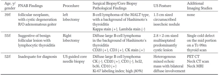
The first patient was a 39-year-old woman who had an incidentally found thyroid nodule when undergoing a medical screening health examination. US showed a 1.5 cm sized circumscribed isoechoic mass in the right thy- roid (Fig. 1). The second woman was a 55-year-old fe- male patient who was referred to the Endocrine Department from a local clinic for further evaluation of incidentally found nodules in the thyroid gland. Her US showed partially ill-defined, multi-septated predomi- nantly cystic nodules (Fig. 2). The incidentally detected nodules were initially thought to be follicular neoplasm and benign cystic nodule. The FNA findings were non- specific, so these two patients underwent surgical exci- sion and the pathology revealed B-cell lymphoma. They were treated with adjuvant chemotherapy and further imaging studies revealed no other systemic involve-
ment. They had no previous significant medical histo- ries or family histories of thyroid problems.
The third 52-year-old female patient had developed mild hoarseness for 3 months and a palpable neck mass for 1 year prior to undergoing US imaging. The patient was originally treated with antibiotics for one month without improvement. She also had moderate dyspha- gia to solid food while her hoarseness continued to worsen and the neck mass became larger. US of the thy- roid showed a diffusely enlarged thyroid with heteroge- nously mixed echogenicity together with a non-visual- ized infrathyroidal portion due to the small window.
The bilateral thyroid lobules were replaced by a hetero- geneous mass that extended from the level of the thy- roid cartilage to the thoracic inlet and the mass was wrapped posteriorly around the trachea (Fig. 3). Due to the inadequate specimen on thyroid FNA, US-CNB was
Table 1. The FNAB and Pathological Results with the Summarized US Features of Thyroid Lymphoma Age, y/
FNAB Findings Procedure Surgical Biopsy/Core Biopsy
US Feature Additional
gender Pathological Findings Imaging Studies
39/f follicular neoplasm, left B-cell lymphoma of the MALT type, 1.5 cm sized none with cystic degeneration lobectomy with a background of Hashimoto’s circumscribed
R/O adenomatous goiter thyroiditis isoechoic nodule
Kappa stain (+), Lambda stain (-)
55/f Suggestive of benign Right Diffuse large B-cell lymphoma 2.8×2 cm sized Single cold defect follicular lesion with lobectomy in the background of Hashimoto’s multiseptated on the mid portion
lymphocytic thyroiditis thyroiditis predominantly on a Tc-99m
CD20 (+), CD3 (+), CK stain (+) cystic lesion thyroid scan 52/f Inadequate for diagnosis US guided core Diffuse large B cell lymphoma Heterogenous PET CT
needle biopsy CK (-), CD20 (+), CD3 (-), bcl2, mixed echoic Neck CT scan bcl6, CD10 (+) mass with bilateral Neck MRI Ki-67 labeling index; high (80%) diffuse involvement
A B
Fig. 1. Screening ultrasound of the thyroid of a 39-year-old woman demonstrated a 1.5 cm sized circumscribed oval isoechoic nod- ule on the left thyroid gland on the transverse sonogram (arrows, A). The cytology results of this nodule confirmed follicular neo- plasm with cystic degeneration, which led to surgical excision and the pathology confirmed B-cell lymphoma of the MALT type (B), with a background of Hashimoto’s thyroiditis.
performed to rule out thyroid malignancy and the mass was diagnosed as diffuse B-cell lymphoma. She was treated with chemotherapy (cyclophosphamide, mitox- antrone, vincristine and prednisone) and radiation ther- apy without any further surgical procedures.
Discussion
The US features of thyroid lymphoma have been re- ported to be homogenous low echogenecity in the cases with solitary nodules and extremely hypoechoic masses intermingled with echogenic structures in the cases of diffusely involved lymphoma with underlying thyroidi- tis (2, 3). In our case, when thyroid lymphoma appeared as a focal nodule, it can also appear as an isoechoic solid nodule and it can show a cystic appearance. When thy- roid lymphoma appears as the diffuse heterogenous
type, the echogenicity can be variable due to the in- volvement of thyroiditis. Ota et al. reported on 79 cases of pathology confirmed thyroid lymphoma and they classified the US patterns into three types, which are the nodular type, the diffuse type and the mixed type based on the internal echoes, the borders and the posterior echoes (3). By using their classification system, one of our cases can be classified into the nodular type of lym- phoma, where the hypoechoic nodule was limited to one lobe and it resembled a follicular tumor or an ade- nomatous nodule (Fig. 1) with the FANB findings show- ing a follicular neoplasm. One of our cases showed find- ings like that of the pseudocystic type, where the nodule is solid, but it appears cystic on US (Fig. 2). A pathologic confirmation is always necessary because the imaging findings are not specific enough to differentiate thyroid lymphoma from other malignancies. For the diffuse
A B
C D
Fig. 2. Transverse sonogram of the right thyroid gland shows a partially ill defined pseudocystic septated hypoechoic mass (arrows, A). Color Doppler showed faintly increased power Doppler flow on the periphery of the nodule (arrows, B). On the technetium 99 m thyroid scan, a cold defect was visualized on the lower pole of the right thyroid (arrow, C). The surgical specimen confirmed dif- fuse large B-cell lymphoma in the background of Hashimoto’s thyroiditis; the tumor measured 4.5×3×1.5 cm, and it was a solid nodule. The cut surface shows a gray-tan fish-flesh like appearance and no area of the cystic portion was demonstrated on the gross specimen (D).
type, Ota et al. suggested that enhancement of the poste- rior echoes can be used as a US profile for findings that are suspicious of lymphoma when all the other clinical
findings are supportive (3). But in our case, it was diffi- cult to differentiate the diffuse type lymphoma from se- vere thyroiditis and multi nodular goiter with internal
A B C
D E
H
F G
Fig. 3. Transverse scan of a 52-year-old woman who had a progressively enlarging mass for the previous 1 year with a feeling of dysphagia and voice change, and the scan demonstrated heterogenous mixed echoic thyroid parenchyma with diffuse bilateral en- largements (A, B and C are the right lobe, isthmus and left lobe, respectively). The neck CT scan (D is the axial image and E is the coronal image) and the MRI (F is the Gd-enhanced T1 weighted axial image and G is the coronal image) demonstrated the relation- ship of the infrathyroidal extension and the displacement of the trachea, esophagus and bilateral carotid complex. PET CT demon- strated a bilateral enlarged huge hypermetabolic mass (p-SUV = 37.6) that extended to the aortic arch level of the superior medi- astinum (H). The mass was compressing the bilateral common carotid arteries, the bilateral internal jugular veins, the trachea, esophagus and the SVC. Also, distant metastasis was visualized on the right posterior lower neck, the right liver S6, the right humerus head, the left iliac wing and the jejunum on the lower upper quadrant of the abdomen and the right lower quadrant of the ileum.
hemorrhage.
There is controversy in the previously published pa- pers as to whether making a reliable diagnosis of thyroid lymphoma is possible on the basis of a FNA. None of our cases showed that FNA result was sufficient to diag- nose the lymphoma even with performing immuno- chemical staining. The samples demonstrated scanty cells with poor morphology, and it was difficult to dis- tinguish a lymphoma from the lymphoid infiltrate found in Hashimoto’s thyroiditis.
US guided core biopsy is not used as a routine diagnos- tic tool for thyroid nodules, but for the cases that are in- determinate on cytology, it can be considered as an al- ternative biopsy method because the role of surgery is limited for treating thyroid lymphoma (4). In our case, the patient referred for CNB showed sufficient tissue for the pathological confirmation and it was performed without any complication with accurate and satisfactory
results. In conclusion, our cases show that US shows various different morphological features for primary thyroid lymphoma and in one case, the US-CNB was helpful for diagnosing thyroid lymphoma.
References
1. Derringer GA, Thompson LD, Frommelt RA, Bijwaard KE, Heffess CS, Abbondanzo SL. Malignant lymphoma of the thyroid gland: a clinicopathologic study of 108 cases. Am J Surg Pathol 2000;24:623-639
2. Takashima S, Morimoto S, Ikezoe J, Arisawa J, Hamada S, Ikeda H, et al. Primary thyroid lymphoma: comparison of CT and US as- sessment. Radiology 1989;171:439-443
3. Ota H, Ito Y, Matsuzuka F, Kuma S, Fukata S, Morita S, et al.
Usefulness of ultrasonography for diagnosis of malignant lym- phoma of the thyroid. Thyroid 2006;16:983-987
4. Kwak JY, Kim EK, Ko KH, Yang WI, Kim MJ, Son EJ, et al.
Primary thyroid lymphoma: role of ultrasound-guided needle biopsy. J Ultrasound Med 2007;26:1761-1765
대한영상의학회지 2009;60:391-395
갑상선의 원발성 악성 임파종의 영상학적 소견: 증례 보고1
1성균관대학교 의과대학 강북삼성병원 영상의학과
2성균관대학교 의과대학 강북삼성병원 병리과
김지나∙최윤정∙김동훈2
갑상선의 원발성 악성 임파종은 매우 드문 악성 종양이다. 최근 5년 동안 저자 등이 원발성 악성 임파종 3예 (나이 39-55, 여성) 를 임상적 소견과, 초음파를 포함한 영상소견, 초음파 유도하 총생검의 진단적 유용성을 보고하는 바 이다. 한 명의 환자는 빠르게 커지는 경부 종괴와 이로 인한 압박증상을 보였으나 다른 두 명은 선별검사에서 우연히 발견된 갑상선 결절을 주소로 내원하였다. 조직학적 소견에서 2예는 Hashimoto’s thyroditis 에 동반된 diffuse B-cell lymphoma 로 진단되었고 나머지 한 예는 B-cell lymphoma MALT type 으로 진단되었다. 갑상선 주위 로의 침윤은 한 예에서 관찰되었다. 두 환자는 수술만을 시행하였고, 다른 한 환자는 화학요법과 방사선치료를 병행 하였다.