pISSN 1598-9992 eISSN 2233-6869
CASE REPORT
위 내시경 점막 절제술 후 발생한 기흉
고명석, 장진석, 차재황
동아대학교병원 소화기내과
Pneumothorax Following Gastric Endoscopic Mucosal Resection
Myeongseok Koh, Jin Seok Jang and Jae Hwang Cha
Division of Gastroenterology, Department of Internal Medicine, Dong-A University Hospital, Busan, Korea
An endoscopic mucosal resection (EMR) is used widely as an alternative treatment to a surgical resection for early gastric neo- plastic lesions. Among the unusual complications of gastric EMR, perforation is usually manifested as a pneumoperitoneum. This paper reports a patient with a left-side pneumothorax without pneumoperitoneum as a complication of gastric EMR. The patient developed a left side pneumothorax after gastric EMR in the gastric fundus and recovered without further complications after conservative treatment, including endoscopic clipping. (Korean J Gastroenterol 2020;76:83-87)
Key Words: Endoscopic mucosal resection; Complication; Perforation; Pneumothorax
Received March 27, 2020. Revised May 19, 2020. Accepted May 25, 2020.
CC This is an open access article distributed under the terms of the Creative Commons Attribution Non-Commercial License (http://creativecommons.org/licenses/
by-nc/4.0) which permits unrestricted non-commercial use, distribution, and reproduction in any medium, provided the original work is properly cited.
Copyright © 2020. Korean Society of Gastroenterology.
교신저자: 차재황, 49201, 부산광역시 서구 대신공원로 26, 동아대학교병원 소화기내과
Correspondence to: Jae Hwang Cha, Division of Gastroenterology, Department of Internal Medicine, Dong-A University Hospital, 26 Daesingongwon-ro, Seo-gu, Busan 49201, Korea. Tel: +82-51-240-2798, Fax: +82-51-240-5070, E-mail: [email protected], ORCID: https://orcid.org/0000-0001-6297-102X
Financial support: None. Conflict of interest: None.
서 론
위에서 발생하는 악성 종양 중 림프절 전이와 관계없이 암 세포가 점막 또는 점막하층에 국한된 경우 조기 위암(early gastric cancer, EGC)으로 정의하며, 최근의 내시경 기술의 발전으로 내시경적 절제술(endoscopic resection, ER)은 EGC에 대한 최소 침습적인 치료로 인정받고 있다. 1974년 점막하 주입을 통해 병변을 융기시켜 절제하는 내시경적 점막 절제술(endoscopic mucosal resection, EMR)이 처음으로 소개되었고, 이후 점막하 박리를 통해 병변의 크기와 상관없 이 완전 절제가 가능한 내시경적 점막하 박리술(endoscopic submucosal dissection)이 소개되었다. 이러한 내시경적 절 제술 기법이 발전함에 따라 EGC에서 ER의 적응증이 점차 확대되고 있다.1
이러한 ER의 중요한 합병증으로는 출혈, 천공 등이 있으 며, 특히 천공은 중요한 합병증으로, 주로 기복증과 동반된
복막염으로 발견된다.2 ER 후 발생하는 기흉은 빈도를 파악하 기 힘들 정도로 드물고 일부 증례를 통해 보고되고 있으며, 대체로 기복증과 동반하여 기흉 발생이 보고되고 있다.3저자 들은 EMR 후 기복증 없이 좌측 기흉이 발생하여 내시경적 봉합 및 보존적 치료를 통해 회복한 증례를 경험하여 문헌고 찰과 함께 보고한다.
증 례
45세 여자가 위 선종으로 진단받고 내원하였다. 환자는 다 른 질환 및 수술 병력은 없었으며, 타 병원에서 건강검진을 통해 위 기저부(fundus of stomach) 부위의 단일 편평 미란 이 발견되었고 해당 부위에 시행한 조직 검사는 고도 이형성 관샘종으로 보고되었다. 시술 전 촬영한 복부 전산화단층촬영 에서 위벽의 비정상적인 비후, 주변 림프절의 이상 소견은 관 찰되지 않았다.
Fig. 1. Endoscopic finding of the patient before the procedure. The lesion (tubular adenoma, type IIa+IIc, 10×10 mm in diameter) was located at the fundus of the stomach.
A
A BB CC
Fig. 2. Endoscopic findings of the patient during the procedure. (A) After confirming the lesion boundary, marking was performed using an endoscopic dual knife. (B) After the endoscope equipped with the endoscopic band ligation equipment was inserted to allow the entire lesion to fit inside the rubber band, the lesion was completely ligated into the band after inhalation. (C) Resection was performed using a snare.
A B
Fig. 3. Endoscopic findings of the patient after the procedure. (A) No definite perforating hole was seen immediately after the endoscopic mucosal resection, but a deep muscular site was exposed, and there was bleeding from vascular exposure. (B) Endoscopic clipping was performed at the suspicious micro-perforated site.
입원 후 EMR이 시행되었다. 장경과 단경 약 10×10 mm의 표면함몰형 병변이 위 기저부에 위치해 있었다(Fig. 1). 시술은 내시경(EPK-i7010 with EG29-i10, Pentax Medical, Tokyo, Japan), 내시경 절개도(KD-650U, Olympus Medical, Tokyo, Japan), 올가미(CleaGrasp Snare, 15 mm, Finemedix, Daegu, Korea), 내시경 밴드 결찰술 장비(Steigmann-Goff® Bandito® Endoscopic Ligator, Conmed, New York, USA)를 사용해 진행되었다. 시술은 EMR-L method로 진행되었으며, 표지 후 생리식염수, 인디고칼민, 에피네프린 혼합액의 주입하여 병변 부 주변을 융기시키고, 내시경 밴드 결찰술 장비를 장착한 내시 경을 삽입하여 병변 전체가 고무밴드 안에 들어올 수 있도록 하여 흡입 후 병변을 완전히 밴드로 결찰하였다(Fig. 2A, B).
이후 선단부의 내시경 밴드 결찰술 장치를 탈착하고 올가미 로 밴드 결찰 하부를 잡은 후 절제하는 과정으로 진행되었다 (Fig 2C). 시술 직후 혈관 노출에 의한 출혈이 발생하였고, 미세천공이 의심되어 출혈 부위 및 천공이 의심되는 근육 노출 부위에 내시경 클립(Hx-110LR with HX-610-135s, Olympus
A
A BB CC
Fig. 4. Plain chest and abdominal radiographs. (A) Plain chest radiography performed immediately after the endoscopic mucosal resection (EMR). This panel shows the left-sided pneumothorax and no free air at both sub-diaphragm. (B) Plain abdominal radiography performed immediately after the EMR. This panel shows no gas within the peritoneal space. (C) Plain chest radiography taken the day after EMR. This photograph shows an improved pneumothorax without a chest tube.
A B
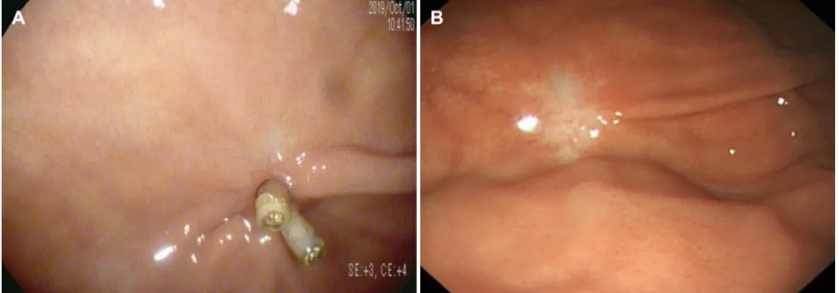
Fig. 5. Follow-up endoscopy findings. (A) The micro-perforated site had completely healed with a scar and two hemo-clips two months later.
(B) After six months, the post- endoscopic mucosal resection site area had completely healed with scars.
Medical)을 4회 시행하였다(Fig. 3). 시술은 약 27분간 시행되 었다.
시술 후 환자는 호흡곤란, 흉부 불편감, 복통 및 복부 팽만 감을 포함한 어떠한 증상도 호소하지 않았고, 활력징후는 혈 압 135/92 mmHg, 맥박 61회/분, 호흡 18회/분, 체온 36.7℃, 산소 포화도 96%로 안정적이었다. 신체검진상 청진 시 폐 좌 측의 호흡음 감소가 있었다. 증상 발생 후 시행한 단순 흉부 및 복부 방사선 촬영에서 좌측 기흉 소견이 발견되었고, 다른 이상 소견은 관찰되지 않았다(Fig. 4A, B). 즉시 비강을 통해 산소를 투여하고 흉부외과에 협진을 의뢰하였으며, 기흉의 양 이 적고 환자의 증상이 없으므로 흉관 삽관 없이 경과 관찰하 기로 결정하였다. 시술 당일 금식이 시행되었고, 3세대 세팔 로스포린 항생제 및 양성자 펌프 억제제 등이 정맥 내로 투여 되었다. 또한 동반된 복막염의 가능성을 고려하여 익일까지 금식을 처방하였다.
시술 1일 후 흉부 방사선 촬영을 시행하였으며, 좌측 기흉 의 소실 및 좌측 하부 폐야의 선형 무기폐 외 양측 폐실질은
정상 소견으로 기관지 확장증, 폐기종은 관찰되지 않았고 횡 격막의 저명한 결함은 없었다(Fig. 4C). 환자는 열, 복통 등의 추가 증상은 발생하지 않았고 경과가 양호하여 시술 3일 후 식이를 진행하였고 시술 5일 후 퇴원하였다.
절제된 조직 검사 결과는 고분화의 EGC로 크기는 5×4×0.3 mm 였으며, 침윤의 깊이는 점막하층을 100 μm 침범하였지만(stage pT1b) 절제면의 암종 침범은 없어 추가적인 조치 없이 경과 관찰을 결정하였다. 시술 2개월 후 외래에서 시행된 추적 내 시경상 위 기저부의 이전 EMR 병소에서 내시경 클립 2개가 관찰되고, 주변 조직은 반흔 변화되어 있으며, 6개월 후 추적 내시경상 반흔 변화 외 특이소견은 관찰되지 않았고, 국소 재 발의 징후는 관찰되지 않았다(Fig. 5).
고 찰
암의 조기 발견의 중요성이 널리 확산되고, 암 선별 검사에 대한 관심 증가와 내시경 기술의 발달로 인해 EGC의 발견이
증가하고 있다.4 수술적 절제 가능한 병기의 위암의 치료는 위 절제술과 림프절 절제술이 표준 치료이다. 내시경을 통한 절제술은 림프절 전이에 대한 정보를 파악할 수 없는 단점이 있으나, 절제된 수술 조직의 병리 검사 자료가 누적되면서 많 은 EGC가 림프절 전이를 동반하지 않음이 보고되었고,5,6 수 술 후 합병증과 이로 인한 삶의 질 감소로 인해 림프절 전이의 가능성이 낮은 일부의 EGC에 대해 내시경 치료가 시도되어 왔으며, EGC의 림프절 전이에 대한 자료 축적, 내시경 기기 및 술기의 발달로 인해 최근 내시경을 이용한 치료가 늘고 있는 추세이다.2,7
EGC에 대한 ER은 수술적 치료와 비교하여 대체로 시술 시간과 입원 기간이 짧고, 시술 후 위의 기능이 보존될 수 있 다는 장점이 있고 전체 생존율이 수술과 비슷하다.8 하지만 ER 후 드물게 출혈, 천공 등의 합병증이 발생할 수 있으며, 그중 천공은 복막염, 패혈증, 쇼크 그리고 사망에 이를 수 있 는 심각한 합병증이다.7,9 ER로 인한 천공은 내시경이나 시술 시 사용되는 절개도에 의한 고유근층의 직접 손상 혹은 올가 미에 의해 고유근층이 포획되어 병변부와 함께 절개됨으로 인 한 직접적, 물리적 손상을 입거나, 전기응고술 같은 추가 치료 적 시술 혹은 과도한 공기주입으로 인한 압력 손상에 의해 발생할 수 있다.10 문헌에 따라 차이가 있으나, 천공의 빈도는 EMR 후 약 0.5%가량으로 보고되며, 내시경적 점막하 박리술 후 3-6% 정도로 보고된다.11 종양의 위치(위 상체부, 근위부), 섬유화의 존재, 긴 시술 시간(2시간 이상), 고령(81세), 병변의 침습 깊이(점막근층 이하) 등의 위험인자들이 ER 후 천공 발 생과 연관이 있다는 보고가 있다.11위 천공은 전통적으로 수 술적 복원이 주된 치료 방법이었으나, ER 후 발생한 천공의 경우 내시경 클립을 적용하여 치료 성공한 결과들이 보고된 다.12 천공이 의심되면 내시경을 통한 공기 유입을 최소화 하 면서 명확한 시야를 확보하려고 노력해야 한다. 하지만 명확 한 시야를 위해 물로 병변을 세척하는 것은 복막염의 우려가 있으므로 주의한다.12 내시경 클립을 적용하여 병변부를 성공 적으로 봉합한 경우 대부분 금식, 정맥 내 항생제 및 양성자 펌프 억제제 투여를 포함한 보존적 치료를 통해 성공적으로 호전된다.10,12-14 위 내용물을 제거하고 복강 내로의 확산을 막 기 위해 비위관을 삽입 또한 고려할 수 있으나, 반드시 필요한 조치는 아니므로 경험에 따라 이러한 상황을 적절히 관리해야 한다.12 천공 환자에서 수술 혹은 내시경 치료의 선택은 천공 손상의 종류와 크기, 장관의 전처치 정도, 병변의 종류, 천공 후 진단까지 걸린 시간 그리고 환자의 임상적 상태 등을 고려 하여 신중히 결정되어야 한다. 만약 환자가 내시경 클립 적용 후에도 임상적 악화(발열, 복막 자극 징후의 지속)를 보일 경 우 외과 협진을 통해 응급 수술을 고려해야 한다.12,14,15 일반 적으로 EMR을 시행 받은 환자는 대개 24시간 동안 물, 미음
형태의 식이만 허용하며, 다음 3일가량 죽 형태의 식이가 허 용된다. 하지만 본 증례의 환자는 미세천공이 우려되어, 시술 3일 후 식이를 진행하게 되었다.12,16
위장의 ER 후 발생하는 기흉은 빈도를 파악하기 힘들 정도 로 드물고, 진단 및 치료적 대장 내시경, 내시경적 역행성 담 췌관 조영술(ERCP) 후 기흉이 발생한 일부 증례들이 보고되 고 있다.17,18 이러한 증례들은 대체로 기복증 또는 후복막공기 증과 동반하여 기흉 발생이 보고되고 있는데, 기흉 발생의 기 전을 후복막강과 종격동 공간 사이의 해부학적 연결을 통해 설명하고 있다. 천공 부위 또는 비교적 저항이 낮은 부위를 통해 공기가 후복막강으로 유입되고, 복막, 피하조직, 종격동, 흉막, 흉강 내로 이동할 수 있다는 것이다. 횡격막 자체의 결 함, 내시경 검사 및 시술 중에 흉강 내 압력이 상승하여 폐포 가 파열되었을 가능성 또한 제시하고 있다.17,18
본 증례에서 발생한 기흉은 기복증 없이 기흉만 발생한 경우 로, 앞서 소개한 증례들과는 차이점이 있다. 본 증례에서는 시술 부위의 해부학적 위치가 위 기저부로 횡격막에 직접 맞닿 아 있어, 시술 동안 횡격막의 직접적인 손상의 가능성이 있으 며, 긴 시술 동안 지속적으로 주입된 공기로 인해 위장관 내압 이 상승하여 미세 천공을 통해 복막강이 아닌 횡격막을 통해 흉강으로의 직접적인 공기 누출 가능성을 생각할 수 있다.
본 증례처럼 ER 후 기흉이 발생한 경우, 산소 투여 및 흉부 외과와의 즉각적인 상의가 필요하다. 기흉의 증상은 무증상으 로부터 생명을 위협하는 정도까지 다양한 양상으로 나타나며, 저산소증, 저혈압, 경정맥 확장 소견, 병변측 폐의 호흡음 감 소를 보일 수 있다. 흉부 방사선 검사를 통한 기흉 소견의 확 인이 진단에 중요하며, 기흉은 흉부 방사선 검사에서 내장 흉 막의 가장자리가 얇고 예리한 흰색 선으로 관찰되며, 이 선의 주변으로 폐실질의 모습이 보이지 않는다. 흉강의 가장자리는 주변보다 방사선 투과성을 보이며, 폐가 완전히 허탈될 수도 있다. 증상이 없는 작은 크기의 기흉은 경과 관찰이 가능하지 만 혈역학적으로 불안정하거나 호흡곤란 등의 증상이 있을 때 그리고 기흉의 크기가 큰 경우(폐문 위치에서 폐 가장자리와 흉벽과의 거리가 2 cm 이상)에는 흉관 삽관이 필요하다. 흉관 삽입 후에도 기흉이 호전되지 않는 경우에는 수술적 치료(흉 강경하 쐐기 절제술 등)를 고려한다.19,20본 증례의 환자는 방 사선 사진에서 기흉의 크기가 작고 환자의 활력 징후가 안정 적이며, 관련 증상이 없었으므로 흉관 삽입 없이 산소 공급을 통한 보존적 치료로 호전되었다.
기흉은 EMR의 합병증으로 매우 드물게 발생하지만, 위 기 저부의 ER을 시행하는 환자에 있어서는 수술 후 기복증 외에 기흉의 발생 가능성도 염두에 두어 시술 후 호흡곤란, 활력징 후의 변화, 산소 포화도를 면밀히 감시하며, 흉부 및 복부 단 순 방사선 촬영을 반드시 확인하여 기흉의 발생 가능성을 확
인해야 하겠다. 또한 신체 검진 및 영상기법을 통한 신속한 진단과 함께 임상 상황에 맞는 적절한 치료가 필요할 것이다.
REFERENCES
1. Ko WJ, Song GW, Kim WH, Hong SP, Cho JY. Endoscopic resection of early gastric cancer: current status and new approaches.
Transl Gastroenterol Hepatol 2016;1:24.
2. Oda I, Gotoda T, Hamanaka H, et al. Endoscopic submucosal dis- section for early gastric cancer: technical feasibility, operation time and complications from a large consecutive series. Dig Endosc 2005;17:54-58.
3. Lee YR, Heo J, Jung MK, et al. A case of pneumothorax following gastric endoscopic submucosal dissection. Korean J Med 2015;88:54-59.
4. Kang KJ, Lee JH. Characteristics of gastric cancer in Korea-with an emphasis on the increase of the early gastric cancer (EGC).
J Korean Med Assoc 2010;53:283-305.
5. Choi KS, Jung HY. Endoscopic resection of early gastric cancer.
J Korean Med Assoc 2010;53:299-305.
6. Gotoda T, Yanagisawa A, Sasako M, et al. Incidence of lymph node metastasis from early gastric cancer: estimation with a large number of cases at two large centers. Gastric Cancer 2000;3:
219-225.
7. Choi KD. Endoscopic resection of early gastric cancer. Korean J Med 2011;81:40-46.
8. Meng FS, Zhang ZH, Wang YM, Lu L, Zhu JZ, Ji F. Comparison of endoscopic resection and gastrectomy for the treatment of early gastric cancer: a meta-analysis. Surg Endosc 2016;30:
3673-3683.
9. Saito I, Tsuji Y, Sakaguchi Y, et al. Complications related to gastric endoscopic submucosal dissection and their managements.
Clin Endosc 2014;47:398-403.
10. Soetikno RM, Gotoda T, Nakanishi Y, Soehendra N. Endoscopic mucosal resection. Gastrointest Endosc 2003;57:567-579.
11. Yoo JH, Shin SJ, Lee KM, et al. Risk factors for perforations as- sociated with endoscopic submucosal dissection in gastric le- sions: emphasis on perforation type. Surg Endosc 2012;26:
2456-2464.
12. Lee DW, Jeon SW. Management of complications during gastric endoscopic submucosal dissection. Diagn Ther Endosc 2012;
2012:624835.
13. Ono H, Kondo H, Gotoda T, et al. Endoscopic mucosal resection for treatment of early gastric cancer. Gut 2001;48:225-229.
14. Kim GJ, Park SM, Kim JS, Ji JS, Kim BW, Choi H. Risk factors for additional surgery after iatrogenic perforations due to endo- scopic submucosal dissection. Gastroenterol Res Pract 2017;
2017:6353456.
15. Kim YJ, Park DK. Management of complications following endo- scopic submucosal dissection for gastric cancer. World J Gastrointest Endosc 2011;3:67-70.
16. Mannath J, Ragunath K. Endoscopic mucosal resection: who and how? Therap Adv Gastroenterol 2011;4:275-282.
17. Neofytou K, Petrou A, Savva C, et al. Pneumothorax following ERCP: report of two cases with different pathophysiology. Case Rep Med 2013;2013:206564.
18. Pourmand A, Shokoohi H. Tension pneumothorax, pneumo- peritoneum, and cervical emphysema following a diagnostic colonoscopy. Case Rep Emerg Med 2013;2013:583287.
19. Loiselle A, Parish JM, Wilkens JA, Jaroszewski DE. Managing ia- trogenic pneumothorax and chest tubes. J Hosp Med 2013;8:
402-408.
20. MacDuff A, Arnold A, Harvey J; BTS Pleural Disease Guideline Group. Management of spontaneous pneumothorax: British thoracic society pleural disease guideline 2010. Thorax 2010;65 Suppl 2:ii18-ii31.