www.e-arms.org 79 Patients with rheumatoid arthritis are predisposed to
developing chronic skin ulcers that could prolong hospitalization, lead to amputation of limbs, and result in increased mortality.1 Moreover, patients with rheumatoid arthritis usually have poor healing capacity. These wounds persist or enlarge despite treating moisture and bacterial balance by performing limited debridement, pain control, and using many systemic medications. The treatment for skin ulcers in patients with rheumatoid arthritis includes anti-inflammatory drugs or immunomodulatory agents, wound coating agents, and surgical procedures such as skin grafting. Herein, we report our experience with the use of the dorsal metacarpal artery perforator (DMAP) flap for coverage of rheumatoid ulcers on the fingers.
CASE REPORT
A 64-year-old woman presented with painful swelling on both the hands and a refractory wound on the third finger of her left
hand (Fig. 1). She had been diagnosed with rheumatoid arthritis 3 years previously, and hypertension and diabetes 5 years previously. On the third finger of her left hand, erythematous painful swelling with inflammation was observed, suspected to be due to cellulitis. She was treated with frequent dressing and intravenous antibiotics injection for 2 weeks after determining the causative bacterial culture. After treatment, swelling and discharge from the wound decreased; we realized that the patient would require operational coverage of this wound.
Several options were considered, but this patient had extremely limited options owing to the underlying diseases; therefore, we choose the DMAP flap to provide stable coverage of the wound.
During surgery, after sufficient debridement of the unhealthy granulation tissue around the wound, the defect was observed to be 3.5×1.0 cm in size (Fig. 1). A hand-held Doppler was used to trace the DMAP on the second web space of the left hand. An elliptical flap was designed by pinching the skin over the donor site as a means to determine if the donor site could
Using the Dorsal Metacarpal Artery Perforator Flap for Reconstruction of Rheumatoid Ulcers
Min Choi, Kyung Min Son*, Woo Young Choi, Ji Seon Cheon, Jeong Yeol Yang
Department of Plastic and Reconstructive Surgery, Chosun University College of Medicine, Gwangju, Korea
CC This is an open-access article distributed under the terms of the Creative Commons Attribution Non-Commercial License (http://creativecommons.org/licenses/by-nc/4.0) which permits unrestricted noncommercial use, distribution, and reproduction in any medium, provided the original work is properly cited.
Copyright © 2015 by the Korean Society for Microsurgery. All Rights Reserved.
Received November 23, 2015 Accepted November 24, 2015
*Correspondence to: Kyung Min Son Department of Plastic and Reconstructive Surgery, Chosun University College of Medicine, 365 Pilmun-daero, Dong-gu, Gwangju 61452, Korea
Tel: +82-62-220-3978 Fax: +82-62-225-0996 E-mail: [email protected]
Financial support: This work was supported by the Chosun University Hospital.
Conflict of interest: None.
Rheumatoid arthritis is a long lasting autoimmune disorder that primarily affects joints, and patients with rheumatoid arthritis are predisposed to development of chronic skin ulcers. In addition, skin ulcers with rheumatoid arthritis tend to persist despite treatment because of sustained inflammation and poor healing capacity. Treatment of skin ulcers involves medications, wound coating agents, and surgical procedures including skin grafting, however, wound dressing or skin grafts are generally excluded because of excessive cost and time and poor intake rate. The dorsal metacarpal artery perforator (DMAP) flap, a vascular island flap for coverage of soft tissue defects on the fingers, provides promising results including matched quality and color. We experienced a case of DMAP flap for reconstruction of a rheumatoid ulcer, and a DMAP flap may be considered as a good faithful option for treatment of patients with rheumatoid ulcer.
Key Words: Dorsal metacarpal artery perforator flap, Rheumatoid ulcer
ARMS
Archieves of Reconstructive MicrosurgeryCase Report
pISSN 2383-5257 eISSN 2288-6184 Arch Reconstr Microsurg 2015;24(2):79-81 http://dx.doi.org/10.15596/ARMS.2015.24.2.79
Arch Reconstr Microsurg Vol. 24. No. 2. November 2015
80
be closed primarily (Fig. 2). The flap was then elevated in the proximal to the distal direction by using a No. 15 blade and bipolar cautery. Suprafascial dissection was performed proximal to the distal edge of the junctura tendinea, and we confirmed that the perforator arises immediately distal to the junctura tendinum (Fig. 3). After elevating the flap, it was rotated 180o on its pedicle in a propeller fashion to cover the defect. A silastic drain was placed under the flap to avoid hematoma, and the flap was inset. The donor site was closed primarily.
The second and third fingers were splinted in abduction in order to avoid excessive pressure on the flap. Distal marginal necrosis due to venous congestion was observed at the end of the first postoperative week; it was treated with secondary intention healing. Apart from this, the postoperative course was uneventful. The sutures were removed after 2 weeks, and the patient was allowed to resume normal activities. At the 3-month follow-up visit, the flap remained soft and showed no evidence of contracture or wound breakdown (Fig. 4).
DISCUSSION
The most basic steps for treating skin ulcers are cleaning, disinfection, and antimicrobial agents, but some ulcers recur after these procedures. Skin ulcers, especially in patients with rheumatoid arthritis, tend to heal slowly owing to prolonged inflammation,2 and the drugs used to control this systemic inflammatory disease decrease host resistance and prevent chronic wounds from healing at the expected rate.3 In general, treatment of skin ulcers in patients with rheumatoid arthritis
should not include wound dressings or skin grafts because wound dressing results in excessive cost and time and skin grafts have poor uptake rates. According to Pun et al.,1 repeated skin grafting is required in more than 50% of the patients with skin ulcers, with a total healing rate of only approximately 43%. Wood and Davies4 concluded that, in the presence of vascular diseases, split-skin grafting is not an effective treatment for chronic leg ulceration. Therefore, we performed flap coverage for the treatment of ulcers in this patient with rheumatoid arthritis.
Numerous flaps have been described for fingertip coverage.
The utilization of local flaps results in donor site morbidity, and this must be considered before the flap is elevated. Although homodigital flaps have the advantage that the finger can remain viable on a single digital artery alone, this flap requires skin grafting for covering the donor site. The Littler neurovascular island flap and cross finger flap provide reliable flap coverage.
However, the use of these flaps, similar to the use of the homodigital flap, is very complicated, requires more time, and has poor wound healing capacity; therefore, we need skin grafts to cover the donor site. Patients with rheumatoid arthritis require flap coverage that is simple, reliable, and had lesser donor site morbidity.
The DMAP flap is a vascular island flap raised on the dorsum of the hand for coverage of soft-tissue defects in the fingers. It was first reported in 1990 by Lu and Wan in the Chinese literature and by Quaba and Maruyama in the English literature.5 The DMAP flap is popular because it provides a reliable skin paddle that is well matched in quality and color.
Moreover, the location of this perforator allows the flap to be
Fig. 1. A 64-year-old woman presented with painful swelling on both the hands and a refractory wound, approximately 3.5×1.0 cm, on the left third finger.
Fig. 2. The design of the elliptical dorsal metacarpal artery perforator (DMAP) flap, and the black arrow shows the location of the DMAP.
Min Choi, et al. Using the Dorsal Metacarpal Artery Perforator Flap for Reconstruction of Rheumatoid Ulcers
www.e-arms.org 81
quite useful in local reconstruction, with the ability to cover defects on the dorsum of the hand and fingers to the proximal interphalangeal joint, as well as the distal palm and web spaces.6 For the anatomical base, the DMAP flap relies on the distal cutaneous perforator of the dorsal metacarpal artery that arises at the level of the metacarpal neck to the junctura tendinea in the second to fourth intermetacarpal spaces. The first through third dorsal metacarpal arteries are present in more than 95% of individuals, while the fourth and fifth arteries are more variable, originating from the palmar system when the dorsal system is not present.7 Even if the dorsal system is absent, the DMAP flap can be raised because the perforator arises directly from one of the branches of the deep palmar arch.8 This continuity of the perforator makes the DMAP flap very reliable.
The major drawback of the DMAP flap is that movement of the flap is achieved by twisting the pedicle. This can lead to kinking and occlusion of the vessels, especially the veins, which are a low-pressure system.6 However, previous experimental studies suggested that twisting of less than 180o has no effect on flap survival.9,10 In our case, the patient developed distal marginal necrosis due to venous congestion at the end of the first postoperative week, which was treated with secondary intention healing.
Herein, we report a patient with an unfavorable condition of poor healing, with rheumatoid arthritis and ulcers as well as a complex wound with an exposed tendon of the finger. The DMAP flap was a good reliable option for treatment, with minimal donor site morbidity and robust skin coverage even for
the rheumatoid ulcers.
REFERENCES
1. Pun YL, Barraclough DR, Muirden KD. Leg ulcers in rheumatoid arthritis. Med J Aust 1990;153:585-7.
2. Sibbitt WL Jr, Williams RC Jr. Cutaneous manifestations of rheumatoid arthritis. Int J Dermatol 1982;21:563-72.
3. Coelho S, Amarelo M, Ryan S, Reddy M, Sibbald RG. Rheumatoid arthritis-associated inflammatory leg ulcers: a new treatment for recalcitrant wounds. Int Wound J 2004;1:81-4.
4. Wood MK, Davies DM. Use of split-skin grafting in the treatment of chronic leg ulcers. Ann R Coll Surg Engl 1995;77:222-3.
5. Bailey SH, Andry D, Saint-Cyr M. The dorsal metacarpal artery perforator flap: a case report utilizing a quaba flap harvested from a previously skin-grafted area for dorsal 5th digit coverage.
Hand (N Y) 2010;5:322-5.
6. Sebastin SJ, Mendoza RT, Chong AK, Peng YP, Ono S, Chung KC, et al. Application of the dorsal metacarpal artery perforator flap for resurfacing soft-tissue defects proximal to the fingertip.
Plast Reconstr Surg 2011;128:166e-78e.
7. Omokawa S, Tanaka Y, Ryu J, Kish VL. The anatomical basis for reverse first to fifth dorsal metacarpal arterial flaps. J Hand Surg Br 2005;30:40-4.
8. Olave E, Prates JC, Gabrielli C, Mandiola E. Perforating branches: important contribution to the formation of the dorsal metacarpal arteries. Scand J Plast Reconstr Surg Hand Surg 1998;32:221-7.
9. Demir A, Acar M, Yldz L, Karacalar A. The effect of twisting on perforator flap viability: an experimental study in rats. Ann Plast Surg 2006;56:186-9.
10. Demirseren ME, Yenidunya MO, Yenidunya S. Island rat groin flaps with twisted pedicles. Plast Reconstr Surg 2004;114:1190-4.
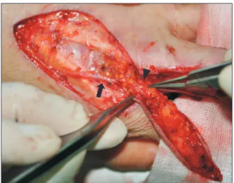
Fig. 3. Intraoperative image. The black arrow shows the dorsal meta- carpal artery, and the black arrowhead shows the dorsal metacarpal artery perforator.
Fig. 4. Image obtained 3 months after the operation.