Kor. J. Fertil. Steril., Vol. 31, No. 4, 2004, 12
클라인펠터 증후군 환자에서 착상전 유전진단의 결과
성균관대학교 의과대학 삼성제일병원 산부인과1, 불임연구실2, 유전학 연구실3, 비뇨기과4
김진영1・임천규2・전진현2・박소연3・서주태4・차선화1・궁미경1・강인수1
Outcome of Preimplantation Genetic Diagnosis in Patients with Klinefelter Syndrome
Jin Yeong Kim
1, Chun Kyu Lim
2, Jin Hyun Jun
2, So Yeon Park
3, Ju Tae Seo
4, Sun Hwa Cha
1, Mi Kyoung Koong
1, Inn Soo Kang
11
Department of Obstetrics and Gynecology,
2Laboratory of Reproductive Biology and Infertility,
3
Laboratory of Genetics,
4Department of Urology, Samsung Cheil Hospital, Sungkyunkwan University School of Medicine
Objectives: Klinefelter syndrome is the most common genetic cause of male infertility and presents with 47,XXY mainly or 46,XX/47,XXY mosaicism. It is characterized by hypogonadism and azoospermia due to testicular failure, however, sporadic cases of natural pregnancies have been reported.
With the development of testicular sperm extraction (TESE) and intracytoplasmic sperm injection (ICSI), sperm can be retrieved successfully and ART is applied in these patients for pregnancy. It has been suggested that the risk of chromosome aneuploidy for both sex chromosome and autosome is increased in the sperms from 47,XXY germ cells. Considering the risk for chromosomal aneuploidy in the offspring, preimplantation genetic diagnosis (PGD) could be applied as a safe and more effective treatment option in Klinefelter syndrome. The aim of this study is to assess the outcome of PGD cycles by using FISH for sex chromosome and autosome in patients with Klinefelter syndrome.
Materials and Methods: From Jan. 2001 to Dec. 2003, PGD was attempted in 8 cases of Klinefelter syndrome but TESE was failed to retrieve sperm in the 3 cases, therefore PGD was performed in 8 cycles of 5 cases (four 47,XXY and one 46,XY/47,XXY mosaicism). In one case, ejaculated sperm was used and in 4 cases, TESE sperm was used for ICSI. After fertilization, blastomere biopsy was performed in 6~10 cell stage embryo and the chromosome aneuploidy was diagnosed by using FISH with CEP probes for chromosome X, Y and 17 or 18.
Results: A total of 127 oocytes were retrieved and ICSI was performed in 113 mature oocytes. The fertilization rate was 65.3±6.0% (mean±SEM) and 76 embryos were obtained. Blastomere biopsy was performed in 61 developing embryos and FISH analysis was successful in 95.1% of the biopsied blastomeres (58/61). The rate of balanced embryos for chromosome X,Y and 17 or 18 was 39.7±6.9%.
The rate of aneuploidy for sex chromosome (X and Y) was 45.9±5.3% and 43.2±5.8% for chromosome
주관책임자: 강인수, 우) 100-380 서울특별시 중구 묵정동 1-19, 삼성제일병원 산부인과 Tel: (02) 2000-7581, Fax: (02) 2000-7790, e-mail: ikang6 @ users.unitel.co.kr
연 락 저 자: 김진영, 우) 100-380 서울특별시 중구 묵정동 1-19, 삼성제일병원 산부인과 Tel: (02) 2000-7582, Fax: (02) 2000-7790, e-mail: jinkim223@yahoo.co.kr
*
본 연구는 2002년도 제일의료장학재단 연구비 지원에 의해 이루어진 것임.
17 or 18, respectively. Embryo transfer was performed in all 8 cycles and mean number of transferred embryos was 2.5±0.5. In 2 cases, clinical pregnancies were obtained and normal 46,XX and 46,XY karyotypes were confirmed by amniocentesis, respectively. Healthy male and female babies were delivered uneventfully at term.
Conclusion: The patients with Klinefelter syndrome can benefit from ART with TESE and ICSI.
Considering the risk of aneuploidy for both sex chromosome and autosome in the sperms and embryos of Klinefelter syndrome, PGD could be offered as safe and more effective treatment option.
Key Words: PGD, Klinefelter syndrome, Chromosome, TESE, ICSI
클라인펠터 증후군은 남성의 성선기능저하증 (hy- pogonadism)의 가장 흔한 원인으로 그 빈도는 0.1~
0.2% 정도이며 불임남성에서 3.1% 정도로 보고된 다.1,2 염색체 핵형은 대부분 47,XXY이며, 그 외 high grade aneuploidy (48,XXXY; 49,XXXXY 등)나 47,XXY/46,XY 모자이시즘, X의 구조적 이상 등으 로 나타날 수 있다. 임상적 양상은 작고 단단한 고 환, 여성형 유방, 성선기능저하로 인한 남성불임 등 이며,3 고환 부전으로 인한 무정자증을 나타내나, 드물게 임신을 하는 경우가 있으며,4 특히 수술적 고환조직내 정자채취술 (testicular sperm extraction, TESE) 및 세포질내 정자주입술 (intracytoplasmic sp- erm injection, ICSI)의 발전으로 47,XXY/46,XY 모자 이시즘이나 일부 47,XXY 핵형의 환자에서 채취된 정자로 보조생식술을 이용한 임신의 시도와 성공이 보고되고 있다.5~10 그러나 이들에서 채취되는 정자 에서 성염색체 및 상염색체 이수성 빈도가 높다는 우려와 이러한 정자의 세포질내 정자주입술을 통해 수정된 배아 및 태아의 염색체 이수성의 위험도 제 기되고 있다.11,12
착상전 유전진단 (preimplantation genetic diagnosis, PGD)은 착상전의 배아에서 염색체 또는 유전자 진 단을 하여, 정상 염색체 및 유전질환에 이환되지 않 은 건강한 임신을 할 수 있는 방법으로 알려져 있 으며, 염색체 이수성이나 유전질환을 가진 태아의 출생 및 자연 유산을 예방할 수 있는 방법으로 보 조생식술에 접목되어 널리 시도되고 있다. 따라서 클라인펠터 증후군 환자에서도 보조생식술을 시행 하는 경우, 정자나 수정란의 염색체 이수성 위험을 고려하여 보다 안전한 방법으로서 착상전 유전진단 의 시행이 보고되고 있다.13~15
이에 본 저자 등은 클라인펠터 증후군 환자에서
사정이나 수술적 고환조직내 정자채취술로 얻어진 정자로 세포질내 정자주입술을 시행하여 수정된 수 정란에서 형광직접보합법 (fluorescent in situ hybri- dization, FISH)을 이용한 착상전 유전진단을 시행한 결과를 분석하여 그 효용성을 알아보고자 하였다.
연구 대상 및 방법
1. 연구 대상
2001년부터 2003년 12월까지 8예의 클라인펠터 증후군 환자에서 착상전 유전진단을 시도되었으며, 이 중 수술적 고환조직내 정자채취술 시도 후 정자 가 획득되지 못한 3예를 제외한 5예의 클라인펠터 증후군 환자 (47,XXY 4예 및 47,XXY/46,XY 1예)에 서 8주기의 착상전 유전진단이 시행되었다. 이 중 1 예 (47,XXY)에서는 사정된 정자 (ejaculated sperm) 을, 나머지 4예에서는 수술적 고환조직내 정자채취 술로 채취된 정자를 이용하여 세포질내 정자주입술 을 시행한 후 착상전 유전진단을 시행하였다. 배우 자의 평균 나이는 29.6±1.2세 (mean ± SEM)였으며, 여성측의 불임요인은 자궁내막종이 1예에서 있었으 며 그 외 특별한 원인은 없었다.
2. 난자의 채취 및 수정
과배란 유도는 Gonadotrophin-releasing hormone (GnRH) agonist (Superfact®, Serono, USA)와 recom- binant FSH (Puregon®, organon, 또는 Gonal F®, Se- rono, USA) 또는 human menopausal gonadotropin (Pergonal®, Serono, USA)를 이용한 황체기 중간 장 기요법을 이용하였다. 초음파로 난포의 크기를 관 찰하여 최소한 18 mm 이상의 난포가 3개 이상 되 었을 때 human chorionic gonadotropin (hCG) 10,000
IU를 주사하였고, hCG 주사 36시간 후 질식 초음 파를 이용하여 난자채취를 하였다. 채취된 난자는 10% synthetic serum substitute (SSS; Irvine Sci, Sant Ana, CA, USA)가 첨가된 HTFM 배양액 또는 Ga- mete (Vitrolife, Sweden) 배양액에 넣어 37℃, 5%
CO2, 95% 공기 중 배양기에서 3~4시간 배양하였 다. 배양 후 성숙된 난자에서 세포질내 정자주입술 (ICSI)을 시행하였으며, 16~18시간 후에 수정을 확 인하였다.
3. 정자의 준비
사정된 정자의 경우 Percoll gradient centrifugation 방법으로 운동성이 좋은 정자를 회수하였으며, 수 술적 고환내 정자채취술은 국소 마취 후 음낭 및 초막 (Tunica vaginalis)을 절개한 후 백막 (tubica albuginea)를 절개하여 세정관 (seminiferous tubule) 을 추출하였고 여분의 세정관은 동결 보관하였다.
신선 고환이나 동결-융해된 고환에서 추출된 세정 관은 0.4% BSA가 첨가된 Ham's F10 medium (Sigma) 내에서 겸자로 압착하여 정자를 획득하고 ICSI 시 행 전까지 37℃, 5% CO2 배양기에서 보관하였다.
4. 할구의 분리와 핵의 준비
수정이 확인된 수정란을 48시간 배양한 후 6~10 세포기에 도달했을 때, 1~2개의 할구를 분리하였다.
배아를 EB10 또는 G-PGD (Vitrolife) 배양액 내에서 내경 20 µm의 pipette과 acidified Tyrode 용액으로, 투명대의 일부를 제거하였다. 투명대 제거 후 동일 한 pipette으로 핵이 뚜렷한 할구를 분리하였다. 할 구의 분리 후 할구에서 핵을 관찰할 수 없거나 할 구의 핵이 2개 이상 관찰되었을 경우에는 한 개의 할구를 더 분리하였다. 할구가 분리된 배아는 G2 배양액으로 세척한 후 10% SSS가 첨가된 HTFM 배양액 또는 G1/G2 (Vitrolife) 배양액에서 배양하였 다. 분리해낸 할구는 0.6% BAS가 첨가된 0.5% triso- dium citrate 저장액에 5분간 처리하였다. Slide glass 위에 저장액 소적을 만들고 할구를 옮겼다. 현미경 하에서 할구의 세포막이 터지는 것과 할구 핵의 위 치를 확인하였다. 저장액이 완전히 증발하도록 공 기중에서 건조시킨 후 현미경 하에서 관찰하면서 -20℃에 보관했던 고정액을 떨어뜨려 세포질을 제
거하면서 할구의 핵을 고정하였다. 할구의 세포질 이 완전히 제거되면 고정액을 완전히 건조시킨 후, 70, 85, 100% ethyl alcohol을 각각 2분씩 처리하여 탈수시켰다.
5. 할구 세포에서 FISH의 시행과 결과 확인 및 배아의 이식
FISH probe로는 X,Y 및 17 또는 18번 염색체에 대한 CEP (chromosome enumeration probe)를 이용하 여 FISH를 시행하였다. Probe 혼합액을 12 mm 원형 cover glass (Fisher Scientific)에 넣고, 할구의 핵이 준비된 Slide glass에 올려놓고 rubber cement로 밀 봉하였다. 75℃ hot plate에서 5분간 denaturation시 킨 후 humidified chamber에서 6~16시간 동안 hy- bridization 시켰다. Hybridization 후 50% formamide/
2X SSC, 2X SSC, 2X SSC/0.1% NP-40 buffer에서 각 각 10분, 10분, 5분씩 세척하여, 잔여 probe를 제거 하였다. 세척 후 125 ng/ml DAPI가 첨가된 antifade mounting solution (Vysis)으로 mounting하고 형광현 미경 (Optiphot-2, Nikon)으로 FISH signal을 확인하 였다. 결과 확인 시 두 개 이상의 signal이 근접해 있을 경우에 두 signal의 거리가 signal 크기의 2배 이상이면 두 개의 signal로, 2배 이하이면 한 개의 signal로 판정하였다. 정상으로 판정된 배아를 난자 채취 4일째에 자궁 내 이식하였다.
결 과
착상전 유전진단의 임상적 결과는 Table 1과 같다.
얻어진 총 난자의 수는 127개로 이 중 세포질내 정 자주입술을 시행하여 76개의 수정란 (2 pronucleus) 을 얻었으며, 평균 수정율은 65.3±6.0% (mean ± SEM)이었다. 배아의 발달이 불량한 경우 할구 생 검을 시행하지 못하였으며, 61개의 수정란에서 할구 생검을 시행하여 모두 성공적으로 생검이 이루어졌 고, 이 중 58개의 수정란에서 FISH를 이용한 진단 이 가능하여 95.1%의 FISH 성공율을 보였다. 각 주 기에서 배아 이식이 가능한 정상 배아의 비율은 평 균 39.7±6.9% (mean ± SEM)로 나타났다. 8주기 모 두에서 배아 이식이 가능하였으며, 평균 2.5±0.5개 의 배아를 이식하였다.
X,Y 및 17 또는 18번 염색체에 대한 FISH를 시 행하여 할구의 이수성을 진단 하였으며 (Figure 1), 비정상 배아로 진단된 경우들을 염색체에 따라 분 석해 보면, X,Y염색체에 대하여 이상을 갖는 배아 의 비율은 45.9±5.3% (mean ± SEM)였으며, 17 또 는 18번 염색체에 대한 이수성 비율은 43.2±5.8%
(mean ± SEM)로 성염색체뿐 아니라 상염색체의 이 수성 확률도 증가됨을 알 수 있었다. 그 외 chaotic aneuploidy가 나타나는 경우도 많았다 (Table 2). 이
들 중 2예에서 임신에 성공하여 산전 양수 검사상 각각 46,XX 및 46,XY로 정상 염색체 핵형으로 진 단되었으며, 1예에서는 임신 39주에 정상 여아를 질 식 분만하였고, 다른 1예에서는 임신 38주에 전치 태반으로 제왕절개술을 시행하여 정상 남아를 출산 하였다.
고 찰
클라인펠터 증후군은 400~500 출생당 1명의 빈 도로 발생하며 남성불임에서는 약 3.1% 정도 나타 나는 비교적 흔한 염색체 이상 질환이다. 임상 양상 은 사춘기 전에는 명확하지 않은데 고환의 부피가 적고 팔다리가 긴 편이며, 사춘기의 2차 성징도 정 상으로 나타날 수 있다. 사춘기 이후에는 작고 단단 한 고환과 다양한 안드로젠 결핍 증상이 나타날 수 있는데 증상이 명확하지 않은 경우도 있으며, 특히 모자이시즘에서는 고환의 양상도 정상인 경우가 많 다. 종종 가임기 남성에서는 불임에 대한 검사 중 무정자증으로 클라인펠터 증후군이 진단되는 경우 가 많으며, poly-X를 갖는 경우에는 다른 표현형에 서도 비정상을 나타낸다.
47,XXY 염색체 이상의 발생은 생식세포발생 (germ cell development)의 감수분열 단계나 배아초 기 mitosis과정에서 일어날 수 있으며, 다른 상염색
Table 1. Outcome of the PGD cycles in patients with
Klinefelter syndrome
No. of patients 5
No. of cycles 8
Mean age of women (yr) 29.6±1.2 No. of oocytes retrieved 15.9±2.5 Fertilization rate (%) 65.3±6.3 Normal embryo rate (%) 39.7±6.9 Abnormal rate for X, Y (%) 45.9±5.3 Abnormal rate for 17 or 18 (%) 43.2±5.8 No. of transferred embryos 2.5±0.5 Clinical pregnancy rate (%)
per transfer cycle 25% (2/8)
* Values are mean ± SEM
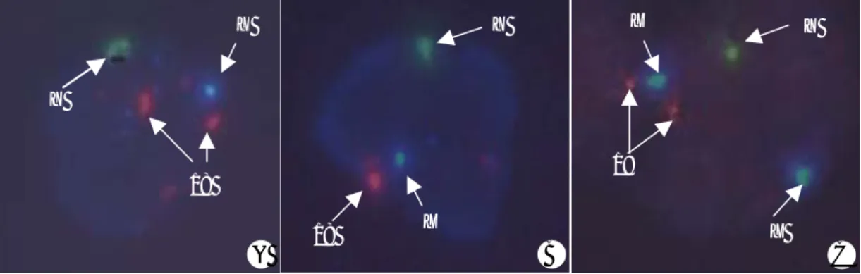
Y
18 X
Figure 1. FISH results in the blastomere of embryos from Klinefelter syndrome by using CEP probe for X (Spectrum
Aqua), Y (Spectrum Green) and 18 (Spectrum Orange). A) FISH signals in the normal embryo (XY1818) showing one spectrum aqua, one spectrum green and two spectrum orange signals. B) FISH signals for aneuploid embryo (XY monosomy 18) showing one spectrum aqua, one spectrum green and one spectrum orange signal. C) FISH signal for aneuploidy embryo (XXY1818) showing two spectrum aqua, one spectrum green and two spectrum orange signals.(×1000)
Y
18 X
18
X
X Y
A B C
체의 이수성이 모계에서 기원하는 경우가 많은 것 과는 달리 47,XXY는 부계에서 기원하는 오류가 절 반 정도로 많은 것으로 알려져 있다.16 즉, 정자의 제1 감수분열 시기에 non-disjunction에 의해 많이 발생한다. 이러한 extra X가 존재함으로써 태아기 고 환에 존재하던 원시정모세포 (primordial germ cell) 가 사춘기 전에 급속히 퇴화되는데 (degeneration), 이는 Sertoli cell과 germ cell간의 communication de- fect로 인한 것으로 생각된다.17 47,XXY를 가진 클 라인펠터 증후군에서는 사정 시 대부분 무정자증을 나타내나, 드물게 정자가 사정되어 자연 임신이 되 고 정상적인 출산이 보고되기도 하며,4,18 특히 46, XY/47,XXY 모자이시즘인 경우에서는 정자과소증 정도로 정자가 채취되는 경우도 많다. 수술적 고환 조직내 정자채취술과 세포질내 정자주입술 기법의 발전으로 클라인펠터 증후군에서도 수술적 고환조 직내 정자채취에 성공한 이래,19 이러한 환자들에서 보조생식술이 시도되고 있으며,6,7 수술적 고환조직 내 정자채취술로 약 21~54.5% 정도에서 성공적으 로 정자가 채취되는 것으로 보고되고 있다.8,20 모자 이시즘을 갖는 클라인펠터 증후군에서 정자가 채취 될 경우에는 정자가 46,XY 정상 germ cell에서만 유래할 것으로 생각되었으나, 47,XXY germ cell로부 터도 정자가 생성되어 염색체 이수성 정자를 생성 할 수 있다고 보고되었다.21,22 또한 정자의 염색체 분석에서 24,XY, 24,XX 등의 hyperploid 정자의 비 율이 정상군의 정자에서의 1.5%에 비해 모자이시즘
에서는 0.9~2.5%,23 47,XXY에서는 2.5~21.7% 정도 로 보고되고 있으며,24,25 Estop 등26은 정상 형태의 정자에서는 17% 및 비정상 형태에서는 61% 정도로 높은 염색체 이수성을 보인다고 하였다. 고환에서 채취된 정자에서는 hyperploidy rate이 6.75~25%로 높게 보고되기도 하였다.22,27 또한 성염색체뿐 아니 라 disomy 21의 정자 비율도 증가되어 trisomy 21의 위험이 증가된다는 보고들도 있어,28 채취된 정자의 이수성에 대한 우려가 있다. 반대로 spermatid나 성 숙 정자에서는 비교적 정상 염색체의 빈도가 높아, 염색체 이상이 있는 정자는 감수분열 과정을 완성 하기가 어렵고 사멸된다고 생각되기도 하며, 실제로 클라인펠터 증후군에서 임신이 되는 경우 태아는 정상 핵형을 보이는 경우가 많아 이를 반영하고 있 다.5,15 그러나 수술적 고환조직내 정자채취술 및 세 포질내 정자주입술을 통해 임신된 삼태 임신 중 정 상 태아와 함께 47,XXY의 태아가 임신된 경우도 보고되어,29 클라인펠터 증후군 환자에서 보조생식 술을 시행할 경우 염색체 이수성을 가진 정자의 수 정으로 인한 이수성 배아의 생성 및 이로 인한 임 신실패나 자연 유산, 염색체 이상을 가진 태아의 출 생 위험을 고려해야 한다.
착상전 유전진단은 유전질환이나 염색체 이수성 에 이환되지 않은 정상 배아만을 이식하여 정상적 인 임신을 유도할 수 있는 방법으로, 염색체 이수 성에 대한 착상전 유전진단으로 자연 유산율을 감 소시키고 착상율 및 임신 유지율을 높이는 등 보조
Table 2. Chromosome abnormalities of embryos diagnosed by FISH in patients with Klinefelter syndrome
Case cycle karyotype Origin of sperm Abnormal FISH results1 1 47,XXY Fresh TESE Monosomy 18, XO
2 1 47,XXY Fresh TESE Monosomy18 / monosomy 18,XO 2 2 47,XXY Fresh TESE Monosomy 18 / multiploidy
3 1 46,XY/47,XXY Fresh TESE Trisomy 17,Y / monosomy 17 / XXY / diploidy 3 2 46,XY/47,XXY Fresh TESE Monosomy 18 / aneusomy 18,XO / trisomy
18,XXX / trisomy 18,XYY 4 1 47,XXY Thawing TESE Chaotic / XYY / monosomy 18
5 1 47,XXY Ejaculated sperm Trisomy 18,XXX / XYY / aneusomy 18,XO 5 2 47,XXY Ejaculated sperm XXY / trisomy 18,XXY / monosomy 18,XO
생식술에 접목되어 중요한 부분을 차지하고 있으 며,30 수정된 배아의 염색체 이상에 대한 정보를 얻 을 수 있다. Silber 등12은 심한 비폐쇄성 무정자증에 서 수술적 고환조직내 정자채취술 및 세포질내 정 자주입술로 수정된 배아에서 착상전 유전진단을 통 해 염색체 이수성을 조사해본 결과, 사정된 정자로 부터 생성된 배아에 비해 정상 염색체의 비율은 더 낮고 (41.8% vs. 22.0%), 이수성 비율은 차이가 없 었으나 모자이시즘의 빈도가 높았는데 (26.5% vs.
53.0%) 특히 chaotic mosaicism이 증가되며, 이는 심 한 무정자증에서 채취된 정자의 centrosome 구조의 문제가 원인일 수 있다고 하였다. 최근 클라인펠터 증후군 환자에서도 염색체 이수성 배아의 위험을 고려하여 착상전 유전진단이 시행되고 있으며, 염색 체 이수성 배아의 비율이 대조군에 비해 높은 것으 로 보고되고 있다.13~15 또한 Bielanska 등31은 46,XY/
47,XXY 모자이시즘의 클라인펠터 증후군에서 정자 및 배아의 염색체 분석결과, 정자의 hyperploidy가 3.9%로 높고 배아에서는 단순히 X나 Y의 이수성이 증가되기 보다는 chaotic embryo가 많이 생성되는 것으로 보고한 바 있다. 실제 무정자증에서 정자 자 체의 염색체 분석에서 이수성 정자의 빈도는 생각 보다 낮은데, 수정된 배아에서 이수성이나 chaotic 모자이시즘은 크게 증가되는 것으로 볼 때, 정자 자체의 이수성 빈도만으로는 설명할 수 없고, 정자 의 centriole의 결함 등으로 인해 초기 mitosis 과정 에 이상을 초래하는 것으로 설명되고 있다.32 본 연 구에서도 진단된 배아 중 정상 배아율은 39.7±6.9%
정도로, 이는 Staessen 등15의 연구에서 정상군에서 의 정상 배아율이 77.2%에 비해 클라인펠터 증후군 에서는 전체적으로 54%, X, Y, 18번 염색체에 대한 정상 배아의 확률은 53%로 보고된 경우보다는 약 간 낮은 경향을 보였다. Kahraman 등14은 염색체 이수성 배아가 39.1%였으며, 이 중 성염색체 이수 성이 55.5% 및 그 외에 haploidy, triploidy, 및 13, 18 번 염색체의 trisomy나 monosomy를 보고하였다.
본 연구에서도 염색체별로는 성염색체의 이수성 (XXY, XXX, XYY 등) 비율이 평균 45.9%, 17 또는 18번의 이수성은 43.2%로 상염색체의 이수성도 증 가됨을 알 수 있었고, multiploidy나 chaotic aneu- ploidy를 갖는 배아도 많았다. 그렇지만 이식하지
않은 배아에서의 전체 할구에 대한 분석은 시행하 지 않았기 때문에 모자이시즘에 대한 분석은 불가 능하였다. 본 연구에서는 XY와 17 또는 18번 염색 체에 대해서만 FISH를 시행하였는데 보다 많은 상 염색체에 대한 PGD를 시행하면 정상 배아율은 더 욱 감소되며, 특히 18번 및 21번 염색체의 이수성 이 유의하게 증가되는 것으로 나타나,15 주요 상염 색체들을 포함한 포괄적인 염색체 이수성의 스크리 닝이 필요할 것으로 생각된다. 한편 무정자증에서 수술적 고환조직내 정자채취술 시행 시 예후인자로 서 시술 전 남성 호르몬 수치, 고환의 초음파, 혈류 저항, 47,XXY의 비율 등이 평가되기도 하였으나 정자채취 여부에 따른 이들 인자간의 유의한 차이 가 없어, 성공을 예측할 수 있는 예후인자가 명확하 지 않으므로,20 클라인펠터 증후군에서는 유전상담 을 비롯한 보조생식술에 대한 상담 후 수술적 고환 내 정자채취술 및 체외수정 시술을 시도하여 임신 의 기회를 최대화하는 것이 바람직할 것으로 생각 된다.
이상의 결과로 클라인펠터 증후군에서 수술적 고 환조직내 정자채취술 및 세포내 정자주입술을 이용 한 체외수정 시술은 임신을 위한 유용한 방법이다.
특히, 이 경우 채취된 정자의 염색체 이수성 또는 정자 자체의 centriole 이상에 의해, 배아의 성염색 체뿐 아니라 상염색체 이수성 및 chaotic 모자이시 즘의 증가 위험이 있어 임신실패나 유산, 태아 염색 체 이수성을 초래할 수 있다. 따라서, 사전 유전 상 담 및 체외수정 시술 시 착상전 유전진단을 적용함 으로써 염색체 이수성을 예방하여 보다 안전하고 효율적인 시술을 할 수 있을 것으로 생각된다.
참 고 문 헌
1. Phillip J, Lundsteen C, Owen D, Hirschhom K. The frequency of chromosome aberrations in tall men with special reference to 47,XYY and 47,XXY. Am J Hum Genet 1976; 28: 404-11.
2. Bojesen A, Juul S, Hojbjerg GC. Prenatal and po- stnatal prevalence of Klinefelter syndrome: a nati- onal registry study. J Clin Endocrinol Metab 2003;
88: 622-6.
3. Klinefelter HF, Reifenstein EC, Albright F. Syn- drome characterized by gynecomastia, aspermato- genesis without Leydigism, increased excretion of follicle stimulating hormone. J Clin Endocrinol 1942; 2: 615-27.
4. Laron Z, Dickerman Z, Zamir R, Galatzer A. Pater- nity in Klinefelter's syndrome- a case report. Arch Androl 1982; 8(2): 149-51.
5. Bourne H, Stern K, Clark G, Pertile M, Speirs A, Baker HW. Delivery of normal twins following the intracytoplasmic injection of spermatozoa from a patient with 47,XXY Klinefelter syndrome. Hum Reprod 1997; 11: 2447-50.
6. Ron-El R, Friedler S, Strassburger D, Komarovsky D, Schachter M, Raziel A. Birth of a healthy neonate following the intracytoplasmic injection of testicular spermatozoa from a patient with Klinefelter's syn- drome. Hum Reprod 1999; 14: 368-70.
7. Friedler S, Raziel A, Strassburger D, Schachter M, Bern O, Ron-El R. Outcome of ICSI using fresh and cryopreserved-thawed testicular spermatozoa in pa- tients with non-mosaic Klinefelter's syndrome. Hum Reprod 2001; 16 (12): 2616-20.
8. Ulug U, Bener F, Akman MA, Bahceci M. Partners of men with Klinefelter syndrome can be benefit from assited reproductive technologies. Fertil Steril 2003; 80(4): 903-6.
9. Komori S, Horiuchi I, Hamada Y, Hasegawa A, Kasumi H, Kondoh N, et al. Birth of healthy neo- nates after intracytoplasmic injection of ejaculated or testicular spermatozoa from men with nonmosaic Klinefelter's syndrome. J Reprod Med 2004; 49 (2):
126-30.
10. Poulakis V, Witzsch U, Diehl W, de Vries R, Becht E, Trotnow S. Birth of two infants with normal karyotype after intracytoplasmic injection of sperm obtained by testicular extraction from two men with nonmosaic Klinefelter's syndrome. Fertil Steril 2001;
76(5): 1060-2.
11. Denschlag D, Tempfer C, Kunze M, Wolff G, Keck C. Assisted reproductive techniques in patients with
Klinefelter syndrome: a critical review. Fertil Steril 2004; 82(4): 775-9.
12. Silber S, Escudero T, Lenahan K, Abdelhadi I, Kilani Z, Munne S. Chromosomal abnormalities in embr- yos derived from testicular sperm extraction. Fertil Steril 2003; 79(1): 30-8.
13. Staessen C, Coonen E, Van Assche E, Tournaye H, Joris H, Devroey P, et al. Preimplantation diagnosis for X and Y normality in embryos from three Kli- nefelter patients. Hum Reprod 1996; 11(8): 1650-3.
14. Kahraman S, Findikli N, Berkil H, Bakircioglu E, Donmez E, Sertyel S, et al. Results of preimplan- tation genetic diagnosis in patients with Klinefelter's syndrome. RBM Online 2003; 7(3): 346-52.
15. Staessen C, Tournaye H, Assche EV, Michiels A, Landuyt LV, Devroey P, et al. PGD in 47,XXY Klinefelter's syndrome patients. Hum Reprod Up- date 2003; 9(4): 319-30.
16. Thomas NS, Hassold TJ. Aberrant recombination and the origin of Klinefelter syndrome. Hum Re- prod Update 2003; 9(4): 309-17.
17. Hunt PA, Worthman C, Levinson H, Stallings J, LeMaire R, Mroz K, et al. Germ cell loss in the XXY male mouse: altered X-chromosome dosage affects prenatal development. Mol Reprod Dev 1998;
49: 101-11.
18. Terzoli G, Lattata F, Lobbiani A. Fertility in 47,XXY patient: assessment of biological paternity by deo- xyribonucleic acid fingerprinting. Fertil Steril 1992;
58: 821-2.
19. Tournaye H, Staessen C, Liebaers I, Van Assche E, Devroey P, Bonduelle M, et al. Testicular sperm recovery in nine 47,XXY Klinefelter patients. Hum Reprod 1996; 11: 1644-9.
20. Westlander G, Ekerhovd E, Granberg S, Hanson L, Hanson C, Bergh C. Testicular ultrasonography and extended chromosome analysis in men with non- mosaic Klinefelter syndrome: a prospective study of possible predictive factors for successful sperm re- covery. Fertil Steril 2001; 75(6): 1102-5.
21. Cozzi J, Chevret E, Rousseaux S, Pelletier R, Benitz
V, Jalbert H, et al. Achievement of meiosis in XXY germ cells: study of 543 sperm karyotypes from an XY/XXY mosaic patient. Hum Genet 1994; 93(1):
32-4.
22. Foresta C, Galeazzi C, Bettella A, Marin P, Rossato M, Garolla A, et al. Analysis of meiosis in intra- testicular germ cells from subjects affected by classic Klinefelter's syndrome J Clin Endocrinol Metab 1999; 84: 3807-10.
23. Chevret E, Rousseaux S, Monteil M, Usson Y, Cozzi J, Pelletier R, et al. Increased incidence of hyper- ploid 24,XY spermatozoa detected by three-colour FISH in a 46,XY/47,XXY male. Hum Genet 1996;
97: 171-5.
24. Okada H, Fujioka H, Tatsumi N, Kanzaki M, Okuda Y, Fujisawa M, et al. Klinefelter's syndrome in the male infertility clinic. Hum Reprod 1999; 14: 946 -52.
25. Foresta C, Galeazzi C, Bettella A, Stella M, Scan- dellari C. High incidence of sperm sex chromoso- mes aneuploidies in two patients with Klinefelter's syndrome. J Clin Endocrinol Metab 1998; 83: 203 -5.
26. Estop AM, Munne S, Cieply KM, Vandermark KK, Lamb AN, Fisch H. Meiotic products of a Kline- felter 47,XXY male as determined by sperm fluore-
scence in-situ hybridization analysis. Hum Reprod 1998; 13(1): 124-7.
27. Bergere M, Wainer R, Nataf V, Bailly M, Gombault M, Ville Y, et al. Biopsied testis cells of four 47, XXY patients: fluorescence in-situ hybridization and ICSI results. Hum Reprod 2002; 17(1): 32-7.
28. Hennebicq S, Pelletier R, Bergues U, Rousseaux S.
Risk of trisomy 21 in offspring of patients with Klinefelter's syndrome. Lancet 2001; 357: 2104-5.
29. Ron-El R, Strassburger D, Gelman-Kohan S, Friedler S, Raziel A, Appelman Z. A 47,XXY fetus concei- ved after ICSI of spermatozoa from a patient with non-mosaic Klinefelte's syndrome. Hum Reprod 2000; 15(8): 1804-6.
30. Gianaroli L, Magli MC, Fiorentio F, Baldi M, Fer- raretti AP. Clinical value of preimplantation genetic diagnosis. Placenta 2003; 24: S77-S83.
31. Bielanska M, Tan SL, Ao A. Fluorescence in-situ hy- bridization of sex chromosomes in spermatozoa and spare preimplantation embryos of a Klinefelter 46, XY/47,XXY male. Hum Reprod 2000; 15(2): 440 -4.
32. Sathananthan AH, Ratnam SS, Ng SC, Tarin JJ, Gianaroli L, Trounson A. The sperm centriole: its inheritance, replication and perpetuation in early human embryos. Hum Reprod 1996; 11: 345-56.