• 검색 결과가 없습니다.

First Multi-Detector Computed Tomography Evidence of Transcatheter Pacing System Migration and Embolization into the Pulmonary Vasculature

N/A
N/A
Protected

Academic year: 2021

Share "First Multi-Detector Computed Tomography Evidence of Transcatheter Pacing System Migration and Embolization into the Pulmonary Vasculature"

Copied!
3
0
0

로드 중.... (전체 텍스트 보기)

전체 글

(1)

310 www.kjtcvs.org

KJTCVS

The Korean Journal of Thoracic and Cardiovascular Surgery

Case Report

First Multi-Detector Computed Tomography Evidence of

Transcatheter Pacing System Migration and Embolization into the Pulmonary Vasculature

Tullio Valente, M.D. 1 , Giorgio Bocchini, M.D. 1 , Maurizio Cappelli Bigazzi, M.D. 2 , Massimo Muto, M.D. 1 , Paolo Golino, M.D. 2 , Giacomo Sica, M.D., Ph.D. 1

1

Division of Radiology, Department of Diagnostic Imaging, Monaldi Hospital;

2

Division of Cardiology, Monaldi Hospital, Vanvitelli University, Naples, Italy

ARTICLE INFO

Received November 13, 2019 Accepted January 27, 2020 Corresponding author Giacomo Sica

Tel 39-081-7062829 Fax 39-081-7062857 E-mail gsica@sirm.org ORCID

https://orcid.org/0000-0002-9518-9744

Transcatheter leadless pacemaker dislodgment is a rare and potentially fatal complication of leadless device implantation. We present the first case of multidetector computed to- mography images of leadless pacemaker migration and embolization in the pulmonary middle lobe artery. The patient was managed by percutaneous retrieval of the dislodged device and re-implantation in the appropriate position.

Keywords: Arrhythmia surgery, Device, Pulmonary embolism, Pacemaker, Computed tomography

Copyright © The Korean Society for Thoracic and Cardiovascular Surgery. 2020. All right reserved.

This is an Open Access article distributed under the terms of the Creative Commons Attribution Non-Commercial License (http://creativecommons.org/licenses/

by-nc/4.0) which permits unrestricted non-commercial use, distribution, and reproduction in any medium, provided the original work is properly cited.

Case report

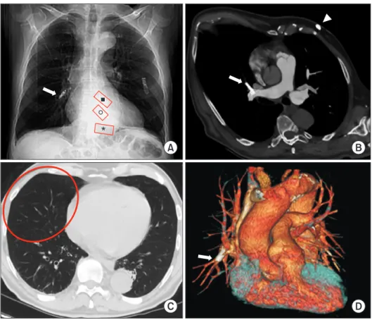
A 64-year-old man in whom a pacemaker was indicated due to bradyarrhythmia and atrial fibrillation presented to Monaldi Hospital for implantation of a transcatheter lead- less pacemaker (TPS). During the device implantation pro- cedure, unacceptable electrical measurements and fluoro- scopic findings indicated possible TPS dislodgement; the procedure was immediately terminated and an emergency computed tomography (CT) scan was requested. Multi-de- tector CT (MDCT) angiography was quickly performed and showed that the TPS had migrated and was embolized in the pulmonary middle lobe artery. The tributary lung territory was hyperlucent due to reduced blood flow and there was no perforation or pericardial effusion on the MDCT findings (Fig. 1). Successful percutaneous retrieval of the dislodged device was readily performed; the device was snared via the proximal retrieval feature of the device and was correctly reimplanted in the next hours (Supple- mentary Videos 1A–G).

No ethical committee approval was required for this case report, because it did not involve studies with human or animal subjects. It was not possible to obtain the patient’s consent for publication, because the patient could not be

traced. All images in this case report are anonymized;

therefore, the confidentiality of personal data is guaran- teed.

Discussion

TPS therapy has recently been introduced into clinical practice to overcome the short- and long-term complica- tions of traditional transvenous pacemakers (including pneumothorax, pocket hematoma/infection, lead dislodge- ment/fractures/infections, endocarditis, and pacemaker syndrome); it shows great promise and its applications are expanding, even though current real-world clinical experi- ence remains limited [1,2]. In patients who require solely single-chamber ventricular pacing, the TPS is implanted directly into the right ventricle (RV) via the femoral vein and affixed near the apex or at the midpoint of the RV sep- tum where the operator attains acceptable electrical mea- surements, eliminating the need for either a lead or subcu- taneous pocket. There are currently 2 commercially available devices: Micra (Medtronic, Dublin, Ireland) and Nanostim (Abbott, Abbott Park, IL, USA) leadless pace- makers. The main differences between these 2 systems re- late to the fixation mechanisms and the size of the delivery

https://doi.org/10.5090/kjtcs.19.085 pISSN: 2233-601X eISSN: 2093-6516

Korean J Thorac Cardiovasc Surg. 2020;53(5):310-312

(2)

311

Tullio Valente, et al. CT Evidence of Leadless Pacemaker Embolism

www.kjtcvs.org

KJTCVS

system used for the implant. Both devices weigh about 2 g.

The Abbott device (Nanostim) is longer and thinner (length, 42 mm; diameter, 5.99 mm; introducer, 18F), and anchors to the myocardium with a helical screw-in active fixation electrode. The Medtronic device (Micra) is shorter and thicker (length, 25.9 mm; diameter, 6.7 mm; introduc- er, 23F), and relies on passive fixation with multiple tiny nitinol tines. The appropriate final position of the TPS is confirmed on the basis of acceptable electrical measure- ments (capture threshold ≤1.0 V at 0.24–0.4 ms, R-wave >6 mV, impedance >500 Ω); usually, a post-procedural chest radiograph confirms the expected device location in the region of the mid-distal RV septum or apex.

In the LEADLESS II trial total cohort of 526 patients, the rate of device-related serious adverse events was 6.5%, in- cluding cardiac perforation in 1.5% of the patients, device dislodgement in 1.1%, and device retrieval due to elevated pacing thresholds in 0.8% [2]. A worldwide post-approval registry of the Micra device reported a 1-year major com- plication rate of 2.7% (95% confidence interval, 2.0%–3.7%) and 1 case (0.06%) of device embolization during implant [3]. As with other implanted devices, there is a learning curve for TPS implantation. A rigorous training program and operator experience may help reduce serious adverse

device events at implantation, defined as any undesirable effect related to the device or implant procedure that re- sults in death, life-threatening illness, prolongation of hos- pitalization, persistent/significant disability, or incapacity [4]. The device cylinder of both currently available systems is anchored to a ventricular wall that is constantly subject- ed to cycles of energetic contraction and relaxation, which entails the possibility of pulmonary anterograde emboliza- tion or retrograde caval embolization. Although infre- quent, these complications appear to be specific to the re- lease systems of these new pacemakers and must be kept in mind.

Conflict of interest

No potential conflict of interest relevant to this article was reported.

ORCID

Tullio Valente: https://orcid.org/0000-0002-8799-0398 Giorgio Bocchini: https://orcid.org/0000-0002-7164-3754 Maurizio Cappelli Bigazzi: https://orcid.org/0000-0002-4332-9426 Massimo Muto: https://orcid.org/0000-0003-3488-9269

A B

C D

*

Fig. 1. MDCT after TPS implanta- tion in a 64-year-old man with bra- dyarrhythmia and atrial fibrillation.

(A) CT scout-view shows TPS dis- lodgement at the right hilum (white arrow); red boxes indicate the ex- pected location of the device in the frontal radiograph at the right ven- tricular apex (*), mid-septal (o), and outflow tract (■). (B) Oblique axial MIP reconstruction shows TPS em- bolization in the middle lobe pul- monary artery (white arrow); in the subcutaneous fat of left ventral chest wall, there is also an ECG loop re- corder (arrowhead). (C) Axial MDCT (parenchymal window) shows the tributary lung territory hyperlucen- cy due to reduced blood flow (red oval). (D) Volume-rendering coronal reconstruction clearly confirms TPS embolization in the middle lobe pulmonary artery (arrow). MDCT, multidetector computed tomogra- phy; TPS, transcatheter leadless pace- marker; CT, computed tomography;

MIP, maximum intensity projection;

ECG, electrocardiography.

(3)

312 www.kjtcvs.org

KJTCVS https://doi.org/10.5090/kjtcs.19.085

Paolo Golino: https://orcid.org/0000-0002-3590-6983 Giacomo Sica: https://orcid.org/0000-0002-9518-9744

Supplementary materials

Supplementary materials can be found via https://doi.

org/10.5090/kjtcs.19.185. Supplementary Videos 1A–G.

Fluoroscopic videos showing the initial Micra retrieval process. The device was hooked and removed from the pe- ripheral pulmonary circulation.

References

1. Magnusson P, Pergolizzi JV Jr, LeQuang JA. Leadless pacemakers.

In: Min M, editor. Cardiac pacing and monitoring: new methods, modern devices [Internet]. London: IntechOpen; 2019 [cited 2019 Nov 2]. Available from: https://doi.org/10.5772/intechopen.83546.

2. El Amrani A, Campos B, Alonso-Martin C, et al. Performance of the Micra cardiac pacemaker in nonagenarians. Rev Esp Cardiol (Engl Ed) 2020;73:307-12.

3. Reddy VY, Exner DV, Cantillon DJ, et al. Percutaneous implantation of an entirely intracardiac leadless pacemaker. N Engl J Med 2015;

373:1125-35.

4. El-Chami MF, Al-Samadi F, Clementy N, et al. Updated performance

of the Micra transcatheter pacemaker in the real-world setting: a

comparison to the investigational study and a transvenous historical

control. Heart Rhythm 2018;15:1800-7.

수치

Fig. 1. MDCT after TPS implanta- implanta-tion in a 64-year-old man with  bra-dyarrhythmia and atrial fibrillation

참조

관련 문서

Nonlinear Optics Lab...

The first time you access to the viewer page, the client viewer software is automatically downloaded and installed in order to allow the user to view the MPEG4 video images of

Images that are required to be recorded include the upper esophagus, middle esophagus, lower esophagus, GEJ, front and reverse images of the fundus, front and reverse images

웹 표준을 지원하는 플랫폼에서 큰 수정없이 실행 가능함 패키징을 통해 다양한 기기를 위한 앱을 작성할 수 있음 네이티브 앱과

_____ culture appears to be attractive (도시의) to the

이하선의 실질 속에서 하악경의 후내측에서 나와 하악지의 내측면을 따라 앞으로 간다. (귀밑샘 부위에서 갈라져 나와

If the volume of the system is increased at constant temperature, there should be no change in internal energy: since temperature remains constant, the kinetic

□ Heat is defined as transferred energy dew to temperature differences through boundary.. - Conduction by molecular