• 검색 결과가 없습니다.

Spinal epidural lipomatosis consists of a pathologic overgrowth of fat tissue in the spinal canal. It is usually secondary to steroid therapy or endocrinopathic diseases.

N/A
N/A
Protected

Academic year: 2021

Share "Spinal epidural lipomatosis consists of a pathologic overgrowth of fat tissue in the spinal canal. It is usually secondary to steroid therapy or endocrinopathic diseases."

Copied!
3
0
0

로드 중.... (전체 텍스트 보기)

전체 글

(1)

Introduction

Spinal epidural lipomatosis consists of a pathologic overgrowth of fat tissue in the spinal canal. It is usually secondary to steroid therapy or endocrinopathic diseases.

Symptomatic spinal epidural lipomatosis is a very rare condition. We describe one case of symptomatic spinal epidural lipomatosis secondary to obesity, and review the relevant literature.

Case Report

A 56-year-old man presented a 6-month history of severe bilateral pain radiating to his feet and a 20-year history of mild low back pain prior to operation. And he also presented neurogenic claudication induced by walking 100 m. Endocrinopathic diseases and chronic steroid therapy were excluded for this patient. But this patient was obese with a mean body mass index of 33.5 kg/m

2

. Radiographs of the lumbar spine demonstrated moderate

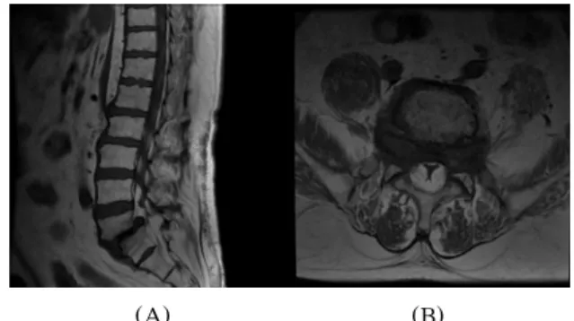
spondylosis with isthmic spondylolisthesis at L5-S1. MRI of the lumbar spine demonstrated a severe disc bulging causing bilateral foraminal stenosis at L5-S1 and a pathologic overgrowth of fat tissue in the spinal canal with a marked impingement of the thecal sac at L3-S1(Fig. 1A-B).

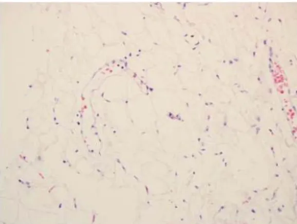
Surgical treatment was performed by L3, L4 and L5 laminectomies, fat debulking, and instrumented posterolateral fusion. At the time of surgery, a large amount of fat which was compressing the thecal sac was found. Histologic examination of the fat revealed normal adipose tissue(Fig. 2).

(A) (B)

Fig. 1A-B. Magnetic resonance imaging sagittal (A) and axial (B) T1-weighted images demonstrate overgrowth of fat tissue in the spinal canal and compressing the thecal sac and nerve roots. Magnetic resonance imaging sagittal image shows also moderate isthmic spondylolisthesis at L5-S1. Axial image (B) through the body of L5 showing the classical Y configuration of the thecal sac secondary to epidural compression by fat.

고신대학교 의과대학 학술지 제 권 제 호23 4 Kosin Medical Journal

Vol. 23. No. 4, pp. 111 113, 2008∼

Dae-Yong Kim, Jin-Wook Kim, Ju-Ho Jeong, Jae-Hoon Cho Department of Neurosurgery, Kosin University College of Medicine, Busan, Korea

――― Abstract ――――――――――――――――――――――――――――――――――――――――

Symptomatic spinal epidural lipomatosis, which consists of a pathologic overgrowth of fat tissue in the spinal canal, is a very rare condition. It has been reported frequently in association with the administration of exogenous steroids.

So, it is a widely recognized complication of excess exogenous glucocorticoids. Here, the authors report on a case of symptomatic spinal epidural lipomatosis secondary to obese, and review the literature.

―――――――――――――――――――――――――――――――――――――――――――――――――

Key words : Epidural lipomatosis, Obesity

교신저자:Dae-Yong Kim

ADD : Amnam-dong, Suh-gu, Busan 602-702, Korea Department of Neurosurgery,

Kosin University Gospel Hospital, 34 TEL : +82-51-990-6705, FAX : +82-51-990-3042 E-mail : [email protected]

(2)

고신대학교 의과대학 학술지 제 권 호 24 4 , 2008

Fig. 2. The microscopic examination shows mature fat cells with thin capillaries, H-E, x200.

The patient showed a improvement in symptoms with a return to daily activities. There was no postoperative complication for 5 months after the operation.

Discussion

Spinal epidural lipomatosis is an uncommon disease in which excess fat tissue deposits around the thecal sac.

Furthermore, the symptomatic one is a truly rare condition as reported in published research. There is no histologic difference between the spinal epidural lipomatosis and normal epidural adipose tissue.

1,2,3)

Clinical manifestations and pathologic mechanism are similiar to patients with spinal stenosis. Therefore, spinal epidural lipomatosis should be considered in the differential diagnosis of lumbar radiculopathy when there is an absence of common causes.

Magnetic resonance imaging is the most useful diagnostic test.

2,4,5)

Additionally, it is possible to distinguish the causes of compression and to differentiate fat growth from the other causes, such as, bone metastasis and discitis;

moreover, epidural fat can be localized clearly. Usually, axial MRI reveals the triangular or "Y" shaped thecal sac.

4,5)

Spinal epidural lipomatosis is usually found in patients with a history of chronic steroid therapy, obesity, and some endocrinopathies. Chronic steroid-induced epidural lipomatosis has been reported in the several literature.

3,6,7,8)

Also, Badami et al.

9)

described symptomatic deposition of epidural fat in the spinal canal in a morbidly obese woman

who had no history of steroid intake, after which a few of similar spinal epidural lipomatosis cases have been reported.

1,2,10)

In one literature, more than 75% of all reported pateints were obese.

2)

Kumar et al.

10)

reported a case presenting with symptomatic epidural lipomatosis secondary to obesity and established three diagnostic crieteria 1) medical history and physical examination : consistent with segmental spinal cord compression 2) epidural fat thickness greater than 7 mm in the region of compression based on MRI or CT 3) a height to weight ratio greater than 27.5 kg/m

2

. In South Korea, Young-Min Han

11)

reported two cases of symptomatic epidural lipomatosis secondary to obese.

In our case, endocrinopathic diseases, and chronic steroid therapy were excluded. But our patient was obese with a mean body mass index of 33.5 kg/m

2

. Therefore, we concluded that our patient was a case presenting with symptomatic epidural lipomatosis secondary to obesity.

The spinal epidural lipomatosis often responds to medical therapy of the underlying disease. Empirically, the first line of treatment for spinal epidural lipomatosis has been weight loss and exercise therapy. However, failure of these measures has resulted in surgical decompression with satisfactory results. Surgical management is recommended with extensive laminectomies and massive fat debulking. An associated instrumented posterolateral fusion is necessary in multilevel laminectomies associated with wide facetectomies that could be the cause of postsurgical instability. In our case, medical therapy came to nothing because of the patient's noncooperation. So the surgical treatment was performed by L3,L4 and L5 laminectomies, fat debulking, and instrumented posterolateral fusion.

Conclusion

We report one case of symtomatic spinal epidural

lipomatosis secondary to obesity. Spinal epidural lipomatosis

may be the cause of neurological symptoms when it is

excessive. Obesity seems to be closely related to the

development of the spinal epidural lipomatosis. Because the

number of obese people is increasing in Korea, spinal

(3)

epidural lipomatosis should be considered when evaluating obese patients with radicular pain and progressive paraparesis.

국문초록

척추 경막상 지방종증은 척추강내에 지방조직이 병적 으로 과도하게 성장하여 침착되는 것을 말하며 매우 드

문 질환이다 이는 장기간 투여한 스테로이드와 연관이 .

있다고 알려져 있으며 일반적으로 증상이 없는 경우가 ,

대부분이다 본 저자들은 비만으로 인해 신경성 파행을 .

동반한 척추 경막상 지방종증 환자를 치험하였기에 문헌 고찰과 함께 보고하고자 한다.

Key words : 경막상 지방종증 비만 ,

References

1) Gero BT, Chynn KY Symptomatic spinal epidural lipomatosis : without exogenous steroid intake. Report of a case with magnetic resonance imaging. Neuroradiology 31 190-192, : 1989

2) Robertson SC, Traynelis VC, Follett KA, Menezes AH:

Idiopathic spinal epidural lipomatosis. Neurosurgery 41:

68-75, 1997

3) Roy-Camille R, Mazel C, Husson JL, Saillant G Symptomatic : spinal epidural lipomatosis induced by a long-term steroid treatment. Review of the literature and report of two additional cases. Spine 16 1365-1371, 1991 :

4) Healy ME, Hesselink JR, Ostrup RC, Alksne JF:

Demonstration by magnetic resonance of symptomatic spinal epidural lipomatosis. Neurosurgery 21 414-415, 1987 : 5) Kuhn MJ, Youssef HT, Swan TL, Swenson LC Lumbar :

epidural lipomatosis : The “Y”sign of thecal sac compression.

Comput Med Imaging Graph 18 367-372, 1994 :

6) Haddad SF, Hitchon PW, Godersky JC Idiopathic and : glucocorticoid-induced spinal epidural lipomatosis. J Neurosurg 74 38-42, 1991 :

7) Ju WI, Ahn JG, Ji C, Park SC, Cho KS, Park CK, et al:

Spinal epidural lipomatosis. Case report. J Korean Neurosurg Soc 28 110-113, 1999 :

8) Lee M, Lekias J, Gubbay SS, Hurst PE Spinal cord : compression by extradural fat after renal transplantation. Med J Aust 1 201-203, 1975 :

9) Badami JP, Hinck VC Symptomatic deposition of epidural fat : in a morbidly obese woman. AJNR Am J Neuroradiol 3:

664-665, 1982

10) Kumar K, Nath RK, Nair CPV, Tchang SP Symptomatic : epidural lipomatosis secondary to obesity. Case report. J Neurosurg 85 348-350, 1996 :

11) Han YM, Ahn MS : Idiopathic Spinal Epidural Lipomatosis.

Case report J Korean Neurosurg Soc 30 795-799, 2001 :

수치

Fig. 1A-B. Magnetic resonance imaging sagittal (A) and axial (B) T1-weighted images demonstrate overgrowth of fat tissue in the spinal canal and compressing the thecal sac and nerve roots
Fig. 2. The microscopic examination shows mature fat cells with thin capillaries, H-E, x200.

참조

관련 문서

It considers the energy use of the different components that are involved in the distribution and viewing of video content: data centres and content delivery networks

After first field tests, we expect electric passenger drones or eVTOL aircraft (short for electric vertical take-off and landing) to start providing commercial mobility

1 John Owen, Justification by Faith Alone, in The Works of John Owen, ed. John Bolt, trans. Scott Clark, "Do This and Live: Christ's Active Obedience as the

Endoscopic spine surgery has become a practical, minimally invasive technique for decompression in patients with spinal disc herniation or stenosis.. However,

Sandberg D, Lavyne M (1999) Symptomatic spinal epidural lipomatosis after local epidural corticosteroid injections:

The students of Gwangju Secondary School had to compete with the students of Gwangju Intermediate School, who were Japanese nationals for access to a

Lumbar discogenic radiculopathy (LDR) is one of the most common spinal disorders which can lead to high medical expenses for the patient, occupational impairment,

With analysis that it will be a chance for children with autism who have difficulty of using languages and usually can't make any relationship with people