997
Utility of Smo as a Prognostic Marker for Human Bladder Tumors
Yun-Sok Ha, Seok-Joong Yun, Yong-June Kim, Sang-Cheol Lee, Wun-Jae Kim
From the Department of Urology, College of Medicine, Chungbuk National Uni- versity, Cheongju, Korea
Purpose: Smoothened (Smo) encodes a 1,024 amino acid transmembrane protein that acts as a transducer of the hedgehog (Hh) signal and maps to 7q31-q32 in humans. In the absence of Hh, Patched (Ptc) prevents Smo from signaling. When M-Hh-N binds to Ptc, however, Smo is free to upregulate downstream genes in the network. Activating mutations in Smo have been identified in sporadic basal cell carcinomas. This study was performed to evaluate the significance of Smo expression in hu- manzbladder cancer.
Materials and Methods: Tumor tissues were obtained from 140 patients with bladder cancer and normal bladder mucosa were acquired from 17 patients without bladder cancer as controls. Smo expression was assessed from paraffin sections of tissues using immunohistochemistry and graded on a scale of 0-12 according to the intensity and rate of staining. Differences of Smo expression between the bladder tumors and normal mucosa were compared. The relationships between their expression and the pathological or clinical characteristics such as tumor stage, grade, recurrence, and progression were also analyzed.
Results: There was no difference in the Smo expression in comparisons between the bladder cancers and the normal tissues (4.96±1.92 vs.4.52±0.87, p=0.111). Superficial bladder tumors had a higher Smo expression com- pared with normal tissues (0.005). Smo expression in the superficial and low-grade bladder tumors were higher than in the invasive and high-grade bladder tumors (p=0.002 and 0.001, respectively). The progression status was correlated with Smo expression but not the recurrence status (p=0.041 and 0.357, respectively). However, the Smo expression levels were not associated with the overall survival of patients (p=0.406).
Conclusions: The results of this study showed that the enhanced ex- pression of Smo was correlated with superficial, low-grade bladder cancer and tumors without progression. These results suggest that Smo is closely correlated with the differentiation and progression of bladder cancer and may, therefore, be useful as a prognostic marker for bladder cancer in the clinical setting. (Korean J Urol 2007;48:997-1003)
Key Words: Bladder tumor, Immunohistochemistry, Smoothened protein
대한비뇨기과학회지 제 48 권 제 10 호 2007
충북대학교 의과대학 비뇨기과학교실
하윤석ㆍ윤석중ㆍ김용준 이상철ㆍ김원재
접수일자:2007년 5월 15일 채택일자:2007년 9월 4일
교신저자: 김원재
충북대학교 의과대학 비뇨기과학교실
충북 청주시 흥덕구 개신동 12번지
361-763
TEL: 043-269-6371 FAX: 043-269-6129 E-mail: wjkim@chungbuk.
ac.kr
서 론
방광암은 비뇨기계 종양 중 가장 높은 빈도로 발생하는 악성종양이다. 방광암의 90% 이상은 이행상피세포암이며,
그 가운데 60% 정도는 저등급 분화도의 표재성 방광암이 다. 하지만 요도경유절제술을 시행한 경우 재발이 빈번하 며 16-25% 정도는 고등급 분화도의 암으로 진행하고 또한 10%의 경우에서는 병기의 진행 및 종양의 전이가 발생한 다.1,2 잦은 재발과 병기의 진행은 환자 및 비뇨기과 의사들
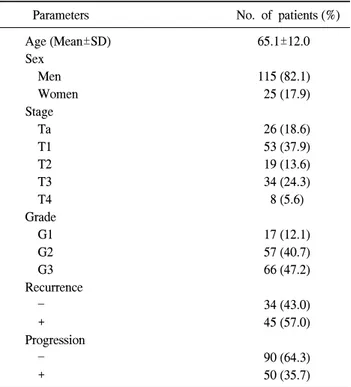
Table 1. Clinico-pathological features of 140 primary bladder transitional carcinomas
Parameters No. of patients (%)
Age (Mean±SD) 65.1±12.0
Sex
Men 115 (82.1)
Women 25 (17.9)
Stage
Ta 26 (18.6)
T1 53 (37.9)
T2 19 (13.6)
T3 34 (24.3)
T4 8 (5.6)
Grade
G1 17 (12.1)
G2 57 (40.7)
G3 66 (47.2)
Recurrence
34 (43.0)
+ 45 (57.0)
Progression
90 (64.3)
+ 50 (35.7)
SD: standard deviation 적인 요소로, 세포에서 분비되면 세포 표면의 수용체 단백
과 결합하여 세포 내 신호전달체계를 작동시킨다.3,4 Hh 전 달체계에는 Patched (Pch) 단백과 Smoothened (Smo) 단백이 주로 관여하는데, Hh 발현이 없는 정상 상태에서는 세포 표 면에 있는 Pch가 세포 내 단백인 Smo와 결합하여 활성화를 억제한다. 하지만 Hh 신호전달체계가 활성화되면 Pch와 Smo는 분리되고 Smo가 활성화되어 Hh 신호전달체계의 하 부 경로 구성요소인 Gli가 표적 세포를 발현시킨다.4,5 최근 여러 가지 종류의 암 발생에 있어 Hh의 중요한 역할들이 밝혀지고 있다. 여러 조직의 줄기세포 유지에 Hh 신호전달 체계가 중요한 역할을 하는데, 돌연변이에 의해 Hh 신호전 달체계가 과도하게 활성화될 경우 줄기세포의 과도한 증식 을 통해 암을 야기할 수 있다는 것이다. 위암, 췌장암, 폐암, 횡문근육종, 기저세포암 등 다양한 암이 Sonic hedgehog (Shh) 신호전달체계에 의해 유도되는 것으로 알려져 있으 며, 신경계에서 발생하는 암 중 Shh 신호전달 체계와 관련 성이 잘 알려진 암은 기저세포암이다.6,7 또한 Smo의 활성화 돌연변이도 속질모세포암과 관련이 있다. 소뇌 과립세포에 활성화된 Smo를 과발현시킨 생쥐의 48%에서 속질모세포 암이 발생하였다.8 즉 성인에서 비정상적으로 Hh이 과도하 게 발현되면 이는 곧 세포 표면에 있는 Pch와 결합하여 Pch 가 더 이상 Smo 활성을 억제할 수 없게 되고, 활성화된 Smo 는 특정 유전자를 과도하게 발현시켜 종양 생성 및 성장을 유도한다.5
Smo는 1,024개의 아미노산으로 구성된 세포 내 단백으로 7개의 소수막과 세포막외 아민 부분과 세포 내 탄산 부분으 로 구성된 G-protein coupled receptor의 Frizzled 군으로 7번 염색체 장완 31-32부위에 위치한다.6,7 최근에는 Smo를 억제 하는 Hh 전달체계 억제제인 cyclopamine이 개발되어, 동물 실험에서 몇몇 암의 발생과 치료에 효과가 있다는 보고가 있었다.9-11
이에 본 저자들은 방광암에서도 종양 생물학으로 유전자 의 발현 차이가 암의 성상에 관여할 것이라는 가정하에 방 광암에서 Smo 단백 발현을 조사하였다. 방광암 환자군의 방광조직에서 면역조직화학염색을 통해 Smo의 발현유무
세포암으로 확진된 방광암 조직 140례와 환자에게 동의를 받은 후 전립선비대증 또는 요실금 등 종양 이외의 양성 질환으로 수술을 시행 받은 후 병리조직학적으로 정상 방 광조직으로 확인된 방광 조직 17례를 대상으로 면역조직화 학염색을 시행하였다. 방광암 환자의 평균 나이는 65.1세 (26-88), 성별로는 남자 115례, 여자 25례였으며, 대조군의 평균 나이는 55.1세 (43-68), 남자 13례, 여자 4례였다. 방광 암의 병기는 표재성 방광암이 79례 (Ta; 26례, T1; 53례), 침 윤성 방광암이 61례 (T2; 19례, T3; 34례, T4; 8례)였으며, 분 화도는 WHO 분류법에 따라 등급 1이 17례, 등급 2가 57례, 등급 3이 66례였다 (Table 1). 방광암의 재발 및 진행에 대한 평균 추적 관찰 기간은 32.8개월 (0.2-110, 중앙값 27.0)이었다.
종양의 병기에 따라 Ta와 T1을 표재성군으로, T2-T4를
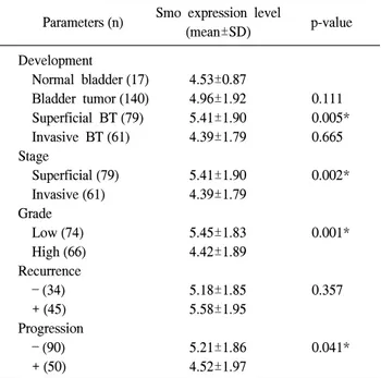
Table 2. Protein expression levels of Smo according to clinico- pathologic features in bladder cancer
Smo expression level
Parameters (n) p-value
(mean±SD) Development
Normal bladder (17) 4.53±0.87
Bladder tumor (140) 4.96±1.92 0.111 Superficial BT (79) 5.41±1.90 0.005*
Invasive BT (61) 4.39±1.79 0.665 Stage
Superficial (79) 5.41±1.90 0.002*
Invasive (61) 4.39±1.79 Grade
Low (74) 5.45±1.83 0.001*
High (66) 4.42±1.89
Recurrence
(34) 5.18±1.85 0.357
+ (45) 5.58±1.95
Progression
(90) 5.21±1.86 0.041*
+ (50) 4.52±1.97
*p<0.05, analyzed by Student's t-test (independent). BT: bladder tumor, SD: standard deviation
Fig. 1. Immunohistochemical staining of Smo. (A) Protein in normal bladder mucosa. (B) Elevated levels of the proteins in TaG1 bladder cancer. (C) Low levels of the proteins in T2G3 bladder.
침윤성군으로 분류하였다. 조직학적 분화도에 따라 등급 1 과 등급 2를 분화도가 좋은 군으로, 등급 3을 분화도가 나쁜 군으로 분류하였다. 추적기간 동안 표재성 방광암에서 수 술 후 병기의 진행 없이 반복하여 종양이 재발한 경우를
‘재발’로 정의하였고, 적절한 치료 후에도 표재성 방광암이 침윤성 암으로 또는 전이성 방광암으로 되거나 침윤성 방 광암이 전이성 방광암으로 되는 경우를 병기의 ‘진행’으로 정의하였다.
2. 면역조직화학염색
환자에게서 얻어진 조직은 파라핀 고정으로 4μm의 초절 편을 만들었고 파라핀을 제거한 후 0.05% hydrogen peroxide 에서 10분간 처리하였다. 비특이적 반응을 제거하기 위해 normal horse serum과 15분간 반응시켰다. 면역화학염색은 Dako-Cytomation Immunostaining kit (Dako, Glostrup, Den- mark)를 사용하였고 1차 항체는 Santa Cruz Biotechnology Inc (Santa cruz Biotechnology Inc, Santa Cruz, USA) 제품 [Smo (H-300):I sc-13943]을 1:100으로 희석하여 사용하였다.
조직 슬라이드는 60oC에서 1시간 동안 가온 후 100% xylene 으로 7분씩 4번 탈파라핀하였고 100%, 100%, 95%, 75% 알 코올로 각각 hydration시킨 다음 3% H2O2 용액에 10분간 유 치하였다. 조직 슬라이드를 10mM Borate buffer (pH 8.0)12에 넣고 전자레인지로 15분간 처리한 후 1차 항체로 60분간 반 응시키고 0.1% Tween13으로 세척한 다음 Envision detection system (anti-mouse/anti-rabbit)으로 20분간 반응시켰다. 다시 충분히 세척 후 3, 3-diaminobenzidine (DiNonA, 서울, 대한 민국)으로 5분간 염색한 후 Meyer's hematoxylin으로 3분간 재차 염색하였다. 면역화학염색 결과 판정은 Sinicrope 등14 이 보고한 방법을 사용하였다. 염색의 강도에 따라 약 (1 점), 중 (2점), 강 (3점)으로 평점하였으며 염색된 세포의 면 적에 따라 5개로 분류하였다. 5% 미만은 0점, 5-25%는 1점,
26-50%는 2점, 51-75%는 3점, 75% 이상은 4점으로 하였다.
염색 강도의 평점과 염색 면적 평점의 값을 곱하여 0점부터 12점까지의 등급으로 구분하여 최종 염색을 평가하는 기준 으로 삼았다. 종양 상피세포와 정상 상피세포의 점막과 세 포질은 갈색으로 염색되나 평활근에는 염색이 되지 않았다
(Fig. 1).
3. 통계학적 분석
통계분석은 SPSS (ver. 10.0)를 이용하여, 종양과 정상 방 광점막의 발현 차이 및 각 임상 지표의 차이를 Student's
Fig. 2. Protein expression levels of Smo. (A) The expression of Smo compared between bladder tumor and normal tissue. No significant difference was found between the two groups (p=0.111), (B) The expression of Smo according to stage. The expression of Smo was significantly higher in the superficial group (5.41±1.90) than in the invasive group (4.39±1.79) (p=0.002), (C) The expression of Smo according to grade. The expression of Smo was significantly higher in the low-grade group (5.45±1.83) than in the high-grade group (4.42±1.89) (p=0.001), (D) The expression of Smo according to progression. The expression of Smo was significantly higher in the group without tumor progression (5.18±1.85) than in the group with tumor progression (5.58±1.95) (p=0.041).
전체 140례의 방광암 조직과 17례의 정상 방광점막과의 비교에서는 Smo의 단백 발현은 방광이행상피세포암에서 는 4.96±1.92, 정상 방광점막에서는 4.53±0.87의 발현을 보 여 방광이행상피세포암에서 Smo 단백 발현이 정상 방광점 막에 비하여 높은 경향을 보였으나 통계적인 의미는 없었
고 등급 3으로 구분한 후 등급 1과 등급 2를 저등급 분화도 로 등급 3을 고등급 분화도로 구분하였다. Smo의 단백 발현 은 고등급 분화도에서 4.42±1.89으로 측정되었고, 저등급 분화도에서 5.45±1.83으로 측정되어 고등급 분화도에 비해 저등급 분화도에서 Smo의 단백 발현이 높았다 (p=0.001).
추적기간 동안 표재성 방광암에서 수술 후 병기의 진행
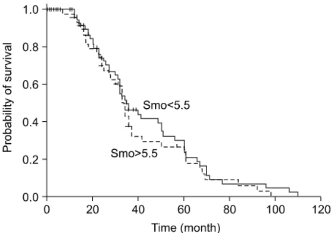
Fig. 3. The Kaplan-Meier curve shows the overall survival of patients with bladder transitional cell carcinoma: there is no statistically significant difference between the two groups (p=
0.406).
없이 반복하여 종양이 재발한 재발군과 비재발군을 비교하 였을 때 Smo의 단백 발현은 재발군에서 5.58±1.85, 비재발 군에서 5.18±1.95로 측정되어 방광암의 재발과는 통계학적 유의성이 관찰되지 않았다 (p=0.357).
적절한 치료 후에도 표재성 방광암이 침윤성 암으로 또 는 전이성 방광암으로 되거나 침윤성 방광암이 전이성 방 광암이 된 진행군과 비진행군의 단백 발현을 비교하였을 때 비진행군에서 5.21±1.86, 병기의 진행군에서 4.52±1.97로 측정되어 비진행군에서 진행군에 비하여 의미있게 높은 Smo 단백 발현을 보였다 (p=0.041).
2. Smo 발현이 방광암 환자의 생존율에 미치는 영향
염색 강도의 평점과 염색 면적 평점의 값을 곱하여 최종 염색을 평가하는 기준으로 한 후 Smo 발현정도의 기준 (cut-off value)을 5.5점으로 사용하였을 때 민감도는 43.3%, 특이도는 81.1% 였다. Smo 발현량이 5.5점보다 높을 경우와 낮을 경우를 나누어 생존곡선 분석결과 Smo 발현량과 생존 율은 유의한 상관관계가 없었다 (p=0.406) (Fig. 3).
고 찰
다른 종류의 암과 마찬가지로 방광암에서도 적절한 시기 에 적절한 치료를 시행하는 것이 중요하다. 이를 위해서는 방광암의 재발 및 진행을 정확히 예측할 수 있는 지표가 매우 중요하며 조직의 분화도와 병기와 같은 임상적 지표 를 보완하면서 치료의 반응을 예측할 수 있는 인자가 필요 하다. 현재까지 세포 주기와 관련하여 세포 주기조절인자, 세포고사과정, 그리고 세포증식에 관여하는 유전자 및 단
백질과 같은 생물학적 종양 표지자에 관한 연구가 많이 진 행되고 있다.
초파리 연구로부터 밝혀지기 시작한 Hh 신호전달체계는 발생과정에서 신경계, 호흡기계, 순환기계, 사지 발생 등 각 종 장기 형성에 중요한 역할을 하는 것으로 알려지고 있다.3 또한 분비된 Hh는 분비세포를 중심으로 단백질 농도경사 를 형성하고, 이 농도경사에 따라, 세포의 운명이나 분화가 달라질 수 있다는 것이다. 이러한 형태발생인자 (morph- ogen)로서의 작용은 초파리에서 생쥐까지, 그리고 팔, 다리 형성, 신경관 형성 등 여러 장기형성에도 그대로 보존되어 있다.3,4 최근에는 Hh 신호전달체계가 성인의 줄기세포 유 지에 관여하며,15-17 Greig cephalopolysyndactyly, Pallister Hall syndrome, VATER syndrome 등의 각종 선천성 질환, 속질모 세포암, 폐암, 위암, 식도암, 췌장암 등의 암 발생과도 관련 성이 있다고 알려지면서 그 임상적 중요성이 부각되고 있 다.18 속질모세포암에서는 Smo의 발현은 초기 소뇌 과립 세 포의 과증식을 일으켰고,5 폐에서는 airway epithelial progen- itor에 관계하는 Hh의 과발현에 의해 소세포암이 발생하였 다.19
Hh 신호전달체계가 활성화되지 않았을 때는 Hh는 Ptc와 결합되어 있으며 heteromeric membrane을 구성하는 7개의 transmembrane으로 구성된 Smo도 역시 발현 하지 않는다.
이 pathway의 요소가 이상 조절되거나, 돌연변이가 발생하 면, Hh 신호전달체계는 활성화되고, 암유발 효과를 가진 다.4,18,20
Ptc는 Hh와 Smo 모두와 상호 작용을 한다고 알려져 있으 나 그 정확한 역할은 불분명하며,21-24 폐의 소세포암에서 Ptc의 돌연변이가 Hh의 과발현을 유도하였다고 밝혀졌다.19 반면에 Smo는 모든 Hh에 반응하여 활성화되는 것으로 알 려져 있다.25 Xie 등6에 의하면 산발성 기저세포암 환자군의 20%에서 Smo의 돌연변이를 확인하였으며, Reifenberger 등7 도 Smo가 산발성 기저세포암 및 속질모세포암의 발생과 관 련이 있다고 보고하였다.Thayer 등26은 Smo의 변이로 인한 Hh의 신호전달체계의 이상이 췌장암을 일으킨다고 하였으 며, Yoshizaki 등27은 위장관기저종양, 평활근종, 신경초종 조직에서 면역조직화학염색을 시행한 결과, 각각 80.6%, 69.2%, 83.3%에서 Smo의 과발현을 보고하였다. Huang 등28 역시 Smo의 억제로 간세포암의 세포 성장의 억제와, 세포 고사의 유도를 증명하였다. 동물 실험 및 임상 실험에서 Smo와 종양발생과의 연관성이 속속 보고되고 있으나 아직 까지 방광암과의 관련성을 조사한 연구보고는 없었다.
본 연구에서는 Smo 발현이 방광암에 미치는 영향을 조사 하기 위하여 면역화학염색법으로 단백질의 발현을 조사하 여 임상적 특징과의 비교를 수행하였다. 그 결과 방광암에
포의 분화 및 진행의 초기 과정이 조절되기 때문이라고 생 각한다. 방광이행상피세포암은 이종 세포 집단의 복합체이 며 병기, 저등급과 고등급, 진행 및 침윤성 방광암에서의 악 성화 가능성에 따라 각각 다른 임상 양상과 분자 생물학적 특성을 가진다.29 따라서 표재성 방광암을 독립된 질환군으 로 인정할 수 있다. 본 연구 결과 Hh 신호전달체계의 하나 인 Smo의 발현은 표재성 방광암과 관련이 있는 것으로 조 사되었고 저등급과 진행하지 않는 군에서 발현이 높았다.
이는 Smo가 높게 발현된 환자에서는 보다 낮은 방광암의 병기 진행률을 잠재하고 있음을 의미하며, 또한 보다 좋은 예후를 갖을 것으로 생각한다. 잦은 재발과 진행, 치료 방법 결정을 위한 논란이 많은 표재성 방광암의 예후 인자들을 찾는 노력의 일환으로 본 연구의 의의를 찾을 수 있고, Smo 를 억제하는 cyclopamine이 몇몇 암 치료에 도입되는 것처 럼,9-11 표재성 방광암의 치료제 개발을 위한 기본 작업이 될 수 있다고 생각한다.
그러나 본 연구는 Smo의 발현과 방광암과 관계를 면역화 학염색법에 의해 측정한 반정량적인 결과이므로 민감도 및 정확도가 향상된 quantitative competitive-polymerase chain reaction (QC-PCR) 방법 등으로 정량적인 확인이 필요하다 고 생각한다. 그리고 본 연구는 Hh 신호전달체계의 하나인 Smo만을 가지고 실험을 하였으나, Hh의 다른 신호 전달 체 계인 Ptc, Gli, Ci, Fu, Cos2 등30을 조사하여 생체관련성을 보는 추가적인 연구가 필요할 것으로 생각한다.
결 론
방광이행상피세포암에서 Smo의 발현은 표재성 및 저등 급이거나 진행이 없을수록 높게 발현되었다. 따라서 Smo의 측정은 암의 분화도 및 병기, 진행을 예측하는데 유용한 예 후 지표로 사용될 수 있을 것으로 생각하며, 더 많은 증례를 통한 확고한 검증 및 QC-PCR을 이용한 정량적 검증이 이 루어진다면 임상 영역에서의 이용 가치가 높을 것이라고 생각한다.
2006;2426-7
3. Ingham PW, McMahon AP. Hedgehog signaling in animal development: paradigms and principles. Genes Dev 2001;15:
3059-87
4. Marigo V, Davey RA, Zuo Y, Cunningham JM, Tabin CJ.
Biochemical evidence that patched is the Hedgehog receptor.
Nature 1996;384:176-9
5. Hooper JE, Scott MP. The Drosophila patched gene encodes a putative membrane protein required for segmental patterning.
Cell 1989;59:751-65
6. Xie J, Murone M, Luoh S, Ryan A, Gu Q, Zhang C, et al.
Activating smoothened mutations in sporadic basal-cell carcinomas. Nature 1998;391:90-2
7. Reifenberger J, Wolter M, Weber RG, Megahed M, Ruzicka T, Lichter P, et al. Missense mutations in SMOH in sporadic basal cell carcinomas of the skin and primitive neuroecto- dermal tumors of the central nervous system. Cancer Res 1998;58:1798-803
8. Hallahan AR, Pritchard JI, Hansen S, Benson M, Stoeck J, Hatton BA, et al. The Smo A1 mouse model reveals that notch signaling is critical for the growth and survival of sonic hedgehog-induced medulloblastomas. Cancer Res 2004;64:
7794-800
9. Stone DM, Hynes M, Armanini M, Swanson TA, Gu Q, Johnson RL, et al. The tumour-suppressor gene patched encodes a candidate receptor for Sonic hedgehog. Nature 1996;384:129-34
10. Chen Y, Struhl G. Dual roles for patched in sequestering and transducing Hedgehog. Cell 1996;87:553-63
11. Alcedo J, Ayzenzon M, Von Ohlen T, Noll M, Hooper JE.
The Drosophila smoothened gene encodes a seven-pass membrane protein, a putative receptor for the hedgehog signal.
Cell 1996;86:221-32
12. Kim SH, Kook MC, Shin YK, Park SH, Song HG. Evaluation of antigen retrieval buffer systems. J Mol Histol 2004;35:
409-16
13. Kim SH, Shin YK, Lee KM, Park SH, Song HG. An improved protocol of biotinylated tyramine-based immunohistochemistry minimizing nonspecific background staining. J Histochem Cytochem 2003;51:129-32
14. Sinicrope FA, Ruan SB, Cleary KR, Stephens LC, Lee JJ,
Levin B. bcl-2 and p53 oncoprotein expression during colorectal tumorigenesis. Cancer Res 1995;55:237-41 15. Bhardwaj G, Murdoch B, Wu D, Baker DP, Williams KP,
Chadwick K, et al. Sonic hedgehog induces the proliferation of primitive human hematopoietic cells via BMP regulation.
Nat Immunol 2001;2:172-80
16. Lai K, Kaspar BK, Gage FH, Schaffer DV. Sonic hedgehog regulates adult neural progenitor proliferation in vitro and in vivo. Nat Neurosci 2003;6:21-7
17. Machold R, Hayashi S, Rutlin M, Muzumdar MD, Nery S, Corbin JG, et al. Sonic hedgehog is required for progenitor cell maintenance in telencephalic stem cell niches. Neuron 2003;39:937-50
18. Beachy PA, Karhadkar SS, Berman DM. Tissue repair and stem cell renewal in carcinogenesis. Nature 2004;432:324-31 19. Watikins DN, Berman DM, Burkholder SG, Wang B, Beachy
PA, Baylin SB. Hedgehog signalling within airway epithelial progenitors and small-cell lung cancer. Nature 2003;422:313-7 20. Marigo V, Davey RA, Zuo Y, Cunningham JM, Tabin CJ.
Biochemical evidence that patched is the hedgehog receptor.
Nature 1996;384:176-9
21. Motoyama J, Takabatake T, Takeshima K, Hui C. Ptch2, a second mouse patched gene, is co-expressed with sonic hedgehog. Nat Genet 1998;18:104-6
22. Carpenter D, Stone DM, Brush J, Ryan A, Armanini M, Frantz G, et al. Characterization of two patched receptors for the vertebrate hedgehog protein family. Proc Natl Acad Sci USA 1998;95:13630-4
23. Zaphiropoulos PG, Unden AB, Rahnama F, Hollingsworth RE, Toftgard R. PTCH2, a novel human patched gene, undergoing alternative splicing and up-regulated in basal cell carcinomas.
Cancer Res 1999;59:787-92
24. Smyth I, Narang MA, Evans T, Heimann C, Nakamura Y, Chenevix-Trench G, et al. Isolation and characterization of human patched 2 (PTCH2), a putative tumour suppressor gene in basal cell carcinomas and medulloblastoma on chromosome 1p32. Hum Mol Genet 1999;8:291-7
25. Hynes M, Ye W, Wang K, Stone D, Murone M, de Sauvage F, et al. The seven-transmembrane receptor smoothened cell- autonomously induces multiple ventral cell types. Nat Neur- osci 2000;3:41-6
26. Thayer SP, di Magliano MP, Heiser PW, Nielson CM, Roberts DJ, Lauwers GY, et al. Hedgehog is an early and late mediator of pancreatic cancer tumorigenesis. Nature 2003;425:851-6 27. Yoshizaki A, Nakayama T, Natio S, Wen CY, Sekine I.
Expressions of sonic hedgehog, patched, smoothened and Gli-1 in human intestinal stromal tumors and their correlation with prognosis. World J Gastroenterol 2006;35:5687-91
28. Huang S, He J, Zhang X, Bian Y, Yang L, Xie G, et al.
Activation of the hedgehog pathway in human hepatocellular carcinomas. Carcinogenesis 2006;27:1334-40
29. Al-Sukhun S, Hussain M. Molecular biology of transitional cell carcinoma. Crit Rev Oncol Hematol 2003;47:181-93 30. Ogden SK, Ascano M Jr, Stegman MA, Robbins DJ.
Regulation of Hedgehog signaling: a complex story. Biochem Pharmacol 2004;67:805-14