대한소아혈액종양학회지
제 11 권 제 1 호 2004 17
책임저자:정낙균, 서울시 영등포구 여의도동 62, 가톨릭대학교 의과대학 소아과학교실, 150-713 Tel: 02-3779-1065, Fax: 02-783-2589, E-mail: [email protected]
CD133 양성세포 분포와 특성
가톨릭대학교 의과대학 소아과학교실
박혜진․정낙균․김선영․정대철․장필상․조 빈․김학기
Distribution and Characteristics of CD133+, CD34+ Cells in Counterflow Centrifugal Elutriation Fraction of Cord Blood and Bone Marrow Hye-Jin Park, M.D., Nak-Gyun Chung, M.D., Sun-Young Kim, M.D., Dae-Chul Jeong, M.D.,
Pil-Sang Jang, M.D., Bin Cho, M.D. and Hack-Ki Kim, M.D.
Department of Pediatrics, College of Medicine, The Catholic University of Korea, Seoul, Korea
Purpose: Many studies for hematopoietic stem cell have investigated CD133, instead of CD34, as a new surrogate stem cell marker. Counterflow centrifugal elutriation (CCE) is a physical separation of a homogeneous cell population through cell sedimentation characteristics. We evaluated the stem cell distribution and hematopoietic function from cord blood (CB) and bone marrow (BM) through CCE. Methods: We obtained total nucleated cells from CB and BM, and separated the cell fractions according to media infusion flow rates (17 mL/min (FR 17), 24 mL/min (FR 24), 29 mL/min (FR 29), and rotor off (R/O)) by CCE. We analyzed the proportion of CD34+ and CD133+ cells in each fraction, and performed methylcellulose-based colony assay. Results: In CB, the cell recovery rates after CCE were 5.9±4.3% in FR 17, 4.2±2.1%
in FR 24, 19.4±11.9% in FR 29, and 61.9±11.7% in R/O. In BM, they were 14.9±8.2% in FR 17, 17.4±13.4% in FR 24, 23.6±6.11% in FR 29, and 27.1±8.9% in R/O. The distributions of CD133+ and CD34+ cells in CB were more abundant in R/O (2.91%, 1.85%) than in other fractions. In BM, CD133+ and CD34+ cell rates in R/O (5.40%, 2.75%) were similar with those in unmanipulated BM (5.48%, 2.78%). In both CB and BM, there was more CFU-GM and BFU-E in R/O than in other fractions. Conclusion: We suggested that the distribution of CD34+ and CD133+ cells might be different between CB and BM. However, the R/O contain- ing relatively large cells could have an effective clonogenicity compared with the unmanipulated sample in both CB and BM. (Korean J Pediatr Hematol Oncol 2004;11:17~25)
Key Words: Counterflow centrifugal elutriation (CCE), CD133+ cells, CD34+ cells, Colo-
ny-forming unit granulocyte-macrophages (CFU-GM), Burst-forming unit-eryth-
roid (BFU-E), Cord blood, Bone marrow
서 론
조혈모세포는 골수뿐 아니라 제대혈, 조혈성장 인자로 가동화된 말초혈 등에 존재하고 있어 이식 에 이용되고 있다. 조혈모세포의 표지자로서 CD34 양성세포가 가장 잘 알려져 왔으나 최근 CD133 항 원이 새로운 표지자로서 인정되고 있다
1,2). Yin 등
2)과 de Wynter 등
3)은 CD133 양성세포를 선택적으 로 분리하여 각각 면양의 태아와 중증복합면역결 핍(NOD/SCID) 마우스에 이식하면서 성공적인 생 착을 관찰하여 CD133 양성세포의 조혈기능을 확 인하고 선택배양을 통해 CD133 양성세포의 장기 세포배양 능력을 확인하였으며 임상적으로는 Koehl 등
4)이 급성백혈병환자에서 CD34 양성세포 대신 CD133 양성세포를 선별하여 자가이식에 성 공하였다.
역류 원심성 세포분리(Counterflow centrifugal elutriation, CCE)는 세포를 손상시키지 않으면서 침강 능력에 따라 특징적인 여러 군으로 분리하 는 물리적인 방법으로 높은 회복성, 분리된 세포 의 우수한 생존성, 신속함 등의 장점이 있고
5,6), 특히 골수의 분리된 분획 중 R/O 분획(rotor off fraction)은 T 세포가 적은 대신 조혈모세포들이 농 축되어 있기 때문에 동종골수이식 후 급성 이식편 대 숙주병의 예방과 골수의 생착을 촉진한다
7~10). CCE로 분리된 세포에 대한 조혈기능에 대한 연 구에서 Jones 등
11)은 장기적인 생착이 가능한 다 능성의 조혈모세포가 작은 림프구형태의 분획에 있다고 하였으며 골수, 제대혈과 말초혈을 CCE로 분리하여 조혈기능을 비교한 Gengozian 등
12)은 말 초혈과 제대혈의 경우 크기가 작거나 중등도의 세포들에서 집락형성능의 회복률이 높고 골수의 경우 크기가 중등도 또는 큰 세포들의 분획에서 집락형성능의 회복률이 높음을 보고하여 조혈모 세포의 공급원에 따라 CCE로 분리된 세포들의 분획의 특성에 차이가 있음을 보고하였다.
본 연구에서는 CCE를 이용하여 제대혈과 골수 세포를 분리하여 각 분획의 CD133 및 CD34 양성
세포의 분포를 알아보고 과립구-대식세포 집락형 성능(colony-forming unit granulocyte-macrophages, CFU-GM)과 적혈구 집락형성능(burst-forming unit- erythroid, BFU-E)을 확인함으로써 제대혈과 골수 의 조혈기능의 특성과 차이점을 비교 분석하고자 하였다.
대상 및 방법
1) 제대혈 및 골수에서의 유핵세포 분리
(1) 제대혈: 보존제가 첨가되지 않은 헤파린을 넣은 백에 만삭아의 분만 시 폐기되는 제대혈을 얻어 Pentastarch (Pentaspan 1,065, Jeil Pharm. Co.
Korea)와 50 ml Falcon tube에 각각 제대혈 40 ml, Pentastarch 5 ml (8:1)의 비율로 잘 섞은 후 1시 간 동안 세워 적혈구를 제거한 상층액을 분리하 여 유핵세포를 얻은 후 phosphate-buffered saline (PBS)로 2회 세척하였다. 분리된 유핵세포들은 CCE배양액(0.9% NaCl, 0.3 mM EDTA, 1% D-glu- cose, 0.5% fetal bovine serum)에 다시 부유시켰다.
(2) 골수: 건강한 골수이식의 공여자로 부터 동 의를 얻어 사용하였으며 골수를 COBE 2,991 cell processor (COBE BCT, Lakewood, USA)로 적혈구 와 혈장을 제거하고 유핵세포를 분리하여 CCE배 양액에 다시 부유시켰다.
2) 역류 원심성 세포분리(Counterflow centrif- ugal elutriation)
JE 5.0 rotor와 5.0 ml 표준 chamber가 장착된
J6MI CCE (Beckman Instrument, Palo Alto, CA)를
이용하였다. CCE의 방법을 간단히 기술하면 다음
과 같은데 소독된 line 전체를 CCE용 배양액으로
미리 채워놓은 다음, 원심분리기의 온도를 20
oC에
맞추고, rotor를 3,000 rpm으로 회전시킨 상태에서
준비된 골수 또는 제대혈 세포부유액(2~3×주입
한 후에 CCE용 배양액의 단계별 주입속도와 각
세포 분획의 수집되는 양을 컴퓨터와 전자저울
(Mettler Instrument, Greifense, Switzerland)로 조절
하여 원하는 세포 분획들을 얻었다. 본 실험에서
0 25 50 75 100 125
17 FR 24 FR 29 FR R/O Total
Fraction
Recovery rate(%)
Cord Bone marrow
0 25 50 75 100 125
17 FR 24 FR 29 FR R/O Total
Fraction
Recovery rate(%)
Cord Bone marrow
Fig. 4. Cell recovery of CCE-fractionated cord blood and bone marrow expressed as percentage of loaded cells. Data are shown as mean with standard deviation.
는 골수세포 및 제대혈세포를 주입할 때 배지의 주입속도를 17 ml/min (17 FR), 24 ml/min (24 FR), 29 ml/min (29 FR)로 하여 각각 200 ml씩을 취한 다음 rotor를 정지시켜 100 ml를 얻었다(R/O). 각 분획의 세포들을 PBS로 두 차례 세척한 후 세포 수와 생존상태를 확인하였고 배양과 유세포분석 에 사용하였다.
3) CD133과 CD34항원 양성세포의 측정
CCE로 세포분리 후 분리된 세포를 Phycoeryth- rin (PE)이 결합된 항 CD133 항체(Miltenyl Biotec, Germany)와 Peridinin Chlorophyll Protein (PerCP)가 결합된 항 CD34 항체(BD Pharmingen, Mountain View, CA, USA), Flourescein Isothiocyanate (FITC) 가 결합된 항 CD45 항체(BD Pharmingen)를 이용 하여 분석하였다. 모든 항체는 각각에 해당되는 isotype control antibody를 이용하여 비교분석에 이 용하였다. 방법을 간단하게 기술하면 골수와 제대 혈에서 분리한 유핵세포와 CCE하여 얻은 17 FR, 24 FR, 29 FR, R/O의 세포들을 각각 1.0×10
6개씩 round tip tube에 넣고 각 항체를 10μl씩 혼합한 다음, 실온상태로 암실에서 20분간 반응시킨 후 2 회 PBS로 세척하고 0.1% bovine serum albumin과 0.04% sodium azide가 함유된 PBS 200μl와 CellFIX (BD Pharmingen) 20μl를 넣은 후 분석할 때까지 4
oC의 암실에서 보관하였다. 유세포 분석 은 EPICS-XII (Beckman Coulter, Miami, FL, USA) 을 이용하여 분석하였다.
4) 집락형성능
CFU-GM과 BFU-E의 측정은 30% fetal calf serum, 1% bovine serum albumin, 10
-4M 2-mer- captoethanol, 5% phytohemagglutinin leukocyte- conditioned medium (PHA-LCM), 2 mM L-gluta- mine 및 3 U/ml rh erythropoietin을 함유하는 Iscove's methylcellulose 배지(MethoCult
TMH4433, StemCell Technologies, Inc. Vancouver, BC, Cana- da)에 배양하여 측정하였다. 이 배지에 골수와 제 대혈에서 분리한 세포와 17 FR, 24 FR, 29 FR,
R/O의 세포를 각각 1.0×10
5nucleated cells/ml의 농도로 한 개의 petri dish당 1.5 ml을 duplicate하여 37
oC, 5% CO
2배양기에서 14일간 배양 후 CFU- GM과 BFU-E의 수를 역상현미경으로 확인하였으 며 40개 이상의 세포가 군락을 형성한 것을 콜로 니로 산정하였다.
5) 통계분석
모든 자료의 통계분석은 SPSS10.0을 사용하여 분석하였으며 제대혈과 골수의 집락수 및 CD34, CD133 양성세포 분포의 차이는 비모수검정인 Mann-Whitney 방법을 사용하여 검정하였다.
결 과
1) 제대혈과 골수 세포의 역류원심성 세포 분리 후 세포회수율
제대혈과 골수를 각각 7개씩 역류 원심성 세포분 리기로 분리하였다. 부하된 제대혈 유핵세포에 대 한 전체 세포의 회수율은 91.3±4.78%였으며 17 FR, 24 FR, 29 FR의 세포 회수율은 각각 5.9±4.3%, 4.2±2.4%, 19.4±11.9%이었고 R/O는 61.9±11.7%
의 세포 회수율을 보였다. 골수세포의 경우 전체 세포의 회수율은 83.0±5.4%였고 17 FR, 24 FR, 29 FR 및 R/O의 세포 회수율은 각각 14.9±8.2%, 17.4
±13.4%, 23.6±6.11%, 27.1±8.9%였다(Fig. 1).
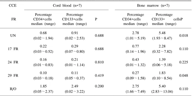
Table 1. Distribution of CD34+ Cell and CD133+ Cells in CCE Fractions
CCE Cord blood (n=7) Bone marrow (n=7)
Percentage Percentage Percentage Percentage
FR CD34+cells CD133+cells P CD34+cells CD133+ cellsP
median (range) median (range) median (range) median (range)
0.68 0.91 2.78 5.48
UN 0.688 0.018
(0.02~1.94) (0.02~2.53) (1.01~5.19) (1.93~8.47)
0.22 0.29 0.77 2.28
17 FR 0.688 0.110
(0.03~0.52) (0.07~0.80) (0.14~1.96) (0.32~7.82)
0.16 0.21 0.43 1.39
24 FR 0.810 0.225
(0.01~0.83) (0.01~1.14) (0.01~1.32) (0.06~5.18)
0.10 0.11 0.27 1.83
29 FR 0.419 0.048
(0.03~0.18) (0.05~0.37) (0.09~1.58) (0.10~8.54)
1.85 2.49 0.200 2.75 5.40
R/O 0.110
(0.05~2.37) (0.02~3.22) (1.66~7.49) (2.83~13.04)
Data reported as the median (range) for seven CCE separation of each tissue.
Abbreviations: CCE; counterflow centrifugal elutriation, UN; unfractionated portion of bone marrow/cord blood, FR;
fraction, R/O; rotor off
0 0.5 1 1.5 2
17 FR 24 FR 29 FR R/O
Fraction
Relative ratio
CB CD34+
CB CD133+
BM CD34+
BM CD133+
0 0.5 1 1.5 2
17 FR 24 FR 29 FR R/O
Fraction
Relative ratio
CB CD34+
CB CD133+
BM CD34+
BM CD133+
Fig. 2. Relative ratio of stem cell markers in cord blood (CB) and bone marrow (BM) cells. The relative ratio of CD34+ and CD133+ cells in each fraction based on the number of CD34+ and CD133+ cells in unfractionated CB and BM cells.
2) CD133과 CD34 양성세포의 분포
역류 원심성 세포분리 후 각 분획의 CD34 양성 세포의 분포는 제대혈의 경우 분리하지 않은 제 대혈이 0.68%로 17 FR, 24 FR 및 29 FR 각각의
0.22%, 0.16%, 0.10% 보다 높았고 R/O의 1.85% 보 다는 낮았다. 골수에서 CD34 양성세포의 분포는 분리하지 않는 군의 2.78%에 비해 17 FR, 24 FR 및 29 FR은 각각 0.77%, 0.43% 및 0.27%로 낮았 고 R/O는 2.75%로 비슷한 분포를 보였다. CD133 양성세포의 경우 제대혈에서 분리하지 않은 군이 0.91%로 17 FR, 24 FR 및 29 FR의 0.29%, 0.21%
및 0.11%에 비해 높았으며 R/O에서 2.49%로 가장 높게 분포하였다. 골수에서의 CD133 양성세포는 분리하지 않은 군과 R/O가 5.48%, 5.40%로 비슷 하게 분포하고 있었으며 17 FR, 24 FR, 29 FR은 각각 2.28%, 1.39%, 1.83%로 낮은 분포를 보였다 (Table 1).
제대혈과 골수 모두 R/O에서 CD34 및 CD133
양성세포가 다른 분획에 비해 높게 분포하고 있
었으며 분리하지 않은 군에 대한 R/O 분획 CD34
양성세포와 CD133양성세포의 상대적인 농축비율
이 제대혈에서 골수보다 높았다(Fig. 2). CD34 양
성세포 중 CD133을 함께 표현하는 경우는 제대혈
Table 2. Percentage of CD133 and CD34 Double Positive Cells Among the CD34 or CD133 Positive Cells from Cord Blood and Bone Marrow According to CCE Fractionations
CCE CD34+CD133+ CD133+CD34+
/CD34+ (%) /CD133+ (%)
Cord blood Bone marrow Cord blood Bone marrow
FR P P
(n=7) (n=7) (n=7) (n=7)
UN 94.0±5.0 91.7±12.4 0.84 62.1±20.5 36.6±8.1 0.51
R/O 87.9±14.2 96.8±2.7 0.53 58.2±18.0 44.8±7.0 0.10
Data reported as the mean±SD for seven CCE separation of each tissue. Abbreviation: See Table 1 Table 3. Number of Colony Forming Units in CCE Fractions of Cord Blood and Bone Marrow
No. of CFU-GM* colonies on day 14 No. of BFU-E* colonies on day 14
CCE FR CB BM CB BM
UN 114.9±67.5 39.3±18.9 19.2±9.1 12.4±5.3
17 FR 8.0±6.3 1.7±1.5 15.0±8.2 0
24 FR 0 0 0 0
29 FR 4.1±4.9 0 1.4±1.6 0
R/O 70.5±35.9 107.4±45.5 16.9±11.8 49.3±32.8
Data was obtained from seven consecutive elutriation of cord blood and bone marrow. Values are presented as mean±SD.
*: total number of colonies/1.5×105 cells after 14 days culture.
Abbreviations: CFU-GM; colony-forming unit for granulcyte/macrophage, BFU-GM; burst-forming unit for erythroid, CCE; counterflow centrifugal elutriation, CB; cord blood, BM; bone marrow, See Table 1
과 골수의 R/O에서 각각 87.9±14.2%, 96.8±2.7%
로 다른 분획들 보다 높은 빈도를 보이고 있었으 며 CD133 양성세포 중 CD34를 함께 표현하는 경 우는 R/O에서 제대혈과 골수의 R/O에서 58.2±
18.0, 44.8±7.0의 낮은 빈도를 보이고 있었다 (Table 2).
3) 집락형성능의 비교
제대혈 및 골수의 집락형성능을 각 분획별로 배양 14일 후에 측정한 결과 1.5×10
5세포 당 CFU-GM의 수는 제대혈에서 분리하지 않은 군이 114.9±67.5개였으며 R/O에서 70.5±35.9개로 17 FR, 24 FR, 29 FR의 8.0±6.3개, 0, 4.1±4.9개에 비하여 많았다. 골수에서는 CFU-GM의 수가 R/O 에서 107.4±45.5개로 분리하지 않은 군의 39.3±
18.9개 보다 많았으며 다른 분획에서는 집락형성 이 거의 관찰되지 않았다.
BFU-E는 제대혈에서 17 FR과 R/O에서 주로 관 찰되었으며 분리하지 않은 제대혈과 비슷한 집락 형성능을 보이고 있었다. 골수에서는 17 FR, 24 FR, 29 FR에서는 집락을 형성하지 않았고 R/O에서 49.3±32.8개로 분리하지 않은 골수의 12.4±5.3개 에 비해 높게 형성되는 것이 관찰되었다(Table 3).
이러한 집락형성능의 결과로 분리하지 않은 제 대혈이나 골수세포군의 총 CFU-GM과 BFU-E의
수를 100%로 하였을 때 각각의 집락수 회복율로 환산하면 제대혈의 경우 17FR, 24FR, 29FR 및 R/O에서 CFU-GM의 회복률은 각각 0.4%, 0%, 0.7% 및 36.4%였고 BFU-E의 회복률은 4.7%, 0%, 1.5% 및 52.2%로 모두 R/O 분획에서 대부분의 집 락형성능이 보존되고 있음을 알 수 있었다. 골수 세포의 경우 17FR, 24FR, 29FR 및 R/O의 CFU- GM의 회복률은 각각 0.5%, 0%, 0% 및 65.1%였고 BFU-E는 R/O에서만 95.0%의 회복률을 보이고 있 었다(Fig. 3).
고 찰
CCE는 세포의 서로 다른 침강속도의 특성을 이용하여 세포의 기능의 소실없이 특정세포집단 으로 분리하는 방법으로 본 연구에서는 제대혈과 골수의 CCE 후 전체 세포수의 회수율은 분리하 기 전 제대혈과 골수의 주입 세포수를 기준으로 제대혈은 91.3%, 골수는 83.0%로 다른 보고들과 비슷한 세포회수율을 보였다
13~17). 본 연구에서는 제대혈과 골수 모두 R/O에서 가장 높은 세포 회 수율을 보이고 있었으나 제대혈은 R/O가 62%, 29 FR이 19%로 주로 중간크기 이상의 세포 회수율 이 약 80%를 차지하고 있어 각 분획별로 15~
27%의 세포회수율을 보인 골수와 CCE의 분획별
0 20 40 60 80 100
17 FR 24 FR 29 FR R/O
Fraction
Recovery rate (%)
CB CFU-GM CB BFU-E BM CFU-GM BM BFU-E
0 20 40 60 80 100
17 FR 24 FR 29 FR R/O
Fraction
Recovery rate (%)
CB CFU-GM CB BFU-E BM CFU-GM BM BFU-E
Fig. 9. Recovery rate of CFU-GM and BFU-E colonies in each fraction based on the number of CFU-GM and BFU-E in unfractionated cord blood and bone marrow.
분포에 차이를 보이고 있었다. 이러한 결과는 Gengozian 등
12)의 제대혈과 골수의 CCE 후 세포 회수율 비교에서 골수는 큰 세포가, 제대혈은 중 간크기의 세포가 모인 분획에서 최고 회수율을 보인 결과와는 차이가 있다.
조혈모세포의 조혈기능은 장기 및 단기배양을 통한 집락형성능과 CD34 양성세포의 수를 측정 함으로써 확인하는데 이러한 조혈기능은 이식 후 생착과 밀접한 관련이 있다
18~21). 최근 CD133은 CD34을 대신할 수 있는 조혈모세포의 표지자로 서 CD34에 비하여 조혈기능이 뛰어나 좀더 초기 단계의 조혈모세포를 표현하는 것으로 알려져 있
는데
2,22,23)본 연구에서는 CD34의 분포와 함께
CD133의 분포를 확인하여 CD34 및 CD133 양성 세포의 빈도가 높은 R/O에서 다른 분획들보다 집 락형성능이 높은 것을 관찰하였으나 세포배양에
는 여러 가지 요소가 작용함으로 CD133과의 직접 적인 연관성을 판단하기는 어렵다.
CCE로 분리한 골수의 R/O 분획은 이식편대 숙 주반응의 문제가 있는 T 세포들의 비율이 적고 조혈모세포는 농축되어 있어서 이를 이용한 주조 직 적합항원 불일치 이식에서 이식편대숙주병 발 생 예방에 매우 효과적이나
8,14,24)이에 반해 거부 반응, 생착의 지연 등이 문제가 되며 이는 이식에 사용하는 R/O 분획이 CCE 과정 중에 조혈모세포
가 다른 분획으로 소실될 수 있기 때문이다. Chang 등
17)은 조혈모세포를 이식편대숙주병을 예방하기 위하여 R/O를 이식하는 경우 생착실패의 요인은 좀더 초기단계의 조혈모세포 소실로 인한 것으로 세포의 크기가 작은 분획에서 좀더 미성숙한 초 기단계의 CD34세포가 있는 것으로 보고하였는데 본 연구에서도 적은 수이기는 하나 17 FR이나 29 FR에서 집락형성능을 관찰할 수 있어 CCE를 통 해 얻은 R/O 이외의 다른 분획으로 조혈모세포의 소실이 있는 것을 확인할 수 있었다. 특히 제대혈 의 경우 17 FR에서 다수의 CFU-GM과 BFU-E 형 성을 확인할 수 있어 크기가 작은 세포에서 초기 단계의 조혈모세포 분포의 가능성을 보여주고 있 다. 한편 R/O 이외의 다른 분획에서의 CD34 및 CD133 양성세포 존재를 통해서도 R/O 이외의 분 획으로 조혈모세포 소실이 있음을 알 수 있다.
Gengozian 등
12)은 CCE를 이용한 말초혈, 제대 혈 및 골수의 조혈모세포연구에서 제대혈과 골수 의 조혈모세포가 주로 존재하는 분획이 차이가 있다고 하였는데 본 연구에서는 제대혈과 골수 모두 R/O에서 대부분의 CFU-GM 및 BFU-E가 회 복되어 큰 차이를 보이지 않았다. 그러나 제대혈 에서 17 FR에서 일부 집락형성능이 관찰되어 제 대혈의 경우 작은 세포분획에서 조혈기능이 이루 어짐을 확인할 수 있었으며 R/O에서는 골수의 CFU-GM 및 BFU-E의 회복률이 제대혈에 비해 높 아 CCE를 통한 골수의 분리에서 R/O에 조혈모세 포가 제대혈에 비해 효율적으로 농축이 됨을 알 수 있었다. R/O를 좀더 세분할 수 있도록 CCE를 시행한다면 본 연구에서 가장 집락형성능이 높았 던 R/O 내에서도 집락형성능과 같은 세포 특성의 차이를 확인할 수 있을 것으로 생각한다.
본 연구에서는 CD34 및 CD133의 빈도가 가장
높은 R/O에서보다 분리하지 않은 제대혈이나 골
수에서 가장 많은 CFU-GM 및 BFU-E 회복률을
보였는데 이는 Wagner 등
15)과 Gengozian 등
16)의
보고에서 CFU-GM 수가 분리하지 않은 골수에서
R/O 분획보다 더 높았던 결과와 일치하며 CFU-
GM이나 BFU-E 형성이 같은 분획 내의 다른 세
포들의 영향을 받기 때문이라고 생각된다. 즉, 같 은 분획 내에 세포성장 억제 인자가 있거나 분리 하지 않은 제대혈 및 골수에서 여러 가지 집락형 성에 도움이 되는 인자들을 분비하는 세포들이 있을 가능성이 있다.
이 연구를 통하여 CCE를 이용하여 분리한 제 대혈 및 골수의 각 분획별 CD133 양성세포의 분 포를 확인할 수 있었으며 R/O 분획이 CD133 및 CD34 양성세포의 빈도가 높고 집락형성능의 회 복률이 가장 높은 것을 알 수 있었다. 특히 골수 의 R/O 분획이 제대혈의 R/O 분획보다 효율적으 로 조혈모세포가 농축됨을 확인할 수 있었으며 향후 각 분획별 CD34, CD133 양성세포 분리 연 구를 통하여 CD34 및 CD133 양성세포의 기능과 조혈기능과의 관계를 알 수 있을 것으로 생각한다.
요 약
목적: CD133항원은 새로이 확인된 혈액줄기세 포 항원으로 CD133 양성세포를 이용한 이식에서 성공적인 생착이 관찰되고 장기세포배양 능력이 확인되어 CD34항원을 대신하는 혈액줄기세포의 표지자로 이용되고 있다. 역류원심성 세포분리 (CCE)는 세포를 손상시키지 않으면서 침강능력에 따라 특징적인 여러군으로 분리하는 물리적 방법 으로 본 연구에서는 CCE를 이용하여 분리한 제 대혈과 골수의 각 분획의 특성을 확인하고 CD34 와 CD133 양성세포의 분포와 조혈기능의 차이를 알아보고자 하였다.
방법: 제대혈과 골수의 유핵세포를 CCE로 배지 의 주입속도에 따라 17, 24, 29, R/O 분획으로 나 누어 세포를 분리하였으며 각 분획의 CD133 양성 세포의 분포를 CD34 양성세포와 비교하고 과립 구-대식세포 집락형성능(CFU-GM)과 적혈구 집락 형성능(BFU-E)을 비교하였다.
결과: 각 분획의 제대혈 유핵세포 회수율은 17, 24, 29 및 R/O 분획에서 각각 5.9±4.3%, 4.2±
2.4%, 19.4±11.9% 및 61.9±11.7%였고, 골수 유핵 세포 회수율은 14.9±8.2%, 17.4±13.4%, 23.6±
6.11%, 27.1±8.9%였다. 제대혈에서 각 분획의 CD133 양성세포는 분리하지 않은 군에서 0.91%
이었으나 R/O 분획에서 2.49%로 다른 분획에 비 하여 높게 나타났으며 CD34 양성세포의 분포도 분리하지 않은 군에서 0.68%이었지만 R/O 분획에 서 1.85%로 가장 많이 분포하고 있었다. 한편, 골 수에서는 CD133 양성세포가 분리하지 않은 군이 5.48%이었고 R/O에서 5.40%로 비슷하였으나 다 른 분획에 비하여 월등히 많이 분포하고 있었고 CD34 양성세포는 분리하지 않은 군과 R/O 분획 이 2.78%와 2.75%로 비슷한 분포를 보였다.
배양 14일 후에 측정한 제대혈의 1.5×10⁵ 유핵 세포 당 CFU-GM의 수는 17, 24, 29 및 R/O 분획 에서 각각 8.0±6.3개, 0, 4.1±4.9개 및 70.5±35.9 개, BFU-E는 각각 15.0±8.2개, 0, 1.4±1.6개 및 16.9±11.8개였다. 골수에서는 CFU-GM이 17, 24, 29 분획에서는 거의 없었으며 R/O 분획에서만 107.4±45.5개로 분리하지 않은 군의 39.3±18.9개 보다 많았고 BFU-E도 R/O 분획에서만 49.3±32.8 개로 분리하지 않은 골수의 12.4±5.3개에 비해 높게 형성되는 것이 관찰되었다.
결론: CCE를 이용하여 분리한 제대혈 및 골수 의 각 분획별 CD133 양성세포의 분포를 확인할 수 있었으며 R/O 분획이 CD133 및 CD34 양성세 포의 빈도가 높고 집락형성능의 회복률이 가장 높은 것을 알 수 있었다. 특히 골수의 R/O 분획이 제대혈의 R/O 분획보다 효율적으로 조혈모세포가 농축됨을 확인할 수 있었다.
참 고 문 헌
1. Miraglia S, Godfrey W, Yin AH, Atkins K, Warnke R, Holden JT, et al. A novel five-transmembrane hema- topoietic stem cell antigen: isolation, characterization, and molecular cloning. Blood 1997;90:5013-21 2. Yin AH, Miraglia S, Zanjani ED, Almeida-Porada G,
Ogawa M, Leary AG, et al. AC133, a novel marker for human hematopoietic stem and progenitor cells.
Blood 1997;90:5002-12
3. de Wynter EA, Buck D, Hart C, Heywood R, Cou- tinho LH, Clayton A, et al. CD34+AC133+ cells
isolated from cord blood are highly enriched in long-term culture-initiating cells, NOD/SCID- repopu- lating cells and dendritic cell progenitors. Stem Cells 1998;16:387-96
4. Koehl U, Zimmermann S, Esser R, Sorensen J, Gruttner HP, Duchscherer M, et al. Autologous transplantation of CD133 selected hematopoietic progenitor cells in a pediatric patient with relapsed leukemia. Bone Marrow Transplant 2002;29:927-30 5. Almici C, Carlo-Stella C, Donnenberg AD, Rizzoli V.
Counterflow centrifugal elutriation: present and future.
Bone Marrow Transplant 1993;12:105-8
6. Noga SJ. Elutriation: new technology for separation of blood and bone marrow. Lab Med 1988;19:234-9 7. Saffran DC, Parsons MF, Singhal SK. Separation of
allostimulatory and natural suppressor/stem cell func- tions of murine bone marrow - implications for bone marrow transplantation. Transplant 1991;52:680-4 8. Noga SJ, Davis J, Schepers K, Eby L, Berenson RJ.
The clinical use of elutriation and positive stem cell selection columns to engineer the lymphocyte and stem cell composition of the allograft. Prog Clin Biol Res 1994;389:317-24
9. De Witte T, Hoogenhout J, de Pauw B, Holdrinet R, Janssen J, Wessels J, et al. Depletion of donor lymp- hocytes by counterflow centrifugation successfully prevents acute graft-versus-host disease in matched allogeneic marrow transplantation. Blood 1986;67:
1302-8
10. Neudorf SM, Rybka W, Ball E, Blatt J, Bloom E, Corey S, et al. The use of counterflow centrifugal elutriation for the depletion of T cells from unrelated donor bone marrow. J Hematother 1997;6:351-9 11. Jones RJ, Wagner JE, Celano P, Zicha MS, Sharkis
SJ. Separation of pluripotent haematopoietic stem cells from spleen colony-forming cells. Nature 1990;347:188-9 12. Gengozian N, Hill RJ, Caudle MR, Panella TJ. Re-
lative sedimentation of hematopoietic progenitors in human cord blood, peripheral blood, and bone marrow as determined by counterflow centrifugal elutriation.
Transplantation 1998;65:939-46
13. Jemionek JF, MacVittie TJ, Byrne PJ, Schein PS, Walden DA. Fractionation of mammalian bone mar- row by counterflow centrifugation-elutriation using a continuous albumin gradient: analysis of granulocyte- macrophage colony forming units. Br J Haematol 1982;50:257-67
14. Noga SJ, Donnenberg AD, Schwarz CL, Lewis C, Strauss LC, Civin CI, et al. Development of a
simplified counterflow centrifugal elutriation proce- dure for depletion of lymphocytes from human bone marrow. Transplant 1986;41:220-9
15. Wagner JE, Donnerberg AD, Noga SJ, Wiley JM, Yeager AM, Vogelsang GB, et al. Separation of rat bone marrow cells by counterflow centrifugal elutri- ation: a model for studying the effects of lymphocyte depletion. Exp Hematol 1988;16:206-12
16. Gengozian N, Legendre AM. Separation of feline bone marrow cells by counterflow centrifugal elutriation.
Identification and isolation of presumptive early and late myeloid/erythroid progenitors. Transplant 1995;60:
836-41
17. Chang Q, Harvey K, Akard L, Thompson J, Dugan MJ, English D, et al. Differences in CD34+ cell subpopulations between human bone marrow and
“mobilized” peripheral blood as determined with counterflow centrifugal elutriation. Exp Hematol 1997;25:423-31
18. Feugier P, Bensoussan D, Girard F, Alla F, Schuh- macher A, Latger-Cannard V, et al. Hematologic recovery after autologous PBPC transplantation:
importance of the number of postthaw CD34+ cells.
Transfusion 2003;43:878-84
19. Fu SQ, Abboud CN, Brennan JK, Ifthikharuddin JJ, Nichols D, Liesveld JL. Impact of mobilized blood progenitor cell quality determined by the CFU- GM/
CD34+ ratio on rapid engraftment after blood stem cell transplantation. Blood Cells Mol Dis 2002;28:
315-21
20. al-Fiar F, Prince HM, Imrie K, Stewart AK, Crump M, Keating A. Bone marrow mononuclear cell count does not predict neutrophil and platelet recovery following autologous bone marrow transplant: value of the colony-forming unit granulocyte-macrophage (CFU-GM) assay. Cell Transplant 1997;6:491-5 21. Kim SK, Im MW, Nahn CH, Kim CS, Seo JY, Fraser
JK. Ex vivo expansion and clonality of CD34 cells from bone marrow, umbilical cord blood and mobil- ized peripheral blood in serum-free media. Korean J Pediatr Hematol-Oncol 2001;8:101-9
22. Pasino M, Lanza T, Marotta F, Scarso L, De Biasio P, Amato S, et al. Flow cytometric and functional characterization of AC133+ cells from human umbil- ical cord blood. Br J Haematol 2000;108:793-800 23. Kobari L, Giarratana MC, Pflumio F, Izac B, Cou-
lombel L, Douay L. CD133+ cell selection is an alternative to CD34+ cell selection for ex vivo ex- pansion of hematopoietic stem cells. J Hematother Stem Cell Res 2001;10:273-81
24. Jeong DC, Han CW, Jin JY, Kim DS, Choi IB, Kim WI, et al. Effectiveness of rotor off fraction in allogeneic murine bone marrow transplantation with complete disparity of major histocompatibility. Exp Hamatol 1999;27:1219-25