DOI:10.4047/jap.2010.2.2.46
46
J Adv Prosthodont 2010;2:46-9
INTRODUCTION
Patients with destroyed dentition have several characteris- tics such as loss of posterior occlusal support, severe occlusal wear of remained teeth, missing of multiple teeth, etc. The loss of posterior occlusal support may cause following problems:
the attrition of anterior teeth, the reduction of vertical dimen- sion (VD), and the alteration of normal occlusal plane. These problems will eventually cause the unesthetic facial appear- ance, the pathologic changes of the temporomandibular joint (TMJ) and masticatory muscles, and the inability of mastication.1 Patients with destroyed dentition require extensive restorative treatment.1In such cases the restoration with implant-supported crowns and fixed partial dentures is preferred. However, in patients with financial, anatomical, and/or medical limitations alternative treatment modalities, such as removable partial den- ture (RPD), should be considered.2
The edentulous patients with compromised esthetic zone can be successfully treated with a rotational path RPD.3-9Rotational path RPD has been often overlooked by the dental profession due to its complex prosthetic design and sensitive laboratory techniques.10With better understanding of the principles of rota- tional path RPD, the dental clinician can deliver excellent esthet- ic outcome in compromised areas in which other treatment options may often be limited.
The rotational path design concept uses a rigid retainer portion of the framework as the retention component.11The prox-
imal plate provides retention through its intimate contact with the proximal tooth surface below the height of contour at a zero-degree tilt. These rigid retentive components have dual paths: the first path to engage into the undercuts and the second rotational path to fully seat the prosthesis.3,10,12There are three basic types of rotational path RPDs: anterior-posterior (AP), posterior-anterior (PA), and lateral. Jacobson and Krol narrowed the rotational paths to two categories.11Category I includes all prosthesis designs that first seat the rest that is associated with the rigid retainer. After the first rest is seated, the second seg- ment rotates into place and conventional clasps are engaged.9 The rotational centers are located at the end of long rests.
Category I includes all PA and AP paths of insertion that replace posterior teeth.10Category II includes all lateral paths and AP paths that replace anterior teeth. The centers of rotation and the rigid retainers are located at the gingival extension of minor connectors.9,11These prostheses use a dual path of insertion: at first RPD is inserted straightly from an incisal or occlusal direc- tion to the rotational centers and then the conventional posterior or contralateral retainer is rotated into position. It is important that the initial straight path of insertion must be parallel with the cingulum rests to permit complete seating. The rotation- al path RPD offers the advantages of improved esthetics by elim- inating anterior clasps, shortened treatment duration, cleanliness, and lower treatment cost over implants or fixed partial den- tures.5,6,11
Full mouth rehabilitation of destroyed dentition with rotational path removable partial denture: a case report
Moon-Hyoung Kim, DDS, Seong-Joo Heo, DDS, MSD, PhD, Seong-Kyun Kim, DDS, MSD, PhD Jai-Young Koak*, DDS, MSD, PhD
Department of Prosthodontics and Dental Research Institute, School of Dentistry, Seoul National University, Seoul, Korea
BACKGROUND. Though implant dentistry is very successful and predictable in treatment of patients with destroyed dentition, there are some cases with limitations to implant therapy. In these cases, alternative treatment modality should be considered. CASE DESCRIPTION. A patient with destroyed dentition was rehabilitated with a lateral rotational path removable partial denture. According to the diagnosis, we determined to raise vertical dimension for esthetic and functional restoration. The final restoration was performed after four months of provisional period.
CLINICAL IMPLICATION. The edentulous patients with compromised esthetics and functions can be successfully treated with a rotational path removable partial denture through adequate treatment planning and precise laboratory procedure. [J Adv Prosthodont 2010;2:46-9]
Corresponding author: Jai-Young Koak
Department of Prosthodontics and Dental Research Institute, School of Dentistry 275-1, Yeongeon-Dong, Jongno-Gu, Seoul, 110-768, Republic of Korea Tel, +82 2 2072 3860: e-mail, [email protected]
Received April 30, 2010 / Last Revison May 20, 2010 / Accepted May 24, 2010 CASE REPORT
KEY WORDS. Esthetic restoration, Occlusal wear, Rotational path RPD, Vertical dimension
ⓒ 2010 The Korean Academy of Prosthodontics
This is an Open Access article distributed under the terms of the Creative Commons Attribution Non-Commercial License (http://creativecommons.org/licenses/by- nc/3.0) which permits unrestricted non-commercial use, distribution, and reproduction in any medium, provided the original work is properly cited.
47 J Adv Prosthodont 2010;2:46-9
CASE STUDY
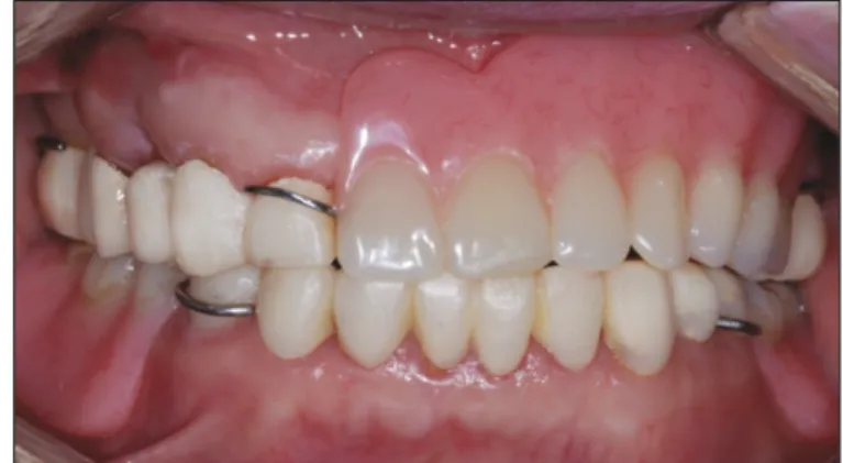
The patient was a 70 year-old female complaining of inabil- ity of mastication and unesthetic facial appearance. She had no specific medical history and dysfunctional habit. Typical features of destroyed dentition were observed in the clinical and radiographic examinations (Fig. 1). However, she had sev- eral problems: severe attrition of almost remained teeth, #14 horizontal root fracture, #13 periapical lesion and buccal fis- tula formation, insufficient interocclusal distance, loss of anterior guidance. In addition, severe ridge resorption result- ed in asymmetric and unsupported upper lips. She had fear of surgery, financial limitations and severe ridge resorption of esthet- ic zone. For these reasons, we decided that not implant pros- thesis but removable prosthesis was indicated for her. In this case, the extremely worn anterior dentition was planned to be restored with fixed prosthesis. All attrited teeth were restored with single porcelain-fused gold (PFG) crowns except four low- er incisors, which were restored with 4-unit splinted PFG fixed dentures due to their very short crown lengths. The upper miss- ing area was to be restored with rotational path RPD. Finally, the lower missing area was to be restored with conventional RPD. The rotational path RPD is the excellent treatment
modality of restoring missing incisors when patients have con- traindications for implants or when patients are financially limited.
Preliminary impression was made and then recording base and wax rim were fabricated on the diagnostic casts for VD deter- mination and centric relation (CR) registration. Anatomic landmarks, facial measurements and the resting positions of mandibular jaw were used to determine appropriate vertical dimension for the patient. As the result, it was decided that VD raising was necessary for esthetic and functional restoration.
This increased vertical dimension was 4 mm from the tips of incisors. After that, CR registration was taken using bilateral manipulation1, and diagnostic wax up was performed to con- firm the need of increasing VD for esthetic and functional reha- bilitation. Prior to teeth preparation, composite resin was filled in the incisal and occlusal surfaces of all attrited teeth.
This conservative correction was for conservation of destroyed tooth structure and ideal tooth preparation.1Teeth were prepared using a omni-vac as the preparation guide. To compensate for short clinical crown lengths, axial surfaces were reduced as par- allel as possible. The provisional restoration was fabricated based on the diagnostic wax up (Fig. 2). While the patient was wearing the provisional restorations for the 4 months, occlusal
Full mouth rehabilitation of destroyed dentition with rotational path removable partial denture: a case report KIM MH et al.
Fig. 1. Initial intraoral status. Fig. 2. Provisional restoration.
Fig. 3. Transfer procedure of provisional vertical dimension to final restoration. A: Stable bite registration with resin coping crowns, B: Lower record- ing base and wax rim were used for posterior bite registration, C: Upper anterior recording base and wax rim were used for anterior bite registration.
A B C
48 J Adv Prosthodont 2010;2:46-9
stability and TMJ were periodically checked. The patient was satisfied with the provisional restoration. Thus, it was decid- ed to reproduce of temporary VD and CR state on the final restoration. It was performed using the recording base, wax- rim and two pattern resin (Dura Lay, Reliance, Illinois, USA) coping crowns. This transfer procedure was described in the Fig. 3.
Final impression was made for PFG crowns. PFG crown cop- ings were tried in the mouth to check for marginal fit. Before glazing, surveying was performed repeatedly for rotational path RPD. All PFG prostheses were cemented with resin modified glass ionomer cement (Fuji-cem, GC, Tokyo, Japan). To fab- ricate RPD framework, upper and lower master casts were made.
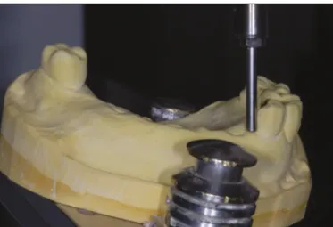
Upper RPD was classified as Class III Mod. 1 RPD and was designed in lateral rotational path RPD with no clasp in the ante- rior region. Lateral or category II rotational path RPD in this report was surveyed in the next two steps (Fig. 4). The first surveying was performed at a zero-degree tilt to identify the mesial surface undercut of the anterior abutments (at least 0.010 inch) and buccal undercuts of the posterior abutments. The diag- nostic cast was then tilled upward until the mesial undercuts on the anterior abutments were eliminated. The cast was again surveyed to determine if the anterior rest seats are
accessible during the initial straight path of insertion. Lower RPD was conventional class I RPD with linguoplate major con- nector (Fig. 5). Both RPD frameworks were tried in the
KIM MH et al.
Fig. 5. A: Upper rotational path RPD, B: Lower conventional RPD.
A B
Full mouth rehabilitation of destroyed dentition with rotational path removable partial denture
Fig. 4. The surveying procedure for lateral rotational path RPD; following the first surveying procedure at a zero-degree tilt, the diagnostic cast was tilled upward until the mesial undercuts on the anterior abutments were eliminated for the second surveying.
Fig. 6. Final prostheses; the stable occlusion was established.
49 J Adv Prosthodont 2010;2:46-9
mouth and adapted with silicone material (Fit-checker, GC, Tokyo, Japan). RPD frameworks were very stable and showed very accurate fit and retention. For lower RPD, altered cast was made. A face-bow transfer was completed, and interocclusal registration was made with wax rim and bite material (O-bite, DMG, Hamburg, Germany). The casts were mounted in a semi- adjustable articulator. Denture teeth were set up to establish the stable and harmonious occlusion as such13: (1) Simultaneous bilateral contacts of opposing posterior teeth in the centric occlu- sion (CO) (2) unilateral balanced occlusion by denture teeth for left working side contacts (3) canine guidance by natural teeth for right working side contacts (4) contacts of opposing anterior teeth in the CO. After obtaining the patient’s approval, RPDs were processed using pink denture resin (Rapidsimplified, Vertex, Zeist, Netherlands). The finished RPDs were placed in the mouth, and the following criteria were evaluated:
adaptation of the clasps and rests, retention of the RPD, esthetics, and occlusion (Fig. 6). Finally the patient was instructed in placing the prosthesis and maintaining oral hygiene.
CONCLUSION
Treatment of patients with destroyed dentition is very difficult clinical procedure and challenging for dental profession. In this case, proper diagnosis and treatment plans are the necessity for not only esthetic and functional restorations but also the sta- bility and adaptation of the neuromuscular system and TMJ.
For compromised patients to implant dentistry, rotational path RPD is an alternative treatment option. It improves esthetics to replace missing teeth without placing a conventional clasp in the esthetic region. It is important that dentists eval- uate each patient carefully and survey casts meticulously to ensure success.
REFERENCES
1. Dawson PE. Functional Occlusion - From TMJ to smile design.
1sted. N.Y.; Mosby; 2007, p. 18-26, 114-29, 430-52.
2. Ancowitz S. Esthetic removable partial dentures. Gen Dent 2004;52:453-9; quiz 460.
3. King GE. Dual-path design for removable partial dentures. J Prosthet Dent 1978;39:392-5.
4. Carreiro Ada F, Machado AL, Giampaolo ET, Santana IL, Vergani CE. Dual path: a concept to improve the esthetic re- placement of missing anterior teeth with a removable partial den- ture. J Prosthodont 2008;17:586-90.
5. Byron R Jr, Frazer RQ, Herren MC. Rotational path removable partial denture: an esthetic alternative. Gen Dent 2007;55:
245-50.
6. Suh JS, Billy EJ. Rotational path removable partial denture (RPD):
conservative esthetic treatment option for the edentulous mandibular anterior region: a case report. J Esthet Restor Dent 2008;20:98-105;discussion 106-7.
7. Grageda E. Achieving an aesthetic anterior-posterior rotation- al path partial denture: case report. Dent Today 2007;26:130, 132- 5.
8. Donovan T. Use of the rotational path removable partial denture concept in a Kennedy Class II patient: a case report. J Esthet Restor Dent 2008;20:294-8.
9. Jacobson TE. Rotational path partial denture design: a 10-year clinical follow-up-Part I. J Prosthet Dent 1994;71:271-7.
10. Halberstam SC, Renner RP. The rotational path removable partial denture: the over-looked alternative. Compendium 1993;14:544, 546-52.
11. Jacobson TE, Krol AJ. Rotational path removable partial den- ture design. J Prosthet Dent 1982;48:370-6.
12. Firtell DN, Jacobson TE. Removable partial dentures with ro- tational paths of insertion: problem analysis. J Prosthet Dent 1983;50:8-15.
13. McCracken WL, Carr AB, McGivney GP. McCracken’s re- movable partial prosthodontics. 10thed. St. Louis; Mosby;
2000, p. 356-60.
Full mouth rehabilitation of destroyed dentition with rotational path removable partial denture: a case report KIM MH et al.