107 A 22-year-old man was hospitalized for right femur fracture
due to a motorcycle accident. Although he had no known car- diac or family history, he felt intermittent chest tightness dur- ing moderate intensity of exercise. His electrocardiography showed patterns of left ventricular strain. The echocardiogra- phy showed left ventricular hypertrophy, mild eccentric mitral regurgitation, and regional wall motion abnormality and thin- ning of left anterior descending (LAD) coronary artery territory with lower normal left ventricular systolic function, in which ejection fraction was 50%. Diastolic flow showing peak veloc- ity of 2.5 cm/sec was observed at interventricular septum, which was suspicious of excessive collateral flow at parasternal short axis view (Fig. 1A). Dilated right coronary artery (RCA) osti- um of 10 mm was observed (Fig. 1B) on parasternal long axis view, whereas left main coronary artery was not detected in typi- cal situs. Notably, an abnormal retrograde shunt flow was de- tected (Fig. 1C, Supplementary movie 1) and a drainage site of abnormal shunt flow was observed at the main pulmonary ar- tery (PA) level of parasternal short axis view (Fig. 1D). Thus, we suspected a congenital anomaly of the coronary arteries.
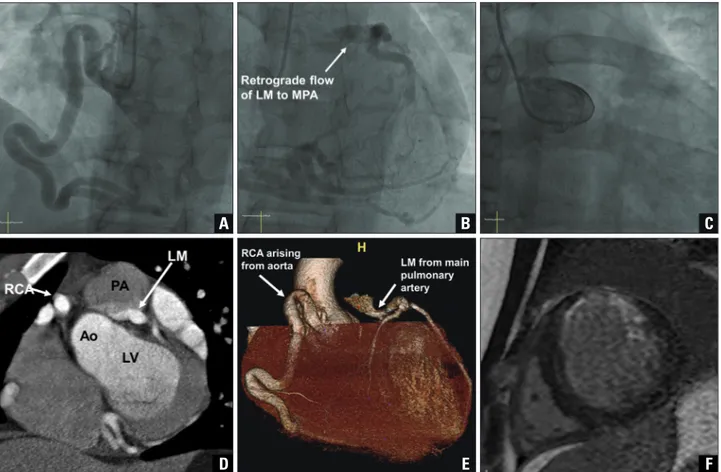
Coronary angiography revealed an enlarged and tortuous RCA with abundant septal collateral flows toward the left coronary artery (LCA). An unusual location of the left main coronary artery opening with an abnormal retrograde shunt flow was observed in the left superior part of aorta, most likely PA (Fig.
2A and B, Supplementary movie 2). However, LCA was not shown in the left coronary cusp (Fig. 2C). To specify the loca- tion of the left main coronary artery opening, cardiac multide- tector computed tomography (CT) was performed and the
pISSN 1975-4612 / eISSN 2005-9655 Copyright © 2017 Korean Society of Echocardiography www.kse-jcu.org https://doi.org/10.4250/jcu.2017.25.3.107
• *Byung Gyu Kim and Sung Woo Cho contributed equally to this work.
• Received: August 7, 2017 • Revised: September 1, 2017 • Accepted: September 1, 2017
• Address for Correspondence: Jong Chun Nah, Division of Cardiology, Department of Internal Medicine, Inje University College of Medicine, Seoul Paik Hospital, 9 Mareunnae-ro, Jung-gu, Seoul 04551, Korea Tel: +82-2-2270-0114, Fax: +82-2-2270-0579, E-mail: [email protected]
• This is an Open Access article distributed under the terms of the Creative Commons Attribution Non-Commercial License (http://creativecommons.org/licenses/by-nc/4.0) which permits unrestricted non-commercial use, distribution, and reproduction in any medium, provided the original work is properly cited.
anomalous origin of left coronary artery from pulmonary artery (ALCAPA) was finally confirmed (Fig. 2D and E). To determine myocardial viability, cardiac magnetic resonance imaging (MRI) was performed and the thinning and subendocardial delayed enhancement of anterior wall from base to middle left ventri- cle was observed, which suggested chronic subendocardial in- farction of LAD territory (Fig. 2F). The patient received beta- blocker and angiotensin converting enzyme inhibitor, and completed femur fracture surgery without any cardiovascular events. He was discharged and is scheduled for an open cardiac surgery for the correction of ALCAPA.
ALCAPA is a very rare congenital heart disease with report- ed incidence rate of 1 in 300000 children, or 0.5% of those with congenital heart disease.1) If untreated, up to 90% of patients with ALCAPA die during the first year of life due to severe myo- cardial ischemia. Only 10–15% of ALCAPA patients reach adulthood depending on the development of sufficient inter- coronary communications.2) Despite abundant collaterals, blood supply to LCA territory can be inadequate, especially to the sub- endocardial region according to the proportion of coronary steal from coronary artery to PA.
For a precise diagnosis and risk stratification of ALCAPA, multimodality imaging studies are needed. Echocardiography is essential and provides important clues for diagnosis of AL- CAPA, as demonstrated in our case. RCA dilatation, collateral coronary artery flow, LCA flow reversal, mitral regurgitation, and left ventricular dysfunction are known as important echo- cardiographic findings of ALCAPA.3) Cardiac CT is useful for visualization of coronary arteries, but is limited in evaluating cor- IMAGES IN CARDIOVASCULAR ULTRASOUND J Cardiovasc Ultrasound 2017;25(3):107-109
Multimodality Imaging of Anomalous Left Coronary Artery from
the Pulmonary Artery
Byung Gyu Kim, MD1*, Sung Woo Cho, MD1*, Dae Hyun Hwang, MD2, and Jong Chun Nah, MD1
1Division of Cardiology, Departments of Internal Medicine, 2Radiology, Inje University College of Medicine, Seoul Paik Hospital, Seoul, Korea
KEY WORDS: Anomalous left coronary artery from pulmonary artery · Coronary malformation · Multimodality imaging.
Journal of Cardiovascular Ultrasound 25 | September 2017
108
onary flow, valvular function, and myocardial viability. Cardiac MRI provides more detailed information on myocardial perfu- sion, fibrosis, and viability for deciding treatment plan. Coro- nary angiography is definitive diagnostic modality for provid- ing anatomic information of anomalous coronary arteries and their flow. There are no current guidelines for the optimal treat- ment of ALCAPA. According to previous literatures, surgical correction has good prognosis and is recommended regardless of age or symptoms in order to prevent adverse outcomes in- cluding myocardial infarction or sudden cardiac death.4)
We present a very rare case of an adult type ALCAPA accom- panied by chronic subendocardial infarction, which was initial- ly suspected by echocardiography and confirmed by multimo- dality imaging studies. The present case also emphasizes the essential role of echocardiography for providing important clues
of ALCAPA.
Supplementary movie legends
Movie 1. Echocardiography: parasternal short axis view at the level of pulmonary trunk.
Movie 2. Coronary angiography.
References
1. KEITH JD. The anomalous origin of the left coronary artery from the pulmo- nary artery. Br Heart J 1959;21:149-61.
2. Moodie DS, Fyfe D, Gill CC, Cook SA, Lytle BW, Taylor PC, Fitzger- ald R, Sheldon WC. Anomalous origin of the left coronary artery from the pulmonary artery (Bland-White-Garland syndrome) in adult patients: long- term follow-up after surgery. Am Heart J 1983;106:381-8.
3. Patel SG, Frommelt MA, Frommelt PC, Kutty S, Cramer JW. Echo- cardiographic diagnosis, surgical treatment, and outcomes of anomalous left Fig. 1. An accelerated diastolic color flow within the interventricular septum indicating a large septal collateral flow with frontal direction from RCA to left anterior descending artery on parasternal short axis view (A). Markedly dilated RCA ostium on parasternal long axis view (arrowheads) (B).
Retrograde diastolic shunt flow toward pulmonary valve at MPA (arrow) (C). Drainage site of abnormal shunt flow at MPA with diastolic reversal flow (D).
MPA: main pulmonary artery, RCA: right coronary artery.
A
C
B
D
Multimodality Imaging of ALCAPA | Byung Gyu Kim, et al.
109 coronary artery from the pulmonary artery. J Am Soc Echocardiogr 2017;
30:896-903.
4. Kottayil BP, Jayakumar K, Dharan BS, Pillai VV, Ajitkumar V, Me-
A
D
B
E
C
F
Fig. 2. Coronary angiography showing an enlarged and tortuous RCA from Ao (A). Retrograde filling of LCA through abundant collaterals from RCA and abnormal shunt flow from left main stem to main PA (B). No visualization of LCA in left coronary cusp (C). Sagittal section and 3D reconstruction images of computed tomography of the LCA originating from the PA with retrograde contrast flow from LCA to PA (D and E). Thinning and subendocardial delayed enhancement of anterior LV wall on cardiac magnetic resonance imaging (F). Ao: aorta, LM: left main artery, LCA: left coronary artery, LV: left ventricle, MPA: main pulmonary artery, PA: pulmonary artery, RCA: right coronary artery.
non S, Sanjay G. Anomalous origin of left coronary artery from pulmonary artery in older children and adults: direct aortic implantation. Ann Thorac Surg 2011;91:549-53.