ABSTRACT
Purpose: Gastric cancer (GC) patients with peritoneal metastasis (PM) have poor prognosis.
Pressurized intraperitoneal aerosol chemotherapy (PIPAC) in combination with systemic chemotherapy is a novel treatment option for patients in stage IV of the disease.
Materials and Methods: Between November 2015 and June 2018, prospective data collection was performed in 24 patients with GC and PM (median age, 57; range, 44–75 years). These patients underwent 46 PIPAC procedures with a median number of 2 interventions per patient (range, 1–6). A laparoscopic access was used and a combined therapy of cisplatin and doxorubicin aerosol was administered.
Results: The median peritoneal carcinomatosis index before the 1st PIPAC was 14 (range, 2–36), and the median ascites volume in patients before the 1st PIPAC was 100 mL (range, 0–6 mL, 300 mL). Eleven patients, who received 2 or more PIPAC procedures, had decreased and stable volumes of ascites, while only 3 patients displayed increasing volume of ascites.
The median overall survival was 121 days (range, 66–625 days) after the 1st PIPAC procedure, while 8 patients who received more than 3 PIPAC procedures had a median survival of 450 days (range, 206–481 days) (P=0.0376).
Conclusions: Our data show that PIPAC is safe and well tolerated, and that the production of ascites can be controlled by PIPAC in GC patients. Patients, who received 2 or more PIPAC procedures, reported a stable overall quality of life. Further studies are required to document the significance of PIPAC as a palliative multimodal therapy.
Trial Registration: ClinicalTrials.gov Identifier: NCT03100708
Keywords: Gastric cancer; Peritoneal metastasis; PIPAC; Palliative chemotherapy
Original Article
Received: Aug 3, 2018 Revised: Dec 3, 2018 Accepted: Dec 3, 2018 Correspondence to Ines Gockel
Department of Visceral, Transplant, Thoracic and Vascular Surgery, University Hospital of Leipzig, Liebigstraße 20, 04103 Leipzig, Germany.
E-mail: [email protected]
*Ines Gockel and Boris Jansen-Winkeln contributed equally to this work.
Copyright © 2018. Korean Gastric Cancer Association
This is an Open Access article distributed under the terms of the Creative Commons Attribution Non-Commercial License (https://
creativecommons.org/licenses/by-nc/4.0) which permits unrestricted noncommercial use, distribution, and reproduction in any medium, provided the original work is properly cited.
ORCID iDs Ines Gockel
https://orcid.org/0000-0001-7423-713X Boris Jansen-Winkeln
https://orcid.org/0000-0002-3996-9391 Matthias Mehdorn
https://orcid.org/0000-0002-0047-368X Yusef Moulla
https://orcid.org/0000-0002-5936-0217
Ines Gockel 1,*, Boris Jansen-Winkeln 1,*, Linda Haase1, Philipp Rhode1, Matthias Mehdorn 1, Stefan Niebisch1, Yusef Moulla 1, Orestis Lyros 1, Florian Lordick 2, Katrin Schierle 3, Christian Wittekind 3, René Thieme 1
1 Department of Visceral, Transplant, Thoracic and Vascular Surgery, University Hospital of Leipzig, Leipzig, Germany
2University Cancer Center Leipzig, University Hospital of Leipzig, Leipzig, Germany
3Institute of Pathology, University Hospital of Leipzig, Leipzig, Germany
Pressurized Intraperitoneal Aerosol Chemotherapy (PIPAC) in Gastric Cancer Patients with Peritoneal
Metastasis (PM): Results of a Single-
Center Experience and Register Study
Orestis Lyros
https://orcid.org/0000-0002-7727-7804 Florian Lordick
https://orcid.org/0000-0001-8591-9339 Katrin Schierle
https://orcid.org/0000-0002-5188-5537 Christian Wittekind
https://orcid.org/0000-0002-3779-9309 René Thieme
https://orcid.org/0000-0002-0537-3979 Trial Registration
ClinicalTrials.gov Identifier: NCT03100708 Funding
This work was supported by a Junior Research Grant to RT and SN, the Clinician Scientist Program to OL, and a scholarship to PR by the Faculty of Medicine, University of Leipzig, Germany.
Author Contributions
Conceptualization: G.I., J.W.B.; Data curation:
J.W.B., H.L., R.P.; Formal analysis: G.I., J.W.B., H.L., R.P., T.R.; Methodology: S.K., W.C.;
Project administration: M.Y., L.O., M.M., L.F.;
Visualization: H.L., R.P., T.R.; Writing - review &
editing: G.I., J.W.B., T.R.
Conflict of Interest
No potential conflict of interest relevant to this article was reported.
INTRODUCTION
Globally, gastric cancer (GC) is the second most common cause of cancer-related death [1]
and although more prevalent in East Asia, it is one of the 5 most common cancers in Europe in terms of incidence and mortality [2]. GC frequently metastasizes hematogenously to distant organs or directly into the peritoneum. Peritoneal metastasis (PM) is the most frequent pattern of metastasis in stage IV GC. Synchronous PM was reported in 10-40% of all GC patients, even in those initially scheduled for curative surgery [3-5]. About 60% of deaths due to GC are related to metachronous PM [3,6]. Life expectancy in patients with GC-induced PM is limited to about 3–9 months [5,7,8]. The effect of systemic therapy on PM is limited, which, in part, has been explained by restricted drug distribution to the peritoneum [9].
Pressurized intraperitoneal aerosol chemotherapy (PIPAC) is a relatively new method used in selected patients with PM who have no indication for cytoreductive surgery (CRS) and hyperthermic intraperitoneal chemoperfusion (HIPEC). In PIPAC, the cytotoxic agents are applied laparoscopically (usually by 2 trocars) using pressurized aerosols. The treatment objective is to alleviate symptoms, in particular to control ascites and to induce regression of PM, leading to a better quality of life (QoL). Before PIPAC can be administered, potentially curative treatment options like CRS and HIPEC have to be excluded and further tumor manifestations, such as distant hematogenous metastases, are to be ruled out.
In December 2015, we initiated a prospective single-center registry study (NCT03100708) to investigate the perioperative morbidity and mortality as well as the safety aspects of PIPAC [10]. In addition, we assessed prospectively its efficacy and patients' prognosis and QoL.
The current publication is dedicated to the cohort of GC patients, comprising the largest population of our study.
MATERIALS AND METHODS
Patients
Patients with a histologically proven GC-induced PM, following a standardized diagnostic laparoscopy, were eligible for this study. The indications for PIPAC were determined in our multidisciplinary tumor board, where also the option of CRS and HIPEC had to be excluded, taking into account the peritoneal carcinomatosis index (PCI) (usually higher than 8–10), comorbidities, and patients' preferences. Patients with progressive disease during systemic chemotherapy and patients with distant metastases other than PM were not eligible for PIPAC.
However, individual interviews were carried out in qualified and low-risk patients who were absolutely interested in this option for symptomatic ascites control. Precondition to undergo surgery was an Eastern Cooperative Oncology Group (ECOG) performance status of 2 or better, which was re-evaluated before each repeated procedure at intervals of about 6 weeks.
The prospective registry study started in December 2015 (NCT03100708) with the 1st GC patient recruited in January 2016, and the last in February 2018, comprising a total of 24 patients undergoing a total of 46 PIPAC procedures. As this study was also open for patients with diagnosis other than GC, this number represented 35% of PIPAC study procedures carried out (131) during the period December 2015–February 2018. Written informed consents were given by all the patients after being properly educated about the experimental nature of the treatment. The treatment involved the “off-label” use of PIPAC in addition to systematic
chemotherapy. Patients, who had undergone repeated PIPAC procedures, received systemic chemotherapy cycles between the PIPAC procedures. The information and the medical enlightenment were provided by specialized surgeons and medical oncologists. The conduct of the study was in accordance with the declaration of Helsinki and the protocol was approved by the local ethics committee of the University of Leipzig (No. of the approval: 106-16-14032016).
Methods
PIPAC procedures have recently been described in detail by our group [10] and others [11,12], and were performed laparoscopically under general anesthesia. The standardized pre-therapeutic diagnostic laparoscopy was video-documented. Both diagnostic laparoscopy and PIPAC strictly followed our internal standardized operational procedures and adhered to the respective checklists. In brief, a pneumoperitoneum of 12 mmHg was induced by an open mini-incision (1 cm) with a 12 mm-trocar inserted consecutively (Kii Fios Advanced Fixation; Applied Medical, Düsseldorf, Germany) under video-optic guidance. An additional 5 mm trocar was placed into the abdominal cavity under direct visualization. If ascites was present, it was completely evacuated by suction and quantified (in mL). First, the accessibility to the abdomen was described (“access” vs. “non-access”) and especially, the adhesion score was assessed and documented if patients have had previous surgeries or adhesions due to tumor manifestation, according to the modified method of Coccolini et al. [13]. PCI according to Sugarbaker was evaluated [14,15] and verified by a second surgeon. A localized peritonectomy of the best accessible areas of the 4 abdominal quadrants was carried out according to our protocol [10], and the samples were sent for pathological analysis. The micropump (Capnomed GmbH, Villingendorf, Germany) was installed into the 12 mm- trocar and fixed under direct vision. After going through the checklist with all the persons present in the odds ratio (OR), the injection pump (Medrad Arterion Mark 7, Leverkusen, Germany) was connected and all the staff left the operating room. The chemo-distribution of aerosol was initiated and controlled from a footswitch in the preparation room, separated by a closed window from the OR. This allowed the monitoring of the laparoscopy tower, the injection pump, and the anesthesia condition of the patients by all the responsible persons. First, cisplatin at a dosage of 7.5 mg/m2 in 150 mL NaCl 0.9%, then doxorubicin at 1.5 mg/m2 body surface in 50 mL NaCl 0.9% were insufflated. The injection pump delivered the chemotherapy at a maximum pressure of 200 psi and a flow rate of 0.5 mL/min to the micropump. Here, the fluid was transformed to aerosol and applied to the abdomen. A constant pressure of 12 mmHg with a zero flow of CO2 ensured that the patient was relaxed and that the aerosol did not escape from the abdomen. After this phase of application, the aerosol was kept in the abdominal cavity for 30 minutes. Finally, the situs was controlled for bleeding and the pump was removed under direct vision, while the rest of the aerosol and CO2 were transferred into the central ventilation system of the clinic (in analogy to the vent of anesthetic gases) through a closed system. The procedure was finalized by removing the trocars (without abdominal drainage) and closing the fascia and the skin. All single-use products and the micropump were disposed, and the multiple-use instruments were cleaned and sterilized [10].
With regard to the postoperative course, complications were classified according to Clavien- Dindo [16].
Histopathological regression was examined in patients undergoing at least 2 PIPAC
procedures [14]. The relative tumor cell covering areas of each biopsy were analyzed (in %) by 2 experienced gastrointestinal pathologists (C.W. and K.S.). The biopsy with the maximum
tumor cell area was used to evaluate peritoneal regression. Briefly, biopsies were fixed in 4%
buffered formalin, embedded in paraffin, cut to 4 µm sections, and stained with hematoxylin and eosin (H&E) using an automated slide stainer (Sakura Tissue Tek Prisma, Tokyo, Japan).
Clinical chemistry (COBAS C-system 8000 and E-module; Roche Diagnostics, Mannheim, Germany) and hematology laboratory values (Sysmex XN 9000 system; Sysmex Europe GmbH, Norderstedt, Germany) were analyzed routinely on day 1 and day 3 postoperatively, according to the guidelines of the German Medical Association (Berlin, Germany).
All the above mentioned intraoperative and postoperative parameters were recorded for each (new and repeated) PIPAC procedure with a special emphasis on the clinical course.
The nutritional risk score (NRS) was assessed at each pre-surgical clinical visit, the QoL was evaluated using the European Organization for Research and Treatment (EORTC) quality of life questionnaire core 30 (QLQ-C30) questionnaires before each PIPAC, and the changes in the volume of ascites were measured. The QLQ survey was introduced to our PIPAC patient cohort in March 2016. Hence, 20 out of 24 patients were interviewed on the quality of their lives before each PIPAC administration. The EORTC QLQ-C30 module was split into items which included: global health status, physical, emotional, role, cognitive, and social functioning. Higher scores represented better functioning and lower scores represented financial difficulties and symptoms of the following items: gastrointestinal tract factors (nausea/vomiting, constipation, diarrhea, and appetite loss), fatigue, pain, and insomnia.
The EORTC QLQ-C30 scores were linearly converted to a 0–100 scale [17]. Of 37, 7 (18.9%) questionnaires were not completed.
Data management and statistical analyses
Demographic, clinical, oncological, intraoperative, and postoperative data of patients with GC undergoing PIPAC were recorded in a special established database in accordance with our prospective registry study (NCT03100708). Data were either expressed as median (range) or mean (±standard error of mean). The course of the intraoperative parameters and the histologically determined tumor cell area were presented on column-tablets (PCI/tumor cell area) and as waterfall blot (ascites). The overall survival (OS) was depicted as Kaplan-Meier curves. Data analyses were performed using GraphPad Prism 6 (GraphPad Software, La Jolla, CA, USA). The statistical analysis methods are displayed in the corresponding figure and table legends.
RESULTS
Twenty-eight patients presented histologically confirmed PM that were attributable to GC and were eligible for PIPAC following the criteria of our multidisciplinary tumor board (Fig. 1). One patient had undergone his 1st–4th PIPAC and another patient his 1st in a different institution (Table 1). In 5 intended procedures, “non-access” abdomens were present, and in 1 patient, aspiration occurred during initiation of intubation for anesthesia and the procedure had to be stopped. Of these 6 prematurely terminated attempts, 4 occurred in the initial, and 2 in the repeated procedure (Fig. 1). Other reasons for different numbers in PIPACs performed are: 2 patients received CRS and HIPEC after their 1st and 3rd PIPAC procedures, respectively, had progressive disease and therefore they were not eligible for PIAPC any more or disease related death. In total, the patients received a median of 2 (range, 1–6) PIPACs. Of 14, 10 patients
(71.4%), who received 2 or more PIPACs, received systemic chemotherapy cycle between each PIPAC procedure.
In total, 46 PIPAC procedures were performed in 24 patients (9 women and 15 men) with a median age of 57 years (range, 44–75 years). Patients had a median ECOG performance status of 1 (range, 0–2) and an NRS of 1 (range, 0–5), which did not change in the course of repeated PIPAC procedures (Table 2).
Patients (n=28) – scheduled procedures (n=52)
1x Aspiration
5x Non-access 3x 1st PIPAC 2x repeated PIPAC
Patients (n=24) − PIPAC procedures (n=46)
Fig. 1. Flow chart of patient inclusion and PIPAC procedures. 28 patients were eligible for PIPAC with 52 scheduled PIPAC procedures. Because of 1 aspiration and 5 “non-access” abdomens, 24 patients received 46 PIPAC procedures.
PIPAC = pressurized intraperitoneal aerosol chemotherapy.
Table 1. PIPAC procedures and post-operative in-hospital stay
1st PIPAC 2nd PIPAC 3rd PIPAC 4th PIPAC 5th PIPAC 6th PIPAC
No. 22 13* 7 2 1* 1*
OP duration mean±SEM (min) 106±4.2 107±7.9 99±3.3 97±19 124 93
Post-OP in-hospital stay (day) 4 (3–7) 4 (2–9) 4 (2–5) 4 (4) 2 4
PIPAC = pressurized intraperitoneal aerosol chemotherapy; OP = operative; SEM = standard error of mean.
*Previous PIPAC procedures N° 1–4 in a different institution.
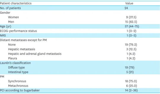
Table 2. Patient characteristics
Patient characteristics Value
No. of patients 24
Gender
Women 9 (37.5)
Men 15 (62.5)
Age (yr) 57 (44–75)
ECOG-performance status 1 (0–2)
NRS 1 (0–5)
Distant metastases except for PM
None 19 (79.2)
Hepatic metastasis 3 (12.5)
Hepatic and adrenal gland metastasis 1 (4.2)
Pleura 1 (4.2)
Laurén's classification
Diffuse type 19 (79)
Intestinal type 5 (21)
PM
Synchronous 18 (75.0)
Metachronous 6 (25.0)
PCI according to Sugarbaker 14 (2–36)
Values are presented as median (interquartile range) or number (%).
ECOG = Eastern Cooperative Oncology Group; NRS = nutritional risk score; PM = peritoneal metastasis;
PCI = peritoneal carcinomatosis index.
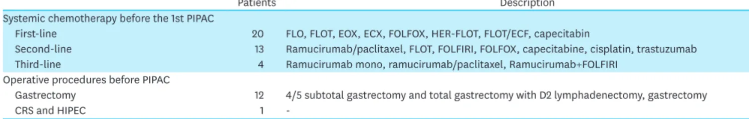
In GC patients treated with PIPAC, synchronous PM was diagnosed in 18 (75%), while 6 (25%) had metachronous PM. The median time delay between 1st diagnosis of GC with synchronous PM and the 1st PIPAC was 5.5 months (range, 2–22 months). In GC patients with metachronous PM, the median time interval between 1st diagnosis of GC to the 1st PIPAC was 48.5 months (range, 15–84 months), and from diagnosis of PM to the 1st PIPAC was 4 months (range, 0–5 months). Patients received their last cycle of systemic chemotherapy 30 days (range, 9–210 days) before PIPAC. Patients were treated with different standard chemotherapy regimens in 1st (20), 2nd (13), and 3rd (4) line therapies before receiving the 1st PIPAC. Four patients with synchronous PM were scheduled for the 1st PIPAC before the 1st systemic chemotherapy cycle. Additionally, 12 of 24 patients had previously undergone a curative intended gastrectomy, and 1 patient underwent CRS and HIPEC before the 1st PIPAC (Table 3). According to Laurén's classification, 19 (79%) patients displayed a diffuse type, and 5 (21%) an intestinal type (Table 2).
In general, patients eligible for PIPAC were considered to have no other distant metastases apart from PM. However, in our series, 5 (20.8%) patients had other forms of systemic metastases apart from PM and were included in this study due to patients' absolute wish and/or indications of symptomatic ascites. Three of the 5 patients had regredient hepatic metastases, 1 had regredient hepatic and adrenal gland metastases, and 1 had a pleura metastasis in the course of the systemic therapy.
The duration of surgery did not increase with repeated PIPAC procedures and was 105 minutes (range, 78–150 minutes) in median. Apart from patients with “non-access-abdomen,”
adhesions were observed only in a minority of the cohort with a median adhesion score of 2.5 (range, 0–23). Only 1 patient who underwent a 2nd PIPAC procedure was diagnosed with an adhesion score of 23, whereas at the 1st PIPAC, an adhesion score of 2 was detected.
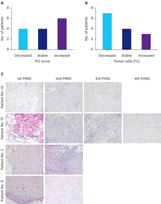
Patients' median PCI according to Sugarbaker at 1st PIPAC was 14 (range, 2–36) and ascites volume was 100 mL (range, 0–6 mL, 300 mL). The patient with a PCI of 2 was offered CRS and HIPEC, but did not want to undergo this more extensive procedure. In patients who received 2 PIPAC procedures or more, the PCI and the development of ascites volume were assessed [14]. In total, 8 (57%) patients displayed a decreased or stable PCI score at repeated PIPACs (Fig. 2A). A median of 5 (range, 3–10) biopsies were taken and evaluated per patient and treatment. Histopathological analysis of the tumor cell portions in biopsies taken before each PIPAC showed a reduction or stabilization of the maximum tumor cell portion in 11 (79%) patients (Fig. 2B). Slides of representative H&E staining of biopsies are depicted in Fig. 2C, exemplary showing patient No. 10 and No. 13 with decreased, and patient No. 3 and
Table 3. Previous treatment before PIPAC
Patients Description
Systemic chemotherapy before the 1st PIPAC
First-line 20 FLO, FLOT, EOX, ECX, FOLFOX, HER-FLOT, FLOT/ECF, capecitabin
Second-line 13 Ramucirumab/paclitaxel, FLOT, FOLFIRI, FOLFOX, capecitabine, cisplatin, trastuzumab
Third-line 4 Ramucirumab mono, ramucirumab/paclitaxel, Ramucirumab+FOLFIRI
Operative procedures before PIPAC
Gastrectomy 12 4/5 subtotal gastrectomy and total gastrectomy with D2 lymphadenectomy, gastrectomy
CRS and HIPEC 1 -
PIPAC = pressurized intraperitoneal aerosol chemotherapy; FLO = 5-fluorouracil+folinsäure+oxaliplatin; FLOT = fluorouracil+folinsäure+oxaliplatin+docetaxel;
ECF = epirubicin+cisplatin+5-fluorouracil; EOX = epirubicin+oxaliplatin+capecitabine; ECX = epirubicin+cisplatin+capecitabine; FOLFOX = leucovorin+5- fluorouracil+oxaliplatin; HER = herceptin; FOLFIRI = folinic acid+fluorouracil+irinotecan; CRS = cytoreductive surgery; HIPEC = hyperthermic intraperitoneal chemoperfusion.
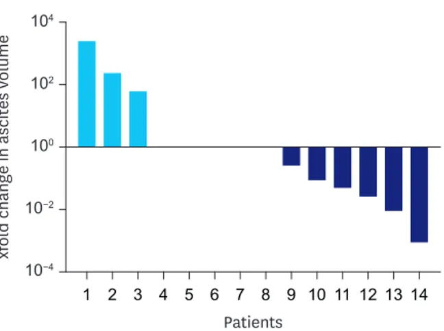
No. 9 with increased tumor cell portions. Of 14 repeatedly-treated patients, 11 displayed decreased or stable ascites volumes, while only 3 showed an increase (Fig. 3). In 10 (71%) patients, the PCI score development correlated with the change in the determined maximum tumor cell portion and in 10 (71%) patients, the PCI score correlated with the ascites volume development. The change in the evaluated maximum tumor cell portion correlated with ascites volume development in 10 (71%) patients. A correlation of all investigated criteria, ascites volume, PCI score, and tumor cell portion, was noted in 8 (57%) patients.
Patient No. 10
1st PIPAC 2nd PIPAC 3rd PIPAC 4th PIPAC
Patient No. 13Patient No. 3Patient No. 9
Decreased Increased
A
C
No. of patients
4 2 0 8 6
Stable Decreased Increased
B
No. of patients
4 2 0 8 6
Stable
PCI score Tumor cells (%)
Fig. 2. PCI and tumor cell portion developments in patients with multiple PIPAC procedures. (A) Number of patients with decreased, stable, and increased PCI, (B) Number of patients with decreased, stable, and increased tumor cell portions in PM biopsies, and (C) representative H&E staining of patients with decreased tumor cell portions (patients No. 10 and No. 13) and increased tumor cell portions (patients No. 3 and No. 9). The biopsies for tumor cell evaluation were taken before each PIPAC.
PCI = peritoneal carcinomatosis index; PIPAC = pressurized intraperitoneal aerosol chemotherapy; PM = peritoneal metastasis; H&E = hematoxylin and eosin stain.
The post-operative in-hospital stay was a median of 4 days (range, 2–9 days) and did not increase with repeated treatment procedures.
We observed only 1 postoperative complication according to Clavien-Dindo (grade I: nausea and emesis). To assess postoperative chemo-toxicity, patients were monitored using 2 standardized blood controls at the 1st and the 3rd postoperative days. There was no patient with postoperative renal failure. Leucocytes and creatinine values were within the reference ranges in median. Seven and 3 patients had higher and lower values, respectively than the reference values for leucocytes (3.5–9.8×10⁹/L). A postoperative increase in C-reactive protein (CRP) was observed. No signs of liver toxicity were documented in any patients. Aspartate amino transferase/alanine amino transferase values were below the reference values at both investigated time points (Table 4).
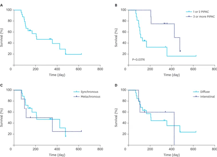
The OS of the 24 investigated patients was a median of 210 days (range, 66–625 days), while the median follow-up was 159 days (range, 66–625 days) after receiving the 1st PIPAC procedure (Fig. 4A). Dividing the patient cohort into those who received 1 or 2 PIPACs [16]
and 3 or more PIPACs [8] yielded a significant (P=0.0376) increase in median OS from 121 days (range, 66–625 days) to 450 days (range, 206–481 days), respectively (Fig. 4B). Stratification to whether patients had synchronous or metachronous PM (Fig. 4C) or to Laurén's classification did not result in different OS (Fig. 4D). QoL was assessed in 20 patients from March 2016 to February 2018 before each PIPAC procedure and a global health score of 52±5.9 was reported before the 1st PIPAC procedure. Patients, who received 2 or more PIPACs, had a stable global health score and stable to slightly decreased functioning and symptom scores, e.g., the physical functioning scale decreased from 78±4.8 at the 1st PIPAC to 66±7.5 at the 2nd PIPAC and was 63±13 at the 3rd PIPAC. No change in gastrointestinal tract item scores (nausea/
vomiting, constipation, and diarrhea) was observed, except in appetite loss, which was slightly increased with repeated PIPAC procedures (Table 5).
1
xfold change in ascites volume
100
10−2
10−4 104
102
2 3 4 5 6 7 8 9 10 11 12 13 14 Patients
Fig. 3. Relative ascites development in patients with multiple PIPAC procedures. The fold changes of decreased (6), stable (5), and increased (3) ascites volumes during multiple PIPAC procedures are blotted as waterfall blot.
PIPAC = pressurized intraperitoneal aerosol chemotherapy.
Table 4. Clinical chemistry and hematology
1-day post-OP 3-day post-OP Reference values
Leucocytes (WBC) (×109/L) 7.15±0.426 5.73±0.396 3.5–9.8
CRP (mg/L) 36.9±5.5 66.8±11.05 <5
Creatinine (µmol/L) 76.8±2.81 73.6±2.68 45–84
WBC = white blood cells; CRP = C-reactive protein; OP = operative.
DISCUSSION
Treatment of patients with PM attributable to GC remains challenging. With the current standard of care, which is the application of systemic chemotherapy in sequential treatment lines, survival outcomes remain limited to 8–12 months (median) in European patients [18].
PM, and especially the presence of ascites, is a feature of poor prognosis for patients with stage IV GC, who receive chemotherapy [19] and even with the contemporary promising
800
200 400 600
0 100
80
60
40
20
Survival (%)
Time (day) A
800
200 400 600
0 100
80
60
40
20
Survival (%)
Time (day)
1 or 2 PIPAC 3 or more PIPAC
B
P=0.0376
800
200 400 600
0 100
80
60
40
20
Survival (%)
Time (day) C
800
200 400 600
0 100
80
60
40
20
Survival (%)
Time (day)
Diffuse Intenstinal Synchronous
Metachronous
D
Fig. 4. Survival curves. (A) Kaplan-Meier survival curves of all 24 patients treated by PIPAC, (B) stratification of patients, who received 1 or 2 PIPACs (16) vs. 3 or more PIPACs (8; P=0.0376), (C) metachronous (6) vs. synchronous (18) PM, and (D) diffuse (19) vs. intestinal (5) type of GC according to Laurén's classification (Log-Rank [Mantel-Cox] test).
PIPAC = pressurized intraperitoneal aerosol chemotherapy; PM = peritoneal metastasis; GC = gastric cancer.
Table 5. EORTC QLQ-C30 EORTC
QLQ-C30 Global health
status Functioning scores Symptom scores Symptoms-single items
QL2 PF EF RF CF SF FA NV PA DI DY AP SL CO FI
1st PIPAC 52±5.9 78±4.8 69±4.8 59±6.9 87±4.7 62±7.6 33±5.2 24±7.3 25±5.0 40±9.8 21±6.0 29±8.9 29±7.4 13±5.1 11±5.1 2nd PIPAC 51±8.3 66±7.5 66±8.3 52±8.0 79±8.2 44±7.7 36±6.2 21±8.2 35±6.6 29±13 33±6.3 21±14 50±11 13±8.8 25±11 3rd PIPAC 48±14 63±13 68±8.5 47±18 73±14 47±18 22±6.1 20±12 37±14 20±13 33±18 47±17 33±0.0 7±6.7 27±6.7 Data are shown as mean±standard error of mean.
EORTC = European Organization for Research and Treatment; QLQ-C30 = quality of life questionnaire core 30; PIPAC = pressurized intraperitoneal aerosol chemotherapy; PF = physical functioning; EF = emotional functioning; RF = role functioning; CF = cognitive functioning; SF = social functioning; FA = fatigue score; NV = nausea/vomiting score; PA = pain score; DI = diarrhea score; DY = dyspnea score; AP = appetite loss score; SL = insomnia score; CO = constipation score; FI = financial difficulties.
developments in the treatment of GC, survival outcomes have not dramatically changed [20].
Symptom control and the maintenance of QoL remain the major treatment objectives in the management of patients with stage IV GC.
Options beyond palliative chemotherapy are limited in patients with stage IV GC and PM.
Hence, PIPAC might enrich the portfolio of palliative treatment options with the overall goal of symptomatic improvement and alleviation of QoL-reducing parameters. Due to its minimal-invasive applicability, low operative risk, and short post-operative in-hospital stay with a good overall tolerability, this additional option in systemic therapy seems acceptable in critically ill patient cohort.
Our data indicate that the majority of patients developed either regredient or at least stable ascites in the course of PIPAC follow-up treatment. Ascites, especially with associated intraabdominal compression syndrome and/or the necessity of repeated paracenteses are a very decisive parameter in determining global and health-related QoL. In more than half of the repeatedly treated patients, the changes in the volume of ascites correlated with PCI and histopathological regression scores (8; 57%). However, in 6 cases (43%), this correlation was not observed. This might be explained by the potential earlier functional response of peritoneal tumor cells to pressurized aerosol chemotherapy and diminished secretion of fluids. This was possibly earlier than the visible macroscopic (PCI) or microscopic (histopathologic scores) regression with regard to repeated therapy in the investigated intervals of only 6 weeks.
Limitations of our data are:
1) A considerable proportion of patients was included with non-peritoneal metastases for symptomatic control of ascites or according to patients' wishes, although this was general- ly determined as contraindication.
2) Heterogeneous pre-treatment regimens and responses to systemic therapy, different biology according to Lauren's classification, and various PCI scores at 1st PIPAC, rendering evaluation of the true impact of PIPAC on changes at follow-up more difficult.
3) The relative small cohort and short follow-up of a non-randomized population do not allow any conclusive judgement of this new intervention option for PM in GC. In addition, volume of ascites and QoL are well known to correlate with each other, as QoL increases in the course of ascites control.
4) We were not able to provide associated improvement of the QoL by using validated scores, as provided by the structured interview using the EORTC QLQ-C30 questionnaire, with decreased ascites volumes. This was possibly due to the subjective and the multifactorial nature of the assessment and the lack of reliability in repeated evaluations.
To our knowledge, there are only limited data of GC patients undergoing PIPAC and documented follow-up, with similar sized patient cohorts assessed at each center [21-25].
A recently published multicenter study by Nowacki et al. [11] of 832 PIPAC procedures in 15 centers, with 280 (41.1%) cases attributable to GC, revealed that PIPAC procedures were homogeneously performed in these established centers with standardized techniques, facilitating current and future international multicenter prospective clinical trials.
In 24 patients with GC and PM and a mean PCI of 16 (±10) with a similar chemo-aerosol regime as ours (cisplatin and doxorubicin), Nadiradze and colleagues described an objective
tumor response in 12 patients (50%), of whom 6 displayed no vital tumor cells, the same number showed major pathological response, and 3 had minor response. In this series, 17 patients underwent repeated PIPAC, of whom 3 had a “non-access” abdomen in a redo procedure. Postoperative adverse events (Common Terminology Criteria for Adverse Events [CTCAE] grade >2) were observed in a relatively high percentage (37.5%; 6 grade 3:
hepatotoxicity, cholangitis; 1 grade 4: allergy, myolysis; 2 grade 5: ascites decompensation and death, progressive small bowel obstruction and death) [25]. Although the dose applied was only 10% of a usual systemic dose, most of these complications were due to a postoperative inflammatory syndrome with elevated CRP, which the authors explained as the consequence of a possible chemical peritonitis. However, acute and cumulative local toxicities of PIPAC were well controlled, and no bowel perforations or gastrointestinal side effects > CTCAE grade 2 were observed. With a follow-up of 248 days (range, 105–748 days), the median survival time was 15.4 months, although this study also included 4 patients (17%) with extraperitoneal metastases, in particular to the pleura and the liver. Simultaneous chemotherapy was administered in 33% [25].
Patients showed a relatively high QoL in a heterogeneous group of 91 patients undergoing PIPAC, including 29 with GC–induced PM except for a transient moderate increase of pain scores [22]. PIPAC did not cause therapy-related QoL deterioration and especially, there were no additional gastrointestinal symptoms associated with this palliative approach. Using the EORTC QLQ-C30 questionnaire, functioning scores and disease-related symptoms were not altered for at least 3 months in the patients able to receive repeated PIPAC [22]. Most publications available containing reasonable cohorts of GC used the 6–8 weeks interval of PIPAC, embedded in multimodal treatment [11,21,23,25,26]. PIPAC combined with associated systemic therapy showed no accelerated renal or hepatic toxicity and proved a safe and well-tolerated procedure [21,24].
Globally, 4 randomized studies are currently examining the impact of D2-gastrectomy and HIPEC [5] (Germany, HIPEC_stomach [NCT01683864]; France, GASTRICHIP
[NCT01882933]; China [NCT02240524]; USA [NCT03092518]). Final results of these studies are not available yet. Patients with “high-risk cytology” and evidence of at least one of the mentioned predictors had a significantly worse survival chance than “low-risk patients” [27].
These new cytologic criteria could possibly contribute to the identification of patients who might benefit from the addition of PIPAC to the current standard of care therapy. Currently, 2 prospective-randomized studies are being conducted for HIPEC in GC. One of these, the Gastric Cancer with Peritoneal Carcinomatosis study (NCT02158988) evaluates the effect of CRS +/− HIPEC embedded in a perioperative chemotherapy regimen and a 2nd study from France investigates the role of adjuvant HIPEC following resection of GC, expecting results in 2020 and 2023, respectively. A current trial by Goetze et al. [28] is evaluating the impact of PIPAC in combination with standard chemotherapy in primarily untreated naive upper GI-adenocarcinomas with peritoneal seeding (phase II/III trial) and will hopefully clarify its oncologic efficacy (EudraCT: 2018-001035-40)
In conclusions, the oncologic and functional impact of PIPAC, in PM attributable to GC is not conclusive yet. In highly-qualified patients, critically indicated within the frame of systemic chemotherapy, PIPAC is not toxic and may lead to a symptom-oriented optimization with regard to ascites reduction, (partial) macroscopic and histopathologic regression of PM and better QoL.
PIPAC has the potential — due to its restricted invasiveness, limited surgical side-effects and risks, and its lack of systemic toxicity — to be used for symptom control in GC patients with disseminated PM, in addition to systemic therapy. However, the predictors of response and the optimum intervals of PIPAC within systemic therapy concepts are yet to be identified in on-going further studies.
Thus, we are not too optimistic that PIPAC will become standard treatment in GC patients with PM in the near future.
REFERENCES
1. Ferlay J, Shin HR, Bray F, Forman D, Mathers C, Parkin DM. Estimates of worldwide burden of cancer in 2008: GLOBOCAN 2008. Int J Cancer 2010;127:2893-2917.
PUBMED | CROSSREF
2. Ferlay J, Steliarova-Foucher E, Lortet-Tieulent J, Rosso S, Coebergh JW, Comber H, et al. Cancer incidence and mortality patterns in Europe: estimates for 40 countries in 2012. Eur J Cancer 2013;49:1374-1403.
PUBMED | CROSSREF
3. Geng X, Liu H, Lin T, Hu Y, Chen H, Zhao L, et al. Survival benefit of gastrectomy for gastric cancer with peritoneal carcinomatosis: a propensity score-matched analysis. Cancer Med 2016;5:2781-2791.
PUBMED | CROSSREF
4. Roviello F, Caruso S, Marrelli D, Pedrazzani C, Neri A, De Stefano A, et al. Treatment of peritoneal carcinomatosis with cytoreductive surgery and hyperthermic intraperitoneal chemotherapy: state of the art and future developments. Surg Oncol 2011;20:e38-e54.
PUBMED | CROSSREF
5. Brandl A, Pachmayr E, Gül-Klein S, Alberto M, Thuss-Patience P, Rau B. Surgical treatment of peritoneal metastases of gastric cancer. Chirurg 2018;89:669-677.
PUBMED | CROSSREF
6. Yonemura Y, Endou Y, Shinbo M, Sasaki T, Hirano M, Mizumoto A, et al. Safety and efficacy of bidirectional chemotherapy for treatment of patients with peritoneal dissemination from gastric cancer:
selection for cytoreductive surgery. J Surg Oncol 2009;100:311-316.
PUBMED | CROSSREF
7. Wagner AD, Unverzagt S, Grothe W, Kleber G, Grothey A, Haerting J, et al. Chemotherapy for advanced gastric cancer. Cochrane Database Syst Rev 2010:CD004064.
PUBMED
8. Koizumi W, Narahara H, Hara T, Takagane A, Akiya T, Takagi M, et al. S-1 plus cisplatin versus S-1 alone for first-line treatment of advanced gastric cancer (SPIRITS trial): a phase III trial. Lancet Oncol 2008;9:215-221.
PUBMED | CROSSREF
9. Minchinton AI, Tannock IF. Drug penetration in solid tumours. Nat Rev Cancer 2006;6:583-592.
PUBMED | CROSSREF
10. Jansen-Winkeln B, Thieme R, Haase L, Niebisch S, Pommer C, Lyros O, et al. Perioperative safety of intraperitoneal aerosol chemotherapy: analysis of our first 111 pressurized intraperitoneal aerosol chemotherapy (PIPAC) procedures. Chirurg 2018. doi: 10.1007/s00104-018-0667-5 [In press].
CROSSREF
11. Nowacki M, Alyami M, Villeneuve L, Mercier F, Hubner M, Willaert W, et al. Multicenter comprehensive methodological and technical analysis of 832 pressurized intraperitoneal aerosol chemotherapy (PIPAC) interventions performed in 349 patients for peritoneal carcinomatosis treatment: an international survey study. Eur J Surg Oncol 2018;44:991-996.
PUBMED | CROSSREF
12. Hübner M, Grass F, Teixeira-Farinha H, Pache B, Mathevet P, Demartines N. Pressurized intraperitoneal aerosol chemotherapy - practical aspects. Eur J Surg Oncol 2017;43:1102-1109.
PUBMED | CROSSREF
13. Coccolini F, Ansaloni L, Manfredi R, Campanati L, Poiasina E, Bertoli P, et al. Peritoneal adhesion index (PAI): proposal of a score for the “ignored iceberg” of medicine and surgery. World J Emerg Surg 2013;8:6.
PUBMED | CROSSREF
14. Jacquet P, Sugarbaker PH. Clinical research methodologies in diagnosis and staging of patients with peritoneal carcinomatosis. Cancer Treat Res 1996;82:359-374.
PUBMED | CROSSREF
15. Harmon RL, Sugarbaker PH. Prognostic indicators in peritoneal carcinomatosis from gastrointestinal cancer. Int Semin Surg Oncol 2005;2:3.
PUBMED | CROSSREF
16. Dindo D, Demartines N, Clavien PA. Classification of surgical complications: a new proposal with evaluation in a cohort of 6336 patients and results of a survey. Ann Surg 2004;240:205-213.
PUBMED | CROSSREF
17. Fayers P, Aaronson NK, Bjordal K, Groenvold M, Curran D, Bottomley A, eds. EORTC QLQ-C30 Scoring Manual. 3rd ed. Brussels: European Organisation for Research and Treatment of Cancer, 2001.
18. Lordick F, Lorenzen S, Yamada Y, Ilson D. Optimal chemotherapy for advanced gastric cancer: is there a global consensus? Gastric Cancer 2014;17:213-225.
PUBMED | CROSSREF
19. Matsumoto H, Kawazoe A, Shimada K, Fukuoka S, Kuboki Y, Bando H, et al. A retrospective study of the safety and efficacy of paclitaxel plus ramucirumab in patients with advanced or recurrent gastric cancer with ascites. BMC Cancer 2018;18:120.
PUBMED | CROSSREF
20. Lordick F, Shitara K, Janjigian YY. New agents on the horizon in gastric cancer. Ann Oncol 2017;28:1767-1775.
PUBMED | CROSSREF
21. Grass F, Vuagniaux A, Teixeira-Farinha H, Lehmann K, Demartines N, Hübner M. Systematic review of pressurized intraperitoneal aerosol chemotherapy for the treatment of advanced peritoneal carcinomatosis. Br J Surg 2017;104:669-678.
PUBMED | CROSSREF
22. Odendahl K, Solass W, Demtröder C, Giger-Pabst U, Zieren J, Tempfer C, et al. Quality of life of patients with end-stage peritoneal metastasis treated with pressurized intraperitoneal aerosol chemotherapy (PIPAC). Eur J Surg Oncol 2015;41:1379-1385.
PUBMED | CROSSREF
23. Alyami M, Gagniere J, Sgarbura O, Cabelguenne D, Villeneuve L, Pezet D, et al. Multicentric initial experience with the use of the pressurized intraperitoneal aerosol chemotherapy (PIPAC) in the management of unresectable peritoneal carcinomatosis. Eur J Surg Oncol 2017;43:2178-2183.
PUBMED | CROSSREF
24. Robella M, Vaira M, De Simone M. Safety and feasibility of pressurized intraperitoneal aerosol
chemotherapy (PIPAC) associated with systemic chemotherapy: an innovative approach to treat peritoneal carcinomatosis. World J Surg Oncol 2016;14:128.
PUBMED | CROSSREF
25. Nadiradze G, Giger-Pabst U, Zieren J, Strumberg D, Solass W, Reymond MA. Pressurized intraperitoneal aerosol chemotherapy (PIPAC) with low-dose cisplatin and doxorubicin in gastric peritoneal metastasis. J Gastrointest Surg 2016;20:367-373.
PUBMED | CROSSREF
26. Giger-Pabst U, Solaß W, Strumberg D, Zieren J, Reymond M. Erste klinische ergebnisse der “pressurized intraperitoneal aerosol chemotherapy” (PIPAC) bei patienten mit magenkarzinom und fortgeschrittener peritonealkarzinose. Z Gastroenterol 2013;51:K364.
CROSSREF
27. Higaki E, Yanagi S, Gotohda N, Kinoshita T, Kuwata T, Nagino M, et al. Intraoperative peritoneal lavage cytology offers prognostic significance for gastric cancer patients with curative resection. Cancer Sci 2017;108:978-986.
PUBMED | CROSSREF
28. Goetze TO, Al-Batran SE, Pabst U, Reymond M, Tempfer C, Bechstein WO, et al. Pressurized intraperitoneal aerosol chemotherapy (PIPAC) in combination with standard of care chemotherapy in primarily untreated chemo naïve upper GI-adenocarcinomas with peritoneal seeding – a phase II/III trial of the AIO/CAOGI/ACO. Pleura Peritoneum 2018;3.