INTRODUCTION
Cancer of an unknown primary site (CUPS) is defined as a bi
opsy proven metastatic malignant tumor whose primary site cannot be identified during the pretreatment evaluation. Early dissemination, the unpredictability of the metastatic pattern, and aggressiveness constitute the fundamental characteris
tics of these tumors [1,2]. CUPS have various heterogenous clinical and pathologic features, yet within this diverse group there are specific clinical and/or pathologic features that can be used to define several subsets of patients who have favor
able prognoses [3]. For these favorable subsets, taxane based chemotherapy was very effective for a subset of woman with papillary serous peritoneal adenocarcinoma. We describe here
an unusual case of CUPS that manifested with large multiple intraabdominal lymph nodes. We successfully treated the pa
tient with taxane based chemotherapy and surgery.
CASE REPORT
A 52 year old woman underwent [18F]FDG PET/CT for rou for rou
tine health screening. She had suffered from vague abdominal dis comfort. She had no significant past medical history. On PET/
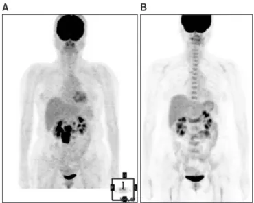
CT, multiple hypermetabolic lymph nodes encasing the inferi
or ve na cava and abdominal aorta were detected (SUVmax, 14.9 in the retrocaval area), and there were no other hypermetabolic abnormalities (Fig. 1A). On physical examination, there was mild tenderness on the midabdomen. The la bo ratory examinations were within normal ranges including complete blood count, renal function tests, and liver function tests. Of the tumor mark
er tests, only CA125 was elevated to 250 U/mL. Neck, chest, and ab dominal CT, pelvic MRI, gastroduodenoscopy and colo
noscopy were performed for further evaluation. The gastro
Case Report
Complete biologic response to taxane based chemotherapy confirmed by [ 18 F]FDG PET/CT and surgery in a cancer of unknown primary site
Jun-Eul Hwang, Ju-Young Yoon, Woo-Kyun Bae, Hyun-Jeong Shim, Ik-Joo Chung
Department of Hematology-Oncology, Chonnam National University Medical School, Gwangju, Korea
Received Dec 13, 2010, Revised Jan 6, 2011, Accepted Feb 9, 2011 Correspondence to Ik-Joo Chung
Department of Hematology-Oncology, Chonnam National University Medical School, 5 HaK-dong, Dong-gu, Gwangju 501-746, Korea. Tel: 82-61-379-7632, Fax: 82-61-379-7628, E-mail: [email protected]
pISSN 2005-0380 eISSN 2005-0399
Copyright © 2012. Asian Society of Gynecologic Oncology, Korean Society of Gynecologic Oncology This is an Open Access article distributed under the terms of the Creative Commons Attribution Non-Commercial License (http://creativecommons.org/licenses/by-nc/3.0/) which permits unrestricted non-commercial use, distribution, and
reproduction in any medium, provided the original work is properly cited.
www.ejgo.org
Cancers of an unknown primary site are heterogenous with respect to their clinical and pathologic features. They are generally very aggressive, but specific favorable subsets have a better prognosis. For these favorable subsets, taxane based chemotherapy is very effective for a subset of woman with papillary serous peritoneal adenocarcinoma. A 52 yearold woman underwent [18F]
FDG PET/CT for routine health screening. On PET/CT, multiple hypermetabolic lymph nodes were detected in the paraaortic spaces, and there were no other hypermetabolic abnormalities. The patient was diagnosed with an unknown primary cancer that probably originated from the ovary or peritoneum, according to clinical studies and biopsy results. This was not a typical case of a favorable subset of cancer of an unknown primary site, but the tumor showed complete biologic response to taxane based chemotherapy as revealed by PET/CT, and necrotic tumor cells were confirmed by surgery.
Keywords: Chemotherapy, Positron emission tomography, Taxane, Unknown primary neoplasms
J Gynecol Oncol Vol. 23, No. 1:65-68 http://dx.doi.org/10.3802/jgo.2012.23.1.65
Jun-Eul Hwang, et al.
http://dx.doi.org/10.3802/jgo.2012.23.1.65 66 www.ejgo.org
duodenoscopy and colonoscopy were negative. Abdominal CT showed about 6 cm sized multiple lymph nodes encasing the abdominal aorta (Fig. 2A). There were no abnormalities in both ovaries by pelvic MRI. She underwent a laparoscopic sur
gi cal biopsy. The results showed metastatic carcinoma and the tumor cells were positive for cytokeratin (CK), CK7, estro gen receptor (ER), CA125 and Wilm’s tumor1 (WT1) on immu
nohistochemical staining (Fig. 3A, B). The other immunohisto
chemical stains were negative, including gross cystic disease fluid protein15 (GCDFP15), CK20 and progesterone receptor (PR). The patient was diagnosed with cancer of an unknown primary site, and it probably originated from the ovary or peri
toneum on the basis of the clinical studies and biopsy results.
We thought that the tumor mass could not be completely re
sected due to encasement of the great vessels, and therefore combination chemotherapy that consisted of paclitaxel and carboplatin was started. The followup CT after 6 cycles of che
motherapy showed marked partial response according to the RECIST criteria, that is, the tumor decreased from 6×5 cm to 2×2 cm, and so we planned a debulking operation (Fig. 2B).
Preoperative PET/CT showed no hypermetabolism in the rem
nant lesion (Fig. 1B). The patient underwent transabdominal Fig. 1. (A) The [18F]FDG PET/CT showed multiple conglo me rated hyper-
metabolic lymph nodes encasing the inferior vena cava and abdominal aorta in the retrocaval, portocaval and paraaortic spaces. (B) The pre- opera tive [18F]FDG PET/CT revealed that there were no hy per metabolic lesions in the remnant lymph nodes.
Fig. 2. (A) The initial CT showed huge intra abdominal lym ph nodes encasing the inferior vena cava and abdominal aorta. (B) The follow-up CT after 6 cycles of taxane based che mo therapy showed a marked partial res ponse according to the RECIST criteria.
Fig. 3. (A) The laparoscopic biopsy results showed the tumor cells that have pleomorphic nuclei and small cytoplasm (H&E, ×100). (B) The immunohistochemical staining results showed the tumor cells were positive for Wilm’s tumor-1. (C) The operative biopsy results showed only extensive necrosis in the remnant lymph nodes without tumor cells (H&E, ×10).
Necrotic unknown primary cancer revealed by PET
J Gynecol Oncol Vol. 23, No. 1:65-68 www.ejgo.org 67
hysterectomy, both salpingooophorectomy and pelvic/para
aortic lymph nodes dissection. The operative biopsy results showed only necrotic cell debris in the lymph nodes without tumor cells, and there were no other tumor cells in the female genital organs (Fig. 3C). An additional 6 cycles of chemotherapy were administered after surgery. She is on followup without evidence of recurrence.
DISCUSSION
For CUPS, the specific favorable subsets are treatable, and initial identification these patients is imperative for their optimal ma
nage ment. At least five of these favorable subsets have clinical features similar to specific cancer types: 1) women with axillary lymph node metastasis, breast cancer; 2) men with blastic bone metastasis and tumor staining or elevated serum levels of pro
state specific antigen, prostate cancer; 3) young men with the extragonadal germ cell syndrome, germ cell tumor; 4) isolated neck nodes involved with squamous cell carcinoma, head and neck primary cancer; 5) woman with papillary serous perito
neal adenocarcinoma, primary peritoneal carcinoma or ovary cancer. Therapy directed at the presumed primary benefits these patients, and this supports the idea that at least some CUPS retain similar sensitivity to therapies that are known to be useful for the known primaries [2]. A woman with papillary serous peritoneal adenocarcinoma has clinical features similar to those for advanced ovarian cancer including ascites, tumor involvement usually limited to the peritoneal surfaces and el
evated serum levels of CA125, yet there is no evidence of pri
mary tumor in the ovaries [4]. These patients should be man
aged as stage III ovarian cancer with taxane/platinum based systemic chemotherapy. The median complete response rate is 20%, the median survival is 16 months and the median long term survival (>2 years) is 16% [3,5].
This was not a typical case of a favorable subset of CUPS. This patient manifested with only huge multiple intraabdominal lym ph nodes, yet we chose taxane based chemotherapy accor
ding to the immunohistochemical results. The main positive markers for ovarian adenocarcinoma are ER, CA125, mesothelin, and WT1, and the diagnosis of primary ovarian tumors is favored by the CK7+/CD20 [6]. The immunohistochemical results of the tumor cells in this case were CK7+/CK20, ER+, CA125+, and WT1+. Besides, the tumors showed complete biologic res pon
se to taxane based chemotherapy as revealed by PET/CT and necrotic tumor cells were confirmed by surgery.
Imaging has a fundamental role in oncology, especially for assessing the tumor response to therapy. The current conven
tional imaging followup is based on morphological criteria
with the changes of the tumor dimensions determining a response or tumor progression. PET/CT provides an indication of the metabolic and proliferative activity within tumors. It is useful in the early evaluation of a tumor response because changes in the gross tumor size are notably delayed, and they substantially lag behind the biological and molecular changes that are known to occur early in responders [7,8]. Monitor
ing the early treatment response will allow modifying and adapting treatment on the basis of the patient’s treatment response. Ultimately, the aim is to improve the outcomes and reduce the acute and late treatmentrelated side effects to achieve the best possible therapeutic gain and quality of life. PET/CT has been recently widely used to evaluate the tumor response to chemotherapy for various cancers such as esophageal cancer, head and neck cancer, lung cancer, breast cancer and lymphoma. It can also be useful for an unknown primary cancer. In this case, we performed PET/CT for evaluat
ing the extent of disease and the treatment response to che
motherapy before the debulking operation.
In conclusion, immunohistochemical evaluation of CUPS to predict the primary sites is very important for the optimal treatment, and PET/CT can also be used for CUPS to evaluate the tumor response to chemotherapy.
CONFLICT OF INTEREST
No potential conflict of interest relevant to this article was reported.
REFERENCES
1. Pavlidis N. Cancer of unknown primary: biological and cli
nical characteristics. Ann Oncol 2003;14 Suppl 3:iii118.
2. Greco FA, Hainsworth JD. Introduction: unknown primary cancer. Semin Oncol 2009;36:67.
3. Hainsworth JD, Fizazi K. Treatment for patients with un
known primary cancer and favorable prognostic factors.
Semin Oncol 2009;36:4451.
4. Fromm GL, Gershenson DM, Silva EG. Papillary serous car
ci noma of the peritoneum. Obstet Gynecol 1990;75:8995.
5. Dalrymple JC, Bannatyne P, Russell P, Solomon HJ, Tattersall MH, Atkinson K, et al. Extraovarian peritoneal serous papillary carcinoma: a clinicopathologic study of 31 cases. Cancer 1989;64:1105.
6. Dennis JL, Hvidsten TR, Wit EC, Komorowski J, Bell AK, Downie I, et al. Markers of adenocarcinoma characteristic of the site of origin: development of a diagnostic algorithm. Clin Cancer
Jun-Eul Hwang, et al.
http://dx.doi.org/10.3802/jgo.2012.23.1.65 68 www.ejgo.org
Res 2005;11:376672.
7. Harry VN, Semple SI, Parkin DE, Gilbert FJ. Use of new ima
ging techniques to predict tumour response to therapy.
Lancet Oncol 2010;11:92102.
8. Pickles MD, Gibbs P, Lowry M, Turnbull LW. Diffusion chan
ges precede size reduction in neoadjuvant treatment of breast cancer. Magn Reson Imaging 2006;24:8437.
Standards for Different Types of Articles
Guidelines for six different types of articles have been adopted by the Journal of Gynecologic Oncology:
1. CONSORT (Consolidated Standards of Reporting Trials) standards for reporting randomized trials 2. PRISMA (Preferred Reporting Items for Systematic Reviews and Metaanalyses) guidelines for
reporting systematic reviews and metaanalyses
3. MOOSE (Metaanalysis of Observational Studies in Epidemiology) guidelines for metaanalyses and systematic reviews of observational studies
4. STROBE (Strengthening the Reporting of Observational Studies in Epidemiology) guidelines for the reporting of observational studies
5. STARD (Standards for Reporting of Diagnostic Accuracy) standards for reporting studies of diag
nostic accuracy
6. REMARK (Reporting of Tumor Markers Studies) guidelines for reporting tumor marker prognos
tic studies
Investigators who are planning, conducting, or reporting randomized trials, metaanalyses of ran
domized trials, metaanalyses of observational studies, observational studies, studies of diagnostic accuracy, or tumor marker prognostic studies should be familiar with these sets of standards and follow these guidelines in articles submitted for publication.
NOW AVAILABLE ONLINE http://www.ejgo.org