10.3988/jcn.2011.7.1.34 J Clin Neurol 2011;7:34-39
Introduction
Guillain-Barré syndrome (GBS) is the most common cause of acute polyneuropathy. The clinical-pathologic spectrum of GBS extends from the classical acute inflammatory demyelinating polyneuropathy (AIDP) to pure axonal variants with (acute mo- tor sensory axonal neuropathy) and without (acute motor axo- nal neuropathy) sensory involvement, and clinical variants such as the Miller Fisher syndrome (MFS). Cranial-nerve involve- ment, dysautonomia, and respiratory insufficiency may be seen during the course of the disease.1,2 Although the cranial nerves are often involved in GBS, the optic nerves are usually spared, presumably because they are part of the central ner- vous system (CNS).3
A few studies have revealed optic-nerve involvement and evoked potential abnormalities in GBS.4-7 In the present study we determined the incidence of visual pathway involvement in GBS with the aid of clinical and electrophysiological assess-
ment, and defined the patterns of visual evoked potential (VEP) abnormalities in GBS.
Methods
Thirty-two patients with a diagnosis of GBS at the Department of Neurology, Ondokuz Mayıs University Health and Research Hospital between April 2005 and December 2009 were includ- ed in the study. All patients met the diagnostic criteria for GBS, as defined previously, based on clinical evaluation, nerve con- duction studies, and cerebrospinal fluid (CSF) investigation.8-10 All patients had symptomatic motor or sensory neuropathy with acute onset. Electrophysiological data were consistent with de- myelinating, axonal, or mixed polyneuropathy; no other etiol- ogy of acute neuropathy was detectable. All patients gave their informed consent to participate in the study. Patients with severe motor, bulbar, or autonomic involvement causing cardiopulmo- nary instability and needing intensive life support and monitor-
Visual Evoked Potentials in Guillain-Barré Syndrome
Levent Güngör,a İnci Güngör,b Hilal Eser Öztürk,b Musa Kazım Onara
aDepartments of Neurology and bOphthalmology, Ondokuz Mayis University School of Medicine, Samsun, Turkey
Received June 17, 2010 Revised August 17, 2010 Accepted August 17, 2010 Correspondence Levent Güngör
Department of Neurology, Ondokuz Mayıs University School of Medicine, 55139 Samsun, Turkey Tel +9-0-362-3121919-3715 Fax +9-0-362-4576757 E-mail ligungor@omu.edu.tr
Background and PurposezzGuillain-Barré syndrome (GBS) is an acute demyelinating poly- neuropathy with various clinical features. Optic neuritis occurs in rare cases. In this study we de- termined the incidence and patterns of visual evoked potential (VEP) abnormality in GBS in as- sociation with ophthalmologic findings.
MethodszzThirty-two patients with a diagnosis of GBS were included in the study. The correla- tion between pathologic VEPs and categories of neurologic deficit and electrophysiological find- ings were examined statistically.
ResultszzThe patients ranged in age from 19 to 77 years. Five cases (16%) had abnormal VEPs.
All five of these patients exhibited increased P100 latency differences between the two eyes. Oth- er abnormalities were prolonged p100 latency, increased interocular amplitude difference, and distorted p100 configuration. Pathologic signs on ophthalmologic examination were observed in 80% of patients with abnormal VEPs. VEP abnormality was never present in pure axonal forms.
There was no significant correlation between pathologic VEP and cerebrospinal fluid protein level or categories of neurologic deficits.
ConclusionszzInvolvement of the optic pathways is not a frequent finding in GBS. When pres- ent it is always asymmetric and generally accompanied with pathologic findings on ophthalmo- logic examination. VEPs may be abnormal in different clinical variants of GBS, and especially in
demyelinating forms. J Clin Neurol 2011;7:34-39
Key Wordszz guillain-Barré syndrome, optic neuritis, visual evoked potentials.
ing, or who died early during the course of illness were excluded.
The neurologic findings of each patient were outlined in six categories with the aid of neurologic examination:
1) Superficial (any deficiency of light-touch, pinprick, or tem- perature sensation) and deep (vibratory sensation or position sense) sensorial loss.
2) Motor deficit (decrease in upper or lower extremity mus- cle strength).
3) Presence of limb ataxia.
4) Cranial-nerve involvement (other than the optic nerve).
5) Autonomic involvement.
Routine blood tests and CSF examination were performed.
The clinical syndrome was defined according to electrophysi- ological and clinical findings.
A pattern reversal-VEP study was carried out for all patients as early as possible when clinical cooperation with the test was technically available. The stimulation source was a black/white full-field checkerboard pattern on a television screen with check size of 14 inches, a reversal rate of 1 Hz, and a Michelson con- trast of 99%. The television screen was positioned 1 m from the eyes. Each eye was tested separately and with the opposite eye occluded. The VEPs were recorded by epicranial surface elec- trodes. The active electrode was placed over the midocciput (Oz) and referred to as the midfrontal lead (Fz). The ground electrode was placed at the vertex (Cz). A bandpass filter (0.1- 1 Hz) was used with a sweep speed of 300 msec.11,12 In total, 375 responses were recorded for each eye and averaged by a computer system (Dantec Keypoint, Medtronic Functional Di- agnostics, Skovlunde, Denmark). Two trials were performed under the same stimulation conditions for each subject to con- firm the reproducibility. The latencies and amplitudes of the N75, P100, and N145 waves, the P100 morphology, and dif- ferences in the latency and amplitude of the P100 wave be- tween the two eyes were evaluated. The latencies and ampli- tudes were evaluated according to normal values from our laboratory obtained from 160 healthy subjects (114 females and 46 males) aged between 19 and 72 years. The following were considered to be abnormal: P100 latencies >2.5 standard deviations above the mean of the normal population (>108 msec for patients younger than 50 years, and >116 msec for males and >109 msec for females older than 50 years), minimal left-to-right amplitude ratio >0.66, and left-right difference of latency >6 msec for the P100 peak.
Each patient underwent a detailed ophthalmologic examina- tion performed by an ophthalmologist who was blinded to the VEP results. Anterior segment evaluation, visual acuity, pres- ence of pupillary light-reflex abnormalities (total loss, anisoco- ria, or relative afferent pupillary defect), fundoscopic findings, and defects of colored vision were recorded. Patients who had diseases that may affect VEP results, such as severe refractive
error, glaucoma, optic media opacity, retinal disease, or previ- ous history of optic neuropathy, were excluded.
Any correlations between the presence of pathologic VEPs and any category of neurologic deficit, electrophysiological data, or CSF protein level were examined. Fisher’s exact, Stu- dent’s t, and Mann-Whitney U tests were used for statistical an- alyses.
Results
The patients ranged in age from 19 to 77 years (mean±SD:
50.13±16.02 years). There were 19 males (59%) and 13 fe- males (41%). The diagnosis was AIDP in 18 patients (56%), MFS in 5 (16%), acute motor sensory axonal neuropathy in 5 (16%), and acute motor axonal neuropathy in 4 (13%), based on clinical and electrophysiological findings. Twenty-one pa- tients (66%) were treated with intravenous immunoglobulin and 4 (13%) were treated with plasmapheresis.
The time interval between the onset of symptoms and the VEP study ranged from 6 to 45 days. VEPs were abnormal in five cases (15.63%) (Fig. 1). The most common abnormality was increased interocular latency difference (7-20 msec), which was present in all five cases. P100 latency was delayed in four cas- es (12.50%), of which two had prolonged P100 latency in both eyes (6.25%). Other abnormalities were distorted (W-shaped) P100 configuration and increased interocular amplitude differ- ence (two cases, 6.25%) (Table 1). N75 and N145 were signifi- cantly prolonged in only one patient (3.13%). Visual examina- tion was abnormal in four of the five patients with abnormal VEPs (80%). Of those with abnormal VEPs, there was decrease in visual acuity in four patients (80%), light-reflex abnormali- ties or afferent pupillary defect in three (60%), dyschromatop- sia in one (20%), and papilledema in one (20%) (Table 1). Am- ong those with normal VEPs, only one patient had light-reflex abnormality (3.13%), and two had unexplained decreases in vi- sual acuity (6.25%). One patient with prolonged P100 in one eye had unexplained decreased visual acuity and dyschroma- topsia in the contralateral eye.
Three of the patients with abnormal VEPs were female (60%), four had a diagnosis of AIDP (80%), and one was diagnosed with MFS (20%) (Table 1). Pathologic VEPs were obtained only in patients with electrophysiological findings suggesting demyelinating peripheral neuropathy, but not axonal forms.
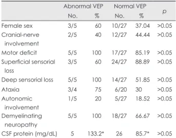
None of the clinical deficits observed on neurologic examina- tion was significantly correlated with VEP pathology (Table 2).
Superficial sensorial deficit (60% versus 88.89%) and cranial- nerve involvement other than the optic nerve (40% versus 44.
44%) appeared to be less prevalent, and autonomic involvement (20% versus 18.52%) slightly more common in GBS patients with abnormal VEPs, but the difference did not reach statistical
significance (p>0.05). All of the patients with pathologic VEPs had deep sensorial loss and motor involvement, while among those with normal VEPs, 51.85% had deep sensorial loss and 85.19% had motor loss (p>0.05). Limb ataxia, which could be evaluated in 24 patients, was more common in those with ab- normal VEPs, but the difference was not statistically significant (75% versus 30%, p>0.05). CSF protein levels tended to be higher in GBS patients with pathologic VEPs, but again the dif- ference did not reach statistical significance (133.2 versus 85.70 mg/dL) (Table 2).
Discussion
GBS is an acute, rapidly progressive, symmetrical polyradicu- loneuropathy that is characterized by weakness, areflexia, sen-
Table 2. Comparison of clinical characteristics and CSF protein lev- els in GBS patients with abnormal and normal VEPs
Abnormal VEP Normal VEP No. % No. % p
Female sex 3/5 60 10/27 37.04 >0.05
Cranial-nerve involvement
2/5 40 12/27 44.44 >0.05
Motor deficit 5/5 100 17/27 85.19 >0.05 Superficial sensorial
loss
3/5 60 24/27 88.89 >0.05
Deep sensorial loss 5/5 100 14/27 51.85 >0.05
Ataxia 3/4 75 6/20 30 >0.05
Autonomic involvement
1/5 20 5/27 18.52 >0.05 Demyelinating
neuropathy
5/5 100 18/27 66.67 >0.05
CSF protein (mg/dL) 5 133.2* 26 85.7* >0.05
*Mean of CSF protein levels.
CSF: cerebrospinal fluid, GBS: guillain-barré syndrome, VEP: visu- al evoked potential.
p100 114.3 Right
Left
p100 126.8
2 µV/D 30 msec/D
Fig. 1. Pathologic VEP of a patient with GBS (no. 3). The P100 la- tency is elongated on the left side, and has a low amplitude (lower trace), while it is normal on the right side (upper trace).
Table 1. Clinical characteristics and findings of patients with abnormal VEPs (pathologic values were given in bolds and italics) No.AgeSexSyndrome
Cranial-nerve involvement Limb ataxia
Autonomic
Ophthalmologic examinationCSF
protein (mg/dL)
VEP RightLeft RightLeftn75Lp100Ln145LAmpCnfn75Lp100Ln145LAmpCnf 171FAIDP---NN103639015710N591101528N 251FAIDP-+-DVA LRADVA92531141616N5310715810N 377MAIDP-+-DVA DCDVA DC73541141727N861271732N 434FMFS++-LRAN74721301528W671121526W 530MAIDP+-+DVA LRA PE
DVA LRA PE
324941532156N921642111W AIDP: acute inflammatory demyelinating polyneuropathy, Amp: amplitude of N75–P100 in microvolts, Cnf: configuration, DC: dyschromatopsia, DVA: decreased visual acuity, L: laten- cies in msec, LRA: light-reflex abnormality, MFS: miller fisher syndrome, N: normal, PE: papilledema,VEP: visual evoked potential, W: W-shaped p100.
sorial loss, and albuminocytologic dissociation in the CSF.
Ataxia and dysautonomia may also be seen.1,6 Cranial-nerve in- volvement may form part of the disease, especially in MFS.
Ophthalmoparesis, facial weakness, or bulbar paralyses are common in patients with GBS, but optic-nerve involvement is less common.
Morley and Reynolds first drew attention to the probability of optic neuritis in GBS in 1966.13 Behan subsequently reported a case of GBS with bilateral decreased visual acuity to 20/70, dyschromatopsia, and optic disc swelling.14 Later clinical series and case reports have identified patients with GBS accompa- nied by optic neuritis. In the study of Mori, which included 45 patients, 42% had mydriasis and light-reflex abnormalities, and approximately 50% had anisocoria.15 Decreased visual acu- ity,7,16,17 total blindness,18 RAPD or other pupillary dysfunc- tions,17,19-22 dyschromatopsia,7,14,20,23 enlarged blind spot,7 cen- trocecal scotoma,16 temporal peripheral field constriction,21,24 and optic disc swelling16,17,25-27 have been described in patients with GBS. Some cases with optic neuritis may also have cere- bral parenchymal abnormalities on MRI.28,29
Pathologies of the optic pathway are best demonstrated by VEP evaluation. The incidence of VEP abnormalities has been investigated in chronic inflammatory demyelinating neuropa- thy, and reportedly varies between 44% and 86%.30-32 Four previous studies examined the incidence of VEP abnormalities in GBS patients. Topçu et al.7 described VEP abnormalities in 33.33% of GBS patients in the pediatric age group. Zgorzale- wicz et al.5 found elongation of P100 or N145 in 5 (17%) of 30 patients diagnosed with GBS between the ages of 8 and 18 ye- ars. In addition to these cases, Wong et al.6 found no VEP abnor- mality in his four pediatric cases, and only one with light-reflex abnormality. Finally, Durand et al. found a VEP abnormality in only one of nine adults with MFS (11%).4
In the present study we found abnormal VEPs in 16% of pa- tients diagnosed with GBS. This rate is similar to that found for a pediatric group and adults with MFS. To our knowledge, the present study is the largest trial demonstrating VEP patholo- gies in adult GBS patients. Previously reported VEP abnormal- ities in GBS patients are absent VEPs,28 P100 elongation,
7,20,24,27,33,34 N145 latency elongation,5 and alterations in P100 morphology.25 P100 latencies differed significantly between the two sides in all of our patients with pathologic VEPs, suggest- ing that if it is present, optic-nerve or postchiasmal involve- ment is always asymmetric. The other VEP abnormalities not- ed among our patients were elongation of the P100 latency in most of those with normal N75 latencies, distorted P100 con- figuration (W shaped), and pathologic differences in ampli- tudes between the two sides.
Nearly all of the patients in our study with abnormal VEPs had abnormal findings in ophthalmologic examinations: decre-
ased visual acuity, RAPD, loss of light reflex, dyschromatop- sia, and papilledema. One of our patients had light-reflex ab- normality but normal VEPs. Fuller et al.35 also described a pati- ent with severe demyelinating GBS and unreactive pupils, but with normal VEPs and a microscope examination revealing no demyelination in the optic nerve. It is thus impossible to con- clude that all patients with pupillary dysfunction have prechi- asmatic optic-nerve involvement. The N75, P100, and N145 wa- ves recorded during our VEP study are known to originate from the striate cortex.36 Light-reflex abnormalities accompanied ab- normal P100 latencies in 60% of our patients, suggesting in- volvement of the optic nerve. For a diagnosis of optic neuritis, light-reflex abnormalities must be accompanied with other oph- thalmologic findings, such as decreases in visual acuity or dys- chromatopsia and VEP abnormality. Our results also show that VEPs may be abnormal in GBS cases without visual compla- ints or ophthalmologic findings, suggesting the presence of cen- tral lesions in the visual pathways; however, the presence of brain lesions was not investigated in the present study.
Involvement of the second cranial nerve in GBS may be re- lated to infectious agents such as Mycoplasma pneumoniae,
17,18,25,34 cytomegalovirus,3 Epstein-Barr virus,28 mumps virus,37 and herpes simplex virus type I.20 Isolated optic neuritis may develop after mycoplasma infection.38 This correlation with in- fectious agents was not investigated in our study.
The cases reported in the literature give the impression that optic-nerve involvement accompanies MFS.7,19,23,24,26,29,39 The human optic nerve contains high levels of sulfated glucuronyl glycolipids and gangliosides such as GD1b, GQ1b, and GT1b.
40,41 The involvement of both peripheral and optic nerves in GBS may result from these shared pathogenic epitopes.21,42 The amount of CNS involvement is greater in MFS than in GBS. Optic-nerve involvement may be part of a spectrum of CNS involvement.43 We were unable to find a significant asso- ciation with optic pathway involvement and the MFS variant of GBS, although deep sensorial loss and limb ataxia were more prevalent among our patients with abnormal VEPs.
None of our patients with axonal GBS had abnormal VEPs, which may be attributable to the low sensitivity of the VEP stu- dy to axonal damage in the optic nerve. Although not statistical- ly significant, the involvement of the motor and autonomic ner- vous systems was more prevalent in our GBS patients with abnormal VEPs, while superficial sensorial loss and involve- ment of other cranial nerves were less common. In addition, there was no significant correlation between CSF protein lev- els and VEP abnormalities. These differences between groups might not have reached statistical significance due to the very small number of patients with pathologic VEPs evaluated in the present study. It should also be emphasized that patients with severe deficits or who are early along the course of the ill-
ness were not included in this study.
VEPs may be abnormal in GBS, but this is not a frequent oc- currence. The findings of our study underline the possibility of visual pathway involvement in GBS. If present, visual pathway involvement is always asymmetric. VEPs may be abnormal in only the demyelinating forms of GBS. VEP studies together with detailed ophthalmologic examinations supply important infor- mation regarding optic-nerve involvement in GBS. However, the clinical correlation between optic-nerve involvement and the prognosis is unclear, and remains an area for future inves- tigation.
Conflicts of Interest
The authors have no financial conflicts of interest.
REFERENCES
1. Hughes RA, Hadden RD, Gregson NA, Smith KJ. Pathogenesis of Guillain-Barré syndrome. J Neuroimmunol 1999;100;74-97.
2. Hartung HP, Kieseier BC, Kiefer R. Progress in Guillain-Barré syn- drome. Curr Opin Neurol 2001;14:597-604.
3. Igarashi O, Fujioka T, Kishi M, Normoto N, Iwasaki Y, Kurihara T.
Guillain-Barré syndrome with optic neuritis and cytomegalovirus infec- tion. J Peripher Nerv Syst 2005;10:340-341.
4. Durand MC, Goulon-Goéau C, Schweitzer A, Chéliout-Héraut F, Ra- phael JC, Gajdos P. [Electrophysiologic study of 10 cases of Miller Fish- er syndrome]. Rev Neurol (Paris) 2001;157:72-79.
5. Zgorzalewicz M, Zielińska M, Kilarski D. [Brain stem auditory and visual evoked potentials in children and adolescents with Guillain-Bar- ré syndrome]. Neurol Neurochir Pol 2004;38:S31-S37.
6. Wong V. A neurophysiological study in children with Miller Fisher syn- drome and Guillain-Barre syndrome. Brain Dev 1997;19:197-204.
7. Topçu M, Ergin M, Nurlu G, Renda Y, Kanra G, Seçmeer G. Evoked potentials in Guillain-Barré syndrome. Turk J Pediatr 1993;35:79-85.
8. Asbury AK, Cornblath DR. Assessment of current diagnostic criteria for Guillain-Barré syndrome. Ann Neurol 1990;27 Suppl:S21-S24.
9. Research criteria for diagnosis of chronic inflammatory demyelinating polyneuropathy (CIDP). Report from an Ad Hoc Subcommittee of the American Academy of Neurology AIDS Task Force. Neurology 1991;
41:617-618.
10. Van der Meché FG, Van Doorn PA, Meulstee J, Jennekens FG; GBS- consensus group of the Dutch Neuromuscular Research Support Cen- tre. Diagnostic and classification criteria for the Guillain-Barré syn- drome. Eur Neurol 2001;45:133-139.
11. Hughes JR, Stone JL, Fino JJ, Hart LA. Usefulness of different stimu- li in visual evoked potentials. Neurology 1987;37:656-662.
12. Delisa JA. Auditory and visual evoked potentials. In: Delisa JA, Lee HJ, Baran EM, Lai K, Spielholz N, Mackenzie K, editors. Manual of nerve conduction velocity and clinical neurophysiology. Philadelphia:
Lippincott Williams and Wilkins,1994:294-304.
13. Morley JB, Reynolds EH. Papilloedema and the Landry-Guillain-Bar- ré syndrome. Case reports and a review. Brain 1966;89:205-222.
14. Behan PO, Lessell S, Roche M. Optic neuritis in the Landry-Guillain- Barré-Strohl syndrome. Br J Ophthalmol 1976;60:58-59.
15. Mori M, Kuwabara S, Fukutake T, Yuki N, Hattori T. Clinical features and prognosis of Miller Fisher syndrome. Neurology 2001;56:1104- 1106.
16. Lüke C, Dohmen C, Dietlein TS, Brunner R, Lüke M, Krieglstein GK.
[High-dose intravenous immunoglobulins for treatment of optic neu- ritis in Guillain-Barré syndrome]. Klin Monbl Augenheilkd 2007;224:
932-934.
17. Nadkarni N, Lisak RP. Guillain-Barré syndrome (GBS) with bilateral optic neuritis and central white matter disease. Neurology 1993;43:842- 18. Pfausler B, Engelhardt K, Kampfl A, Spiss H, Taferner E, Schmutzhard 843.
E. Post-infectious central and peripheral nervous system diseases com- plicating Mycoplasma pneumoniae infection. Report of three cases and review of the literature. Eur J Neurol 2002;9:93-96.
19. Caccavale A, Mignemi L. Acute onset of a bilateral areflexical mydri- asis in Miller-Fisher syndrome: a rare neuro-ophthalmologic disease. J Neuroophthalmol 2000;20:61-62.
20. Hayashi Y, Fukuhara N, Yuki N. [Atypical Guillain-Barré syndrome as- sociated with ophthalmoplegia and visual impairment following herpes simplex virus type 1 infection]. Rinsho Shinkeigaku 1994;34:724-726.
21. Robbins MS, Roth S, Swerdlow ML, Bieri P, Herskovitz S. Optic neu- ritis and palatal dysarthria as presenting features of post-infectious GQ1b antibody syndrome. Clin Neurol Neurosurg 2009;111:465-466.
22. Stevenson VL, Ferguson SM, Bain PG. Bickerstaff’s brainstem enceph- alitis, Miller Fisher syndrome and Guillain-Barre syndrome overlap with negative anti-GQ1b antibodies. Eur J Neurol 2003;10:187.
23. Lolekha P, Phanthumchinda K. Optic neuritis in a patient with Miller- Fisher syndrome. J Med Assoc Thai 2008;91:1909-1913.
24. Chan JW. Optic neuritis in anti-GQ1b positive recurrent Miller Fisher syndrome. Br J Ophthalmol 2003;87:1185-1186.
25. Ginestal RC, Plaza JF, Callejo JM, Rodríguez-Espinosa N, Fernández- Ruiz LC, Masjuán J. Bilateral optic neuritis and Guillain-Barré syndro- me following an acute Mycoplasma pneumoniae infection. J Neurol 2004;251:767-768.
26. Colding-Jørgensen E, Vissing J. Visual impairment in anti-GQ1b pos- itive Miller Fisher syndrome. Acta Neurol Scand 2001;103:259-260.
27. Ropper AH, Chiappa KH. Evoked potentials in Guillain-Barré syn- drome. Neurology 1986;36:587-590.
28. An JY, Yoon B, Kim JS, Song IU, Lee KS, Kim YI. Guillain-Barré syndrome with optic neuritis and a focal lesion in the central white mat- ter following Epstein-Barr virus infection. Intern Med 2008;47:1539- 1542.
29. Ouhabi H, Bourazza A, Rouimi A, Boutaleb N, Mosseddaq R. [Bilat- eral optic neuritis and ponto-mesencephalic involvement shown by MRI in Miller-Fisher syndrome]. Rev Neurol (Paris) 1998;154:780-782.
30. Pakalnis A, Drake ME Jr, Barohn RJ, Chakeres DW, Mendell JR. Evo- ked potentials in chronic inflammatory demyelinating polyneuropathy.
Arch Neurol 1988;45:1014-1016.
31. Stojkovic T, de Seze J, Hurtevent JF, Arndt C, Beaume A, Hache JC, et al. Visual evoked potentials study in chronic idiopathic inflammatory demyelinating polyneuropathy. Clin Neurophysiol 2000;111:2285- 2291.
32. Uncini A, Gallucci M, Lugaresi A, Porrini AM, Onofrj M, Gambi D.
CNS involvement in chronic inflammatory demyelinating polyneurop- athy: an electrophysiological and MRI study. Electromyogr Clin Neu- rophysiol 1991;31:365-371.
33. Carvalho AA, Galvão MD, Rocha MS, Piccolo AC, Maia SC. [Miller fisher syndrome and optic neuritis: case report]. Arq Neuropsiquiatr 2000;58:1115-1117.
34. Cordonnier M, Caspers-Velu LE, Jacquemin C, van Nechel C, Tombroff M. Bilateral optic neuropathy and white dot syndrome following a mycoplasmal infection. Br J Ophthalmol 1993;77:673-676.
35. Fuller GN, Jacobs JM, Lewis PD, Lane RJ. Pseudoaxonal Guillain- Barré syndrome: severe demyelination mimicking axonopathy. A case with pupillary involvement. J Neurol Neurosurg Psychiatry 1992;55:
1079-1083.
36. Hatanaka K, Nakasato N, Seki K, Kanno A, Mizoi K, Yoshimoto T.
Striate cortical generators of the N75, P100 and N145 components lo- calized by pattern reversal visual evoked magnetic fields. Tohoku J Exp Med 1997;182:9-14.
37. Bajaj NP, Rose P, Clifford-Jones R, Hughes PJ. Acute transverse myeli- tis and Guillain-Barré overlap syndrome with serological evidence for
mumps viraemia. Acta Neurol Scand 2001;104:239-242.
38. Milla E, Zografos L, Piguet B. Bilateral optic papillitis following my- coplasma pneumoniae pneumonia. Ophthalmologica 1998;212:344- 39. Toshniwal P. Demyelinating optic neuropathy with Miller-Fisher syn-346.
drome. The case for overlap syndromes with central and peripheral de- myelination. J Neurol 1987;234:353-358.
40. Yoshino H, Maeda Y, King M, Cartwright MJ, Richards DW, Ariga T, et al. Sulfated glucuronyl glycolipids and gangliosides in the optic nerve of humans. Neurology 1993;43:408-411.
41. Chiba A, Kusunoki S, Obata H, Machinami R, Kanazawa I. Ganglio- side composition of the human cranial nerves, with special reference to the pathophysiology of Miller Fisher syndrome. Brain Res 1997;745:
32-36.
42. Sasaki Y, Takubo H, Arai T, Machida Y, Ikebe S, Yuki N, et al. [Atypical Fisher syndrome with optic nerve involvement]. No To Shinkei 2001;53:
571-573.
43. Li H, Yuan J. Miller Fisher syndrome: toward a more comprehensive understanding. Chin Med J (Engl) 2001;114:235-239.