A Case Report of Pontine Infarction as an Initial
Manifestation of Systemic Lupus Erythematosus:
Diagnostic Clues from
MRI and Digital Subtraction Angiography
전신성 홍반성 루푸스의 초기 증상으로 나타난 교뇌경색의 증례 보고: 자기공명영상 및
디지털감산 혈관조영술에서의 진단 단서
Mi Sun Chung, MD , Jun Soo Byun, MD* , Younghee Yim, MD
Department of Radiology, Chung-Ang University Hospital, Chung-Ang University College of Medicine, Seoul, Korea
Brainstem infarction due to vertebrobasilar insufficiency is a rare initial presentation of system- ic lupus erythematosus (SLE) patients and small-vessel dissection as the direct cause of infarc- tion has not been reported. We report the case of a 20-year-old female with acute infarction on the right side of the pons due to a small artery (pontine perforator) dissection, identified on digital subtraction angiography and high-resolution vessel wall MRI (vwMRI). She was diag- nosed with SLE based on the presence of neurologic disorders and relevant laboratory find- ings. The pontine perforator-dissecting aneurysm had occluded and the right distal vertebral artery had resolved on subsequent vwMRI. She had a modified Rankin Scale (mRS) score of 1 at discharge with mild symptom improvement, and exhibited no further aggravation of symptoms at 3 or 12 months, maintaining an mRS score of 1.
Index terms Stroke; Systemic Lupus Erythematosus; Dissecting Aneurysm;
Magnetic Resonance Imaging; Digital Subtraction Angiography
Received February 25, 2021 Revised May 20, 2021 Accepted June 15, 2021
*Corresponding author Jun Soo Byun, MD Department of Radiology, Chung-Ang University Hospital, Chung-Ang University College of Medicine, 102 Heukseok-ro, Dongjak-gu, Seoul 06973, Korea.
Tel 82-2-6299-2643 Fax 82-2-6263-1557
E-mail flightdr61@hanmail.net This is an Open Access article distributed under the terms of the Creative Commons Attribu- tion Non-Commercial License (https://creativecommons.org/
licenses/by-nc/4.0) which permits unrestricted non-commercial use, distribution, and reproduc- tion in any medium, provided the original work is properly cited.
ORCID iDs Mi Sun Chung https://
orcid.org/0000-0003-1141-9555 Jun Soo Byun
https://
orcid.org/0000-0003-3210-9505 Younghee Yim
https://
orcid.org/0000-0002-4224-7832
INTRODUCTION
Systemic lupus erythematosus (SLE) is an autoimmune disorder characterized by micro- vascular inflammation within multiple organ systems secondary to the production of autoan- tibodies. Stroke represents one of the most severe complications of SLE with an incidence rate of 3%–20% (1) and results in increased morbidity and mortality (2). SLE is associated with a twofold higher risk of ischemic stroke compared to the general population and a > 10-fold higher risk in patients < 40 years old (3). There are several potential mechanisms for SLE-relat- ed ischemic stroke, including diffuse cerebral small vessel disease, accelerated atherosclerosis, antiphospholipid syndrome, thrombosis, cerebral vasculitis, dissection, and infection (2, 4).
Ischemic stroke is very rare as the first manifestation of the disease and can often be mis- diagnosed. A few cases have been reported with an acute posterior circulation infarction as the initial manifestation of SLE. These cases showed stenosis, occlusion, or normal appear- ance in the large vessel adjacent to the infarcted region on digital subtraction angiography (DSA), CT, and MRI (5). No studies have reported small vessel dissection as the direct cause of infarction. Here, we report the first case of pontine perforator dissection (identified on DSA and vessel wall MRI [vwMRI]) resulting in acute brainstem infarction as the initial manifesta- tion of SLE.
CASE REPORT
A 20-year-old female presented to the emergency department complaining of headache, dizziness, and blurred vision. She had no history of stroke or autoimmune disease and the family history was unremarkable. There was no evidence of skin lesions, musculoskeletal abnormalities, lymphadenopathy, or splenomegaly. On neurologic examination, there was left-side hypesthesia (admission National Institutes of Health Stroke Scale score was 1). Com- plete blood count, electrolytes, thyroid, and kidney function tests were all normal; however, C-reactive protein (7.7 mg/L) and erythrocyte sedimentation rate (22 mm/hr) were elevated.
Rheumatoid factor was negative and there were no renal abnormalities. However, the patient showed fever (> 38.3°C), thrombocytopenia (34 × 109/L), low complement C3 (78.6 mg/dL), pos- itive antinuclear antibodies (1:320) and anti-dsDNA antibodies (1:320) on examination several days later and was diagnosed as SLE (Total score: 15) (6).
The initial diffusion-weighted imaging (DWI) MRI depicted a subtle high signal intensity area in the right pons, suggesting acute infarction and there was old lacunar infarction in right upper pons (Fig. 1A). Cerebral DSA revealed a vascular structure with a fusiform appear- ance on the right side of the basilar artery, which showed irregular margins and sluggish flow.
Time-of-flight (TOF) MR angiography (MRA) showed two hemispherical aneurysms in non- branching site of right cavernous internal carotid artery and distal vertebral artery (Fig. 1B). At
rebral artery, middle cerebral artery, and external carotid artery were intact on TOF MRA. At day nine follow-up, the vwMRI revealed occlusion of dissecting aneurysm in right pontine perforator by an intramural thrombus as a sequela of the dissection (Fig. 1E). The patient
Fig. 1. Pontine Infraction as an initial manifestation of systemic lupus erythematosus in a 20-year-old female.
A. Initial MRI. DWI (left and middle) and B0 image of DWI sequence (right) depict an acute infarction (black arrows) and an old lacunar infarction (white arrow) in the right pons.
B. Digital subtraction angiography images (left and middle) reveal the dilated vascular structure (black ar- rows) on the right side of the basilar artery. Time-of-flight MR angiography (right) reveals two hemispherical aneurysms in the right cavernous internal carotid artery and distal vertebral artery (white arrows).
C. DWI and vwMRI at day 1 follow-up. DWI (left end) reveals the increased extent of acute infarction in the right pons. Proton density vwMRI images (right series) confirm the dilated vascular structure’s (black arrows) origin from the basilar artery (white arrows), that is, the pontine perforator.
D. Pre and post-contrast MRI images at day 1 follow-up reveal intramural hematoma (double arrow) and wall enhancement in the dissecting aneurysm (arrow) (left two). In the dissecting aneurysm of the right dis- tal vertebral artery, there is a corresponding intimal flap (arrow) on the pre-contrast T1 vwMRI and wall en- hancement (double arrow) on the contrast-enhanced T1 vwMRI (right two).
DWI = diffusion-weighted imaging, vwMRI = vessel wall MRI A
B
C
D
Fig. 1. Pontine Infraction as an initial manifestation of systemic lupus erythematosus in a 20-year-old female.
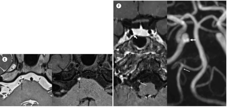
E. The vwMRI at day 9 follow-up depicts occlusion of the dissecting aneurysm. Proton density image vwMRI depicts lumen occlusion (left, ar- row) that is observable as high signal intensity (thrombus) in the lumen of the aneurysm on pre-contrast T1 vwMRI (right, arrow).
F. The vwMRI and time-of-flight MR angiography on day 9 follow-up depict decreased wall enhancement and normalized diameter of the right distal vertebral artery dissection (thin arrows); however, the aneurysm in the right cavernous internal carotid artery is still visible (thick arrows).
vwMRI = vessel wall MRI
E
F
was diagnosed with SLE and dissecting aneurysm in the right pontine perforator could be ex- plained by it. The mural wall thrombus in the pontine perforator likely led to the right pontine infarction. The patient was started on an anti-platelet agent, steroid pulse therapy, and hy- droxychloroquine 400 mg qd. She was given azathioprine 50 mg qd to treat the underlying au- toimmune condition. She had a modified Rankin Scale (mRS) score of 1 at discharge with mild symptom improvement, and she showed no further aggravation of symptoms at 3 months or 12 months, maintaining an mRS score of 1.
Ethics approval and consent to participate. In accordance with the Declaration of Helsinki, informed consent was obtained from the patient.
DISCUSSION
Although it is not completely understood, SLE-related ischemic stroke is known to be caused by various mechanisms including cardioembolism, large artery stenosis (of either non-atherosclerotic or premature atherosclerotic etiology), arterial dissection, hypercoagula- ble states, and cerebral vasculitis (2, 6). Large vessel occlusion due to premature atherosclero-
dissection that might have been caused by underlying vasculitis. Patients with SLE and other connective tissue diseases have a higher risk of vascular dissection than the general popula- tion (8). Multiple factors are involved in the activation of autoimmune inflammation of the arterial walls such as immune complex formation and deposition, intimal damage, and me- dial extracellular matrix damage (9). According to a case report and literature review of 11 patients who presented with ischemic stroke as the initial manifestation of SLE, most pa- tients (7/11), had ischemic infarcts in the vertebrobasilar artery and the pons was the most frequently affected region. The pathophysiology of infarction could be suspected in only three cases (two dissections and one with an inflammatory lesion) but eventually in none of them it was clearly identified. However, most of the cases were reported before 1999 i.e. im- age modalities to identify vasculitis or dissection were very limited. All patients had at least one autoantibody, which probably caused an underlying inflammatory or hypercoagulable state. Arterial occlusion or stenosis was identified in only six of the 11 cases (5).
We suggest vwMRI as diagnostic investigation tools to reveal the direct cause of ischemic infarcts, such as small vessel dissection or underlying vasculitis in the early phase of SLE and DSA can cross-check the lesion depicting an intimal flap, focal vessel wall irregularity, pre- aneurysmal narrowing, and fusiform dilatation. The predominant vwMRI characteristics of dissection are eccentric or combined eccentric/concentric shape, T1 high signal intensity with susceptibility artifact due to intramural hematoma, a double lumen caused by intimal flap, and aneurysmal dilatation with or without wall enhancement. Differential diagnosis of intra- mural hematoma is intraplaque hemorrhage, vasculitis and Moyamoya disease. Intraplaque hemorrhage usually shows irregular eccentric T1 high signal and no change. The main dif- ferentiation point of intramural hematoma are change by time and smooth luminal margin, longer segment than intraplaque hemorrhage. Vasculitis usually shows concentric strong enhancement in the active stage that resolves in the chronic stage but does not show intimal flap or aneurysmal dilatation on vwMRI (10).
In a young patient presenting with acute stroke of unknown origin, it is necessary to inves- tigate for rarer etiologies such as SLE and other autoimmune diseases. High-resolution vwM- RI and DSA might provide important clues.
Author Contributions
Conceptualization, B.J.S.; data curation, all authors; investigation, Y.Y.; methodology, C.M.S.; project administration, B.J.S.; resources, all authors; supervision, B.J.S.; visualization, B.J.S.; writing—original draft, C.M.S.; and writing—review & editing, B.J.S.
Conflicts of Interest
The authors have no potential conflicts of interest to disclose.
Funding None
REFERENCES
1. Saadatnia M, Sayed-Bonakdar Z, Mohammad-Sharifi G, Sarrami AH. The necessity of stroke prevention in patients with systemic lupus erythematosus. J Res Med Sci 2012;17:894-895
2. de Amorim LC, Maia FM, Rodrigues CE. Stroke in systemic lupus erythematosus and antiphospholipid syn-
drome: risk factors, clinical manifestations, neuroimaging, and treatment. Lupus 2017;26:529-536
3. Holmqvist M, Simard JF, Asplund K, Arkema EV. Stroke in systemic lupus erythematosus: a meta-analysis of population-based cohort studies. RMD Open 2015;1:e000168
4. Jennekens FG, Kater L. The central nervous system in systemic lupus erythematosus. Part 2. Pathogenetic mechanisms of clinical syndromes: a literature investigation. Rheumatology (Oxford) 2002;41:619-630 5. Ioannidis S, Mavridis M, Mitsias PD. Ischemic stroke as initial manifestation of systemic lupus erythemato-
sus: a case report and review of the literature. eNeurologicalSci 2018;13:26-30
6. Aringer M, Costenbader K, Daikh D, Brinks R, Mosca M, Ramsey-Goldman R, et al. 2019 European League Against Rheumatism/American College of Rheumatology classification criteria for systemic lupus erythe- matosus. Ann Rheum Dis 2019;78:1151-1159
7. Kwon SU, Koh JY, Kim JS. Vertebrobasilar artery territory infarction as an initial manifestation of systemic lupus erythematosus. Clin Neurol Neurosurg 1999;101:62-67
8. Mitsias P, Levine SR. Large cerebral vessel occlusive disease in systemic lupus erythematosus. Neurology 1994;44:385-393
9. Majeed A, Ribeiro NP, Ali A, Hijazi M, Farook H. A rare presentation of spontaneous internal carotid artery dis- section with Horner’s syndrome, VIIth, Xth and XIIth nerve palsies. Oxf Med Case Reports 2016;10:255-258 10. Pyrpasopoulou A, Chatzimichailidou S, Aslanidis S. Vascular disease in systemic lupus erythematosus. Au-
toimmune Dis 2012;2012:876456
전신성 홍반성 루푸스의 초기 증상으로 나타난 교뇌경색의 증례 보고: 자기공명영상 및
디지털감산 혈관조영술에서의 진단 단서
정미선 · 변준수* · 임영희
척추기저동맥의 혈류공급감소로 인한 뇌간 경색은 매우 드문 전신성 루푸스 환자의 초기 증 상으로, 경색의 직접적인 원인으로 매우 작은 기저동맥 분지인 교뇌공급혈관의 박리성 동맥 류는 보고된 사례가 없다. 이에 저자들은 디지털감산 혈관조영술과 고해상도 혈관벽 자기공 명영상를 이용하여 작은 교뇌공급혈관의 박리성 동맥류의 진단과 추적관찰 중 치유된 20세 여성의 사례를 보고하고자 한다. 전신성 루푸스의 진단은 신경학적장애의 유무와 혈액화학 검사 결과를 바탕으로 하였다. 추적 고해상도 혈관벽 자기공명영상에서 환자의 교뇌천공지 의 박리성동맥류는 폐색되어 있었고 우측 척추동맥의 박리성동맥류는 치유되어 보이지 않