Corresponding author:
Ravindra S. Swamy
Department of Anatomy, Melaka Manipal Medical College, Manipal University, Madhav Nagar, Manipal, Karnataka 576104, India
Tel: +91-820-2922388, Fax: +91-820-257190, E-mail: ravindrammmc@
gmail.com
pISSN 2093-3665 eISSN 2093-3673
Analysis of the morphometry and variations in the extensor digitorum brevis muscle: an anatomic guide for muscle flap and tendon transfer surgical dissection
Srinivasa Rao Sirasanagandla
1, Ravindra S. Swamy
1, Satheesha B. Nayak
1, Nagabhooshana S. Somayaji
1, Mohandas K. G. Rao
1, Kumar M. R. Bhat
21Department of Anatomy, Melaka Manipal Medical College, Manipal University, 2Department of Anatomy, Kasturba Medical College, Manipal University, Manipal, Karnataka, India
Abstract: The extensor digitorum brevis muscle (EDB) is a practical option for use as an island flap or free flap when reconstructing soft tissue defects in the ankle as well as in the entire lower limb. It is frequently used to correct crossover toe deformity and other painful toe disorders. We evaluated the morphometry of the EDB in 44 formalin-fixed limbs. Length and width of the muscles were measured. Surface area was calculated as the product of length and width of the muscle. The length of each tendon was also measured from its origin to the point of distal attachment. Presence of any additional tendons was noted. Mean length, width, and surface area of the muscle were 7.39±0.71 cm, 4.1±0.37 cm, and 30.5±4.78 cm2 on the right side and 7.2±0.84 cm, 3.9±0.37 cm, and 28.4±5.35 cm2 on the left side, respectively. Morphometry of the tendons revealed that the tendon of the great toe had the highest mean length (9.5 cm) and the tendon of the fourth toe had the lowest mean length (6.3 cm). Four of the limbs studied (9.09%) had only three tendons. Three of the limbs studied (6.81%) had five tendons, and in one exceptional case (2.27%), six tendons were detected. These observations have significant value and are applicable to plastic and orthopedic surgery.
Key words: Tendon transfer, Morphometry, Reconstruction, Crossover toe deformity, Extensor digitorum brevis Received January 31, 2013; Revised May 31, 2013; Accepted August 13, 2013
dorsalis pedis artery to insert onto the base of the proximal phalanx of the great toe. This slip is often termed the extensor hallucis brevis (EHB). The other slips attach to the lateral sides of the tendons of the extensor digitorum longus for the second, third, and fourth toes. EDB receives nerve supply from the lateral terminal branch of the deep peroneal nerve.
Functionally, EDB is dispensable, as the toes can be extended by the long extensor muscles. The anatomical position of the EHB muscle belly and its tendon are useful in locating the dorsalis pedis artery and deep peroneal nerve [1-3].
The EDB muscle is often used as an island flap to treat soft tissue defects in the ankle region and distal part of foot [4]. It is also used as a free flap to cover small defects or for
Introduction
The extensor digitorum brevis muscle (EDB) arises from the distal part of the superolateral surface of the calcaneus.
It runs distally across the dorsum of the foot and finally divides into four slips. Usually, the medial part of the muscle ends in a distinct slip that runs distally superficial to the
functional restoration [5]. EDB tendon transfer is frequently used to correct crossover toe deformity and to treat painful toe disorders such as lateral ankle deformity [6-8].
Anatomical variations and morphometry of the EDB have been described previously [9-11]. However, there is a dearth of studies focused solely on the morphometry of the EDB tendons in the existing literature. Recent and ongoing advances in plastic and orthopedic surgeries of the lower limb have created a need for a detailed understanding of ana- tomical variations and morphometry of the EDB muscle. The present study aimed to analyze the morphometry of the EDB muscles and tendons.
Materials and Methods
A total of 44 formalin-embalmed cadavers aged 50 to 60 years were examined. None of the cadavers had a history of malformations of the foot. The study was conducted in the Department of Anatomy, Melaka Manipal Medical College, Manipal University, India. The dorsums of 44 feet of both lateralities (right and left) were neatly dissected, and the proximal and distal attachments of the EDB muscles were carefully analyzed. Anatomical variations in the number of distal slips of the EDBs were recorded and photographed.
The length of the muscle (ML) was measured from its point of origin on the calcaneus to the point of commencement of its tendon to the second toe (Fig. 1). The width of the muscle (MW) was measured at the midpoint between the point of its origin on the calcaneus and the point of commencement of its tendon to the second toe (Fig. 1). The surface area of the muscle (MS) was calculated as the product of length and width (MS=ML×MW). The length of each tendon was measured from its point of origin to the point of its distal attachment (Fig. 1). All distances were measured using calipers. Results are expressed as mean±standard deviation.
Statistical comparisons were made using the student’s t-test and P-values of <0.05 were considered statistically significant.
Results
Minimum, maximum, and mean values of length, width, and surface area of the EDB muscle on both sides (right and left) were measured and tabulated (Table 1). No statistically Fig. 1. Dissection of the dorsum of the right foot showing the proximal
and distal attachments of the extensor digitorum brevis (EDB) muscle.
EDL, extensor digitorum longus; ML, muscle length; MW, muscle width; TL1, length of tendon to the great toe; TL2, length of tendon to the second toe; TL3, length of tendon to the third toe; TL4, length of tendon to the fourth toe.
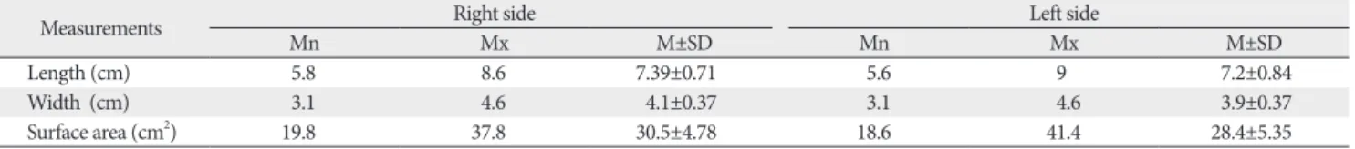
Table 1. Measurements of the length, width and surface area of the extensor digitorum brevis muscle
Measurements Right side Left side
Mn Mx M±SD Mn Mx M±SD
Length (cm) 5.8 8.6 7.39±0.71 5.6 9 7.2±0.84
Width (cm) 3.1 4.6 4.1±0.37 3.1 4.6 3.9±0.37
Surface area (cm2) 19.8 37.8 30.5±4.78 18.6 41.4 28.4±5.35
Mn, minimum; Mx, maximum; M±SD, mean±standard deviation.
Table 2. Measurements of the length of tendons of the extensor digitorum brevis muscle
Tendons Right side (cm) Left side (cm)
Mn Mx M±SD Mn Mx M±SD
TL1 5.6 9.5 7.05±0.86 5 9.4 7.2±0.88
TL2 4.4 8.4 6.3±0.94 4 8.2 6±0.8
TL3 4 6.3 5.2±0.75 3.8 7.8 5.2±1.04
TL4 4.2 7.6 5.5±0.69 4 7.6 5.3±0.94
Mn, minimum; Mx, maximum; M±SD, mean±standard deviation; TL1, length of tendon to the great toe; TL2, length of tendon to the second toe; TL3, length of tendon to the third toe; TL4, length of tendon to the fourth toe.
significant differences in muscle length (P>0.05), width (P>0.05), or surface area (P>0.05) were observed between left and right feet. Mean lengths of each of the EDB tendons were measured and tabulated for all limbs (Table 2). Fur- thermore, no statistically significant differences in tendon length (P>0.05) were observed between left and right feet.
In 36 limbs (81.8%), the EDB presented classically with four tendons for the medial four toes (Fig. 1). In four limbs (9.09%), the EDB had three heads that ended in three tendons for the medial three toes and the tendon for the fourth toe was found to be absent (Fig. 2). In one of these cases, presence of an accessory fasciculus arising from the adjacent sides of the first and second heads of the EDB was observed (Fig. 2). This accessory fasciculus merged with the first dorsal interosseous muscle (Fig. 2). In three cases (6.81%), the four distinct heads of EDB ended in five tendons (Figs. 3, 4). In two of these cases, the second head ended into two tendons for the second toe (Fig. 3), and in the other case, the third head gave rise to two tendons for the third toe (Fig. 4). In one limb (2.27%) the
EDB had four distinct heads but ended in six tendons for the medial four toes (Fig. 5). In this case, the second head divided into three tendons: medial, intermediate, and lateral (Fig. 5).
The medial tendon was attached to the base of the proximal phalanx of the second toe. The intermediate tendon was attached to the fascia over the shaft of the third metatarsal bone. The lateral tendon was inserted into the lateral side of the extensor digitorum longus tendon for the second toe (Fig.
5).
Discussion
The EDB frequently shows variations in its heads, tendons,
Fig. 2. Dissection of the dorsum of the left foot showing the proximal and distal attachments of the extensor digitorum brevis (EDB) muscle.
Note that the number of EDB tendons is only three and the tendon for the fourth toe is absent. MF, muscle fasciculus; T1, tendon to the great toe; T2, tendon to the second toe; T3, tendon to the third toe.
Fig. 4. Dissection of the dorsum of the left foot showing the proximal and distal attachments of the extensor digitorum brevis (EDB) muscle.
Note the additional EDB tendon for the third toe (T3a). T1, tendon to the great toe; T2, tendon to the second toe; T3, tendon to the third toe; T4, tendon to the fourth toe.
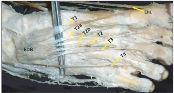
Fig. 5. Dissection of the dorsum of the right foot showing the proximal and distal attachments of the extensor digitorum brevis (EDB) muscle.
Note that the second head (SH) of EDB is divided into three tendons:
medial, intermediate, and lateral. The medial tendon (PP2) is attached to the base of the proximal phalanx of the second toe. The intermediate tendon is attached to the fascia over the shaft of the third metatarsal bone (M3). The lateral tendon is inserted to the lateral side of the extensor digitorum longus tendon for the second toe (EDL2). EHL, extensor hallucis longus; T1, tendon to the great toe; T2, tendon to the second toe; T2a & T2b, additional tendons from the SH of EDB; T3, tendon to the third toe; T4, tendon to the fourth toe.
Fig. 3. Dissection of the dorsum of the left foot showing the proximal and distal attachments of the extensor digitorum brevis (EDB) muscle.
Note the additional EDB tendon for the second toe (T2a). T1, tendon to the great toe; T2, tendon to the second toe; T3, tendon to the third toe; T4, tendon to the fourth toe.
and the presence of additional fascicles. In general, the muscle has four distinct heads that end in four tendons. Occasionally, the EDB may have two or three distinct heads and more rarely the whole muscle may be absent. In cases where two heads are present, the muscle is usually restricted to the first and fourth toes or may be restricted to the second and third toes or the first and little toes. On rare occasions, a double tendon for the second toe has been observed. An accessory fas ciculus taking origin from the bones of the foot may insert to the second or third tendons of the EDB. Accessory fascicles arising between the normal tendons may end on the fifth tendon. Such tendons are frequently found between the great and second toes. Sometimes, these tendons may join to form an interosseous muscle. Rarely, an extra tendon may join the long extensor tendon of the little toe [9-12]. In the present study, four cases (9.09%) had three distinct heads, which were restricted to the first, second, and third toes. An additional fifth tendon was found in three cases (6.81%). In one case (2.21%), the second head of the EDB presented two additional tendons. One of these was attached to the base of the proximal phalanx of the second toe and other tendon was inserted to the fascia over the shaft of the third metatarsal bone.
Occurrence of an additional head or accessory fasciculus and absence of one of the four heads of the EDB can be explained with reference to the embryonic development of the foot muscles. Four fundamental phases have been shown to occur during muscle pattern ontogenesis [13].
Each muscle results from the fusion of muscle primordia from different layers. While this fusion is occurring, some of these muscle primordia disappear due to cell death, although the cells within them can differentiate to myofilaments [14].
Persistence of some muscle primordia in the EDB might result in supernumerary heads or accessory fasciculi. Absence of one of the actual heads may be due to the disappearance of some muscle primordia which otherwise persist and give rise to the usual four-headed pattern [15]. Knowledge relating to the presence of extra tendons or absence of usual tendons is clinically important in plastic and orthopedic surgeries involving EDB tendon transfer.
A previous study of 13 cadavers conducted by del Pinal and Herrero [5], investigated the morphometry of the EDB.
In their study, the mean length was 5.13 cm, mean width was 3.76 cm, and mean surface area was 19.72 cm2. In our study, the mean length, width, and surface area were found to be 7.39±0.71 cm, 4.1±0.37 cm, and 30.5±4.78 cm2 on right side
and 7.2±0.84 cm, 3.9±0.37 cm, and 28.4±5.35 cm2 on the left side, respectively.
In the present study, morphometric analysis of the EDB tendons revealed that the tendon of the great toe had the highest mean length (9.5 cm) and the tendon of the fourth toe had the lowest mean length (6.3 cm). These morphometric findings and information relating to the incidence of supernumerary tendons of the EDB reported in the present study are clinically relevant to surgeries for the treatment of crossover toe deformity and painful toe disorders. Sagittal and/or horizontal plane instability of the second toe is defi- ned as crossover toe deformity. In this deformity, EDB ten- don transfer is the preferred treatment strategy compared with flexor digitorum longus tendon transfer, as it allows preservation of the metatarsophalangeal joint [7, 8]. This surgical protocol also has a reduced risk of postoperative complications such as avascular necrosis, malunion, and stiffness. The EDB tendon is also used to realign medially deviated lesser toes to treat painful toe disorders [6].
Due to a lack of local flaps, soft tissue defects are difficult to treat in the ankle and foot regions. EDB muscle flap is a practical option in such cases as it is easy to rise, has minimal morbidity, and does not require microsurgical skills [16]. In general, fasciocutaneous flaps are not preferable to muscle flaps to treat defects with exposed internal fixation devices.
The use of EDB as a local muscle flap has been previously described, and it has been used as a microvascular free flap in facial re-animation [17, 18]. In 1990, Crocker and Moss [16] demonstrated a new technique for EDB muscle flaps that allows a much larger arc; they suggested that muscle is a viable option to treat defects of the malleoli up to 5 cm in diameter.
They also suggested that the flap can extend up to the lateral malleolar region without any difficulty. Sub sequently, various authors have reported the advantages of ankle and foot soft tissue defect reconstruction using an EDB flap [19-21]. As the clinical applications of EDB free flaps are gaining increasing importance, the morphometric findings of the present study will be of help to plastic and orthopedic surgeons.
Acknowledgements
The authors wish to acknowledge the faculty members of the Department of Anatomy, Melaka Manipal Medical College for their support throughout the study.
References
1. Standring S. Gray’s anatomy: the anatomical basis of clinical practice. 39th ed. Edinburgh: Churchill Livingstone; 2005.
p.1536-7.
2. Moore KL, Dalley AF, Agur AM. Clinically oriented anatomy.
6th ed. Philadelphia: Lippencott, Williams & Wilkins; 2009.
p.612-4.
3. Moore KL, Persaud TV. The developing human: clinically oriented embryology. 6th ed. Philadelphia: W.B. Saunders; 2002.
p.612-4.
4. Baltensperger MM, Ganzoni N, Jirecek V, Meyer VE. The extensor digitorum brevis island flap: possible applications based on anatomy. Plast Reconstr Surg 1998;101:107-13.
5. del Piñal F, Herrero F. Extensor digitorum brevis free flap:
anatomic study and further clinical applications. Plast Reconstr Surg 2000;105:1347-56.
6. Westlin NE, Vogler HW, Albertsson MP, Arvidsson T, Montgomery F. Treatment of lateral ankle instability with transfer of the extensor digitorum brevis muscle. J Foot Ankle Surg 2003;42:183-92.
7. Haddad SL, Sabbagh RC, Resch S, Myerson B, Myerson MS.
Results of flexor-to-extensor and extensor brevis tendon transfer for correction of the crossover second toe deformity. Foot Ankle Int 1999;20:781-8.
8. Lui TH, Chan KB. Technique tip: modified extensor digitorum brevis tendon transfer for crossover second toe correction. Foot Ankle Int 2007;28:521-3.
9. Bergman RA, Afifi AK, Miyauchi R. Extensor Digitorum Brevis (Pedis). Illustrated encyclopedia of human anatomic variation:
Opus I: Muscular system: alphabetical listing of muscles: E.
Anatomy Atalases; [cited 2013 Sep 1]. Available from: http://
www.anatomyatlases.org/AnatomicVariants/MuscularSystem/
Text/E/19Extensor.shtml.
10. Anson BJ. Morris' human anatomy. 12th ed. New York: McGraw-
Hill Book Co.; 1966.
11. Henle J. Handbuch der Muskellehre des Menschen, in Handbuch der systematischen Anatomie des Menschen. Braunschweig:
Verlag von Friedrich Vieweg und Sohn; 1871.
12. Macalister A. Additional observation on muscular anomalies in human anatomy (third series), with a catalogue of the principal muscular variations hitherto published. Trans R Ir Acad 1875;25:
1-134.
13. Cihák R. Ontogenesis of the skeleton and intrinsic muscles of the human hand and foot. Ergeb Anat Entwicklungsgesch 1972;46:5- 194.
14. Grim M. Ultrastructure of the ulnar portion of the contrahent muscle layer in the embryonic human hand. Folia Morphol (Praha) 1972;20:113-5.
15. Kopuz C, Tetik S, Ozbenli S. A rare anomaly of the abductor digiti minimi muscle of the foot. Cells Tissues Organs 1999;164:
174-6.
16. Crocker AD, Moss AL. The extensor hallucis brevis muscle flap.
J Bone Joint Surg Br 1989;71:532.
17. Chattar-Cora D, Pederson WC. Experience with the extensor digitorum brevis muscle flap for foot and ankle reconstruction.
Ann Plast Surg 2006;57:289-94.
18. Landi A, Soragni O, Monteleone M. The extensor digitorum brevis muscle island flap for soft-tissue loss around the ankle.
Plast Reconstr Surg 1985;75:892-7.
19. Leitner DW, Gordon L, Buncke HJ. The extensor digitorum brevis as a muscle island flap. Plast Reconstr Surg 1985;76:777- 80.
20. Martinet X, Forli A, Guinard D, Corcella D, Moutet F. Extensor digitorum muscle flap: its position in ankle and foot coverage.
Report of 15 cases. Ann Chir Plast Esthet 2003;48:159-66.
21. Chateau F, Chabas JF, Niddam J, Guinard D, Legré R. Use of Extensor digitorum brevis flap in routine reconstructive surgery of lower limbs. Report of more than 50 cases. Ann Chir Plast Esthet 2012;57:600-5.