PGHN
Original Article
Characteristics of Upper Gastrointestinal Tract Involvement in Korean Pediatric Crohn’s Disease: A Multicenter Study
Ji Hyoung Park, Hye Na Nam, Ji-Hyuk Lee*, Jeana Hong
†, Dae Yong Yi
‡, Eell Ryoo, In Sang Jeon, and Hann Tchah
Department of Pediatrics, Gachon University Gil Medical Center, Incheon, *Department of Pediatrics, Chungbuk National University Hospital, Cheongju, †Department of Pediatrics, Kangwon National University School of Medicine, Chuncheon, ‡Department of Pediatrics, Chung-Ang University Hospital, Seoul, Korea
Purpose: Crohn’s disease (CD) can involve any site of the gastrointestinal tract (GIT). However, the characteristics of upper GIT involvement in CD are unclear, especially in the Eastern pediatric population. This study aimed to esti- mate the prevalence of upper GIT involvement and identify the clinical features of Korean children with CD.
Methods: This was a retrospective multicenter cohort study that included 52 pediatric patients with CD who underwent esophagogastroduodenoscopy and biopsy. The clinical symptoms and endoscopic and histologic features of the upper GIT were identified according to the presence or absence of upper gastrointestinal symptoms.
Results: Among the 52 patients, upper GIT involvement was noted in 50.0% (26/52). The mean age at CD diagnosis was 14.1±2.1 years. Gastric ulcer was the most common lesion (19.2%) found on upper GIT endoscopy, followed by duodenal ulcers (15.4%). Chronic inflammation was the most common histopathologic feature (75.0%), followed by gastric erosion (17.3%). Granuloma was found in 9.6% of patients. Helicobacter pylori infection was identified in 5.8% of patients. Endoscopic and histologic findings were not significantly different, but the mean values of eryth- rocyte sedimentation rate (60.7±27.1 vs. 43.0±27.6 mm/h, p=0.037) and C-reactive protein (16.5±28.2 vs. 6.62±13.4 mg/dL, p=0.014) were significantly different between patients with and without upper gastrointestinal CD symptoms.
Conclusion: Upper GIT involvement was relatively common in pediatric patients with CD irrespective of upper gastro- intestinal symptoms, and H. pylori infection was relatively uncommon. The results of this study should aid the estab- lishment of regional guidelines for upper GIT examination.
Key Words: Crohn disease, Upper gastrointestinal tract, Granuloma, Helicobacter pylori, Pediatrics
Received:August 26, 2017, Revised:October 1, 2017, Accepted:October 5, 2017
Corresponding author: Eell Ryoo, Department of Pediatrics, Gachon Children’s Hospital, Gachon University College of Medicine, 21 Namdong-daero 774beon-gil, Namdong-gu, Incheon 21565, Korea. Tel: +82-32-460-3224, Fax: +82-32-460-2362, E-mail: [email protected] Copyright ⓒ 2017 by The Korean Society of Pediatric Gastroenterology, Hepatology and Nutrition
This is an openaccess article distributed under the terms of the Creative Commons Attribution NonCommercial License (http://creativecommons.org/licenses/by-nc/4.0/) which permits unrestricted noncommercial use, distribution, and reproduction in any medium, provided the original work is properly cited.
INTRODUCTION
Crohn’s disease (CD) is a chronic inflammatory bowel disease of unknown cause. It occurs primarily in the terminal ileum, but can involve any area of the gastrointestinal tract (GIT), from the mouth to the anus [1]. To date, much research has been conducted to investigate the involvement of the lower GIT, but the involvement of the upper GIT in CD has not been studied sufficiently.
In studies involving Italian and American adults, the prevalence of upper gastrointestinal (GI) CD is reported to be 16-34% [2,3]. In pediatric patients with CD, studies have reported different findings; for example, the upper GIT was involved in 71% of cases in a study conducted in Canada [4], whereas, in a study conducted in the UK, esophagitis involvement was noted in 72%, gastritis in 92%, and duodenitis in 33% [5]. Moreover, in a study conducted in the US, 36% of patients showed symptoms suggestive of up- per GI involvement, and 12% of pediatric patients demonstrated noncaseating granulomas [6]. In a study involving Korean adults who underwent up- per GIT biopsy, 59% of cases had upper GI symptoms.
Erosive gastritis was the most common endoscopic finding, and was noted in 66% of patients. In another study, approximately 70% of patients had an abnor- mal histological finding. However, these studies were based on only a few cases, and moreover, the fo- cus was on abnormal findings in the stomach or Helicobacter pylori [7]. To date, data has been scarce regarding pediatric patients in Korea. In Asians, CD is more male-predominant (1.67:1 to 2.9:1) com- pared to in other races. Moreover, CD of an isolated colonic type is most common in Europeans, whereas CD involving both the small and large bowels is most common among Asians [8-10].
Accordingly, this study aimed to examine whether the presence or absence of upper GI symptoms was related to upper GIT lesions in pediatric patients with CD and to investigate whether the relationship would help determine the need for testing. An addi- tional objective of the study was to examine the clin- ical features, endoscopic and histological findings,
and the prevalence of H. pylori in CD with upper GIT involvement to help create a guideline for upper GIT testing in pediatric CD patients in Korea.
MATERIALS AND METHODS
Study design and subjects
Data from the medical records of pediatric patients (age <18 years) with CD who underwent upper GIT endoscopy and biopsy between January 2001 and August 2016 were collected from 4 Korean tertiary hospitals. The 4 medical centers included Gachon University Gil Medical Center located in Incheon, Chungbuk National University Hospital in the cen- tral region, Kangwon National University School of Medicine in the north region, and Chung-Ang University Hospital in Seoul, South Korea.
Of 64 subjects, 52 were finally enrolled after meet- ing the following criteria: (1) patients who under- went biopsy for abnormal gastric lesions and/or nor- mal-looking gastric mucosa and (2) patients who had no known chronic medical illnesses. Our diag- nostic criteria for CD has been previously described [8,11].
We retrospectively reviewed the patients’ medical records, upper GI endoscopic findings, and histo- pathological findings from the time of CD diagnosis.
The following demographic and clinical information was collected: gender, GI symptoms (diarrhea, lower abdominal pain, weight loss, hematochezia, and poor oral intake), upper GI symptoms (epigastric pain, nausea, vomiting, dyspepsia, burning sensa- tion, epigastric fullness, belching, and melena), per- ianal lesions (skin tag, fissure, abscess, and fistula), and medications ever used for CD (anti-tumor ne- crosis factor-α therapy [infliximab, adalimumab], immunosuppressive therapy [steroid], immunomod- ulators [6-mercaptopurine, azathioprine], 5-amino- salicylic acid [mesalamine, sulfasalazine], and anti- biotics). Laboratory findings such as erythrocyte sed- imentation rate (ESR) and C-reactive protein (CRP) were identified. Disease locations were evaluated ac- cording to the Montreal classification. Disease activ- ity was evaluated with the pediatric Crohn’s disease
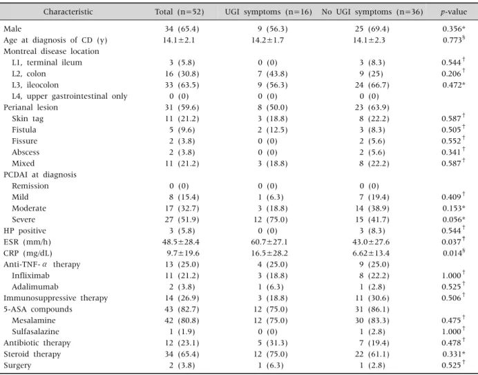
Table 1. Baseline Characteristics of Subjects
Characteristic Total (n=52) UGI symptoms (n=16) No UGI symptoms (n=36) p-value
Male 34 (65.4) 9 (56.3) 25 (69.4) 0.356*
Age at diagnosis of CD (y) 14.1±2.1 14.2±1.7 14.1±2.3 0.773§
Montreal disease location
L1, terminal ileum 3 (5.8) 0 (0) 3 (8.3) 0.544†
L2, colon 16 (30.8) 7 (43.8) 9 (25) 0.206†
L3, ileocolon 33 (63.5) 9 (56.3) 24 (66.7) 0.472*
L4, upper gastrointestinal only 0 (0) 0 (0) 0 (0)
Perianal lesion 31 (59.6) 8 (50.0) 23 (63.9)
Skin tag 11 (21.2) 3 (18.8) 8 (22.2) 0.587†
Fistula 5 (9.6) 2 (12.5) 3 (8.3) 0.505†
Fissure 2 (3.8) 0 (0) 2 (5.6) 0.552†
Abscess 2 (3.8) 0 (0) 2 (5.6) 0.341†
Mixed 11 (21.2) 3 (18.8) 8 (22.2) 0.587†
PCDAI at diagnosis
Remission 0 (0) 0 (0) 0 (0)
Mild 8 (15.4) 1 (6.3) 7 (19.4) 0.409†
Moderate 17 (32.7) 3 (18.8) 14 (38.9) 0.153*
Severe 27 (51.9) 12 (75.0) 15 (41.7) 0.056*
HP positive 3 (5.8) 0 (0) 3 (8.3) 0.544†
ESR (mm/h) 48.5±28.4 60.7±27.1 43.0±27.6 0.037‡
CRP (mg/dL) 9.7±19.6 16.5±28.2 6.62±13.4 0.014§
Anti-TNF-α therapy 13 (25.0) 4 (25.0) 9 (25.0)
Infliximab 11 (21.2) 3 (18.8) 8 (22.2) 1.000†
Adalimumab 2 (3.8) 1 (6.3) 1 (2.8) 0.525†
Immunosuppressive therapy 14 (26.9) 3 (18.8) 11 (30.6) 0.506†
5-ASA compounds 43 (82.7) 12 (75.0) 31 (86.1)
Mesalamine 42 (80.8) 12 (75.0) 30 (83.3) 0.475†
Sulfasalazine 1 (1.9) 0 (0) 1 (2.8) 1.000†
Antibiotic therapy 12 (23.1) 5 (31.3) 7 (19.4) 0.478†
Steroid therapy 34 (65.4) 12 (75.0) 22 (61.1) 0.331*
Surgery 2 (3.8) 1 (6.3) 1 (2.8) 0.525†
Values are presented as number (%) or mean±standard deviation.
UGI: upper gastrointestinal, CD: Crohn’s disease, PCDAI: pediatric Crohn’s disease activity index, HP: Helicobacter pylori, ESR: erythrocyte sedimentation rate, CRP: C-reactive protein, TNF: tumor necrosis factor, 5-ASA: 5-aminosalicylic acid.
*Pearson chi-squared test, †Fisher’s exact test, ‡Student t-test, §Mann-Whitney U-test.
activity index (PCDAI) and was classified as re- mission (PCDAI <10), mild activity (PCDAI 10-27.5), moderate activity (PCDAI 30-37.5), and severe activ- ity (PCDAI >40) [12]. The study protocol was ap- proved by the institutional review boards of Gachon University Gil Medical Center (IRB no. 2016-368).
Upper GIT endoscopy, histologic evaluation, and H. pylori infection assessment
All upper GI endoscopic evaluations were per- formed by expert GIT endoscopists of the participat- ing institutions. Biopsy specimens were obtained for
pathologic evaluation from abnormal gastric lesions and normal-looking gastric mucosa at the endo- scopists’ discretion. Additional biopsies were per- formed at both the gastric antrum and corpus for the rapid urease test. Standard hematoxylin and eosin staining was performed on biopsy specimens. The di- agnosis of H. pylori infection was based on either a positive histopathology plus a positive rapid urease test, or a positive culture. Gastric biopsies were ob- tained for histopathology [13].
The diagnosis of “upper GI involvement of CD”
was based on a combination of compatible endo-
Table 2. Gastrointestinal Symptoms of Pediatric Crohn’s Disease (n=52)
Symptom Case
Diarrhea 33 (63.5)
Low Abdominal pain 32 (61.5)
Weight loss 22 (42.3)
Hematochezia 11 (21.2)
Poor oral intake 9 (17.3)
Upper gastrointestinal symptom 16 (30.8)
Nausea 13 (25.0)
Epigastric pain 10 (19.2)
Vomiting 4 (7.7)
Dyspepsia 1 (1.9)
Melena 1 (1.9)
No symptom 1 (1.9)
Values are presented as number (%).
scopic (ulcerations, erosions, strictures, and aph- thous lesions) and histologic findings (chronic in- flammation, erosion, ulceration, granuloma, and gastric intestinal metaplasia) as described previously [14,15]. Some nonspecific inflammation or inflamm- atory changes explained by other conditions (reflux esophagitis or H. pylori gastritis) were excluded.
After dividing the participants into 2 groups ac- cording to the presence or absence of upper GI symp- toms, we evaluated the differences in demographics, H. pylori infection, treatment, surgery, PCDAI, peria- nal lesions, laboratory findings (ESR, CRP), and en- doscopic and histologic findings.
Statistical analysis
Continuous variables were expressed as means with ranges. Discrete data were expressed as num- bers or percentages or both. Demographics, clinical symptoms, and endoscopic and histologic GIT le- sions of subjects with and without upper GI CD symptoms were compared. For comparative analy- ses, the Mann-Whitney U-test and Student’s t-test were used for continuous variables, while the chi-squared test or Fisher’s exact test were used for categorical variables, where appropriate. All stat- istical analyses were performed using IBM SPSS for Windows ver. 21.0 (IBM Co., Armonk, NY, USA). p<
0.05 was considered statistically significant.
RESULTS
Demographics and disease characteristics Of the total 64 patients, 2 patients did not undergo upper GI endoscopy and 10 patients were excluded from the study because of inadequate data. A total of 52 patients were enrolled. The subjects included 34 men (65.4%), and the mean age at CD diagnosis was 14.1±2.1 years. According to the Montreal classi- fication, the dominant phenotypes were listed as fol- lows: the sites of disease location were L3 (ileocolon, 33/52, 63.5%), L4 (isolated upper GI, 0/52, 0%), L2 (colon, 16/52, 30.8%), and L1 (terminal ileum, 3/52, 5.8%).
Perianal lesions were found in 59.6% of the
patients. Of the lesions, the skin tag was the most common (21.2%, 11/52), followed by fistula (9.6%, 5/52). Overall, a mixed-type lesion was noted in 21.2% (11/52) of patients.
According to PCDAI, severe activity was the most common (51.9%, 27/52). Moderate activity was found in 32.7% (17/52) and mild activity in 15.4%
(8/52), and remission did not occur in any patients.
H. pylori test was positive in 3 patients (5.8%).
The mean CRP value at the time of upper GIT en- doscopy was 9.7±19.6 mg/dL and the mean ESR lev- el was 48.5±28.4 mm/h (Table 1).
GI symptoms
The most common GI symptom was diarrhea (63.5%, 33/52), followed by lower abdominal pain (61.5%, 32/52) and weight loss (42.3%, 22/52).
Hematochezia (21.2%, 11/52) and poor oral intake (17.3%, 9/52) were rare relative to other symptoms.
Upper GI symptoms were noted in 30.8% (16/52).
Of those, nausea was the most common symptom (25.0%, 13/52), followed by epigastric pain (19.2%, 10/52) and vomiting (7.7%, 4/52). There were no complaints of epigastric fullness, belching, or burn- ing sensation (Table 2).
Upper GI endoscopic and pathologic findings The upper GI endoscopic findings were examined
Table 3. Endoscopic Findings between Pediatric Crohn’s Disease Patients with and without Upper Gastrointestinal Symptoms
Total (n=52) UGI symptoms (n=16) No UGI symptoms (n=36) p-value
Endoscopic findings Esophagus
Ulcer 2 (3.8) 2 (12.5) 0 (0) 0.090*
Erosion 4 (7.7) 0 (0) 4 (11.1) 0.299*
Stomach
Erosions 9 (17.3) 4 (25.0) 5 (13.9) 0.431*
Ulcers 10 (19.2) 3 (18.8) 7 (19.4) 1.000*
Aphthous 1 (1.9) 1 (6.3) 0 (0) 0.308*
Stricture 1 (1.9) 0 (0) 1 (2.8) 1.000*
Duodenum
Erosions 3 (5.8) 1 (6.3) 2 (5.6) 0.578*
Ulcers 8 (15.4) 4 (25.0) 4 (11.1) 0.231*
Aphthous 2 (3.8) 1 (6.3) 1 (2.8) 0.525*
Stricture 2 (3.8) 1 (6.3) 1 (2.8) 0.525*
Histologic findings
Chronic inflammation 39 (75.0) 13 (81.3) 26 (72.2) 0.730*
Erosion 9 (17.3) 4 (25.0) 5 (13.9) 0.431*
Ulceration 4 (7.7) 3 (18.8) 1 (2.8) 0.081*
Granuloma 5 (9.6) 3 (18.8) 2 (5.6) 0.163*
Gastric intestinal metaplasia 3 (5.8) 2 (12.5) 1 (2.8) 0.221*
Normal 5 (9.6) 0 (0) 5 (13.9) 1.000*
Values are presented as number (%).
UGI: upper gastrointestinal.
*Fisher’s exact test.
according to the following sites: esophagus, stom- ach, and duodenum.
Gastric ulcer was the most common (19.2%) lesion found on upper GIT endoscopy, followed by duode- nal ulcers (15.4%). In the group with upper GI symp- toms, esophageal ulcer was observed in 12.5% (2/16) of patients, while in the group without upper GI symptoms, esophageal erosion was noted in 11.1%
(4/36) of patients. The difference was not statisti- cally significant.
Regarding gastric findings, gastric ulcer was the most common (19.2%, 10/52). In the group with up- per GI symptoms, gastric erosion was noted in 25.0%
(4/16), while in the group without GI symptoms, gastric ulcer was observed in 19.4% (7/36) of patients. The difference was not statistically significant.
Regarding duodenal findings, duodenal ulcer was the most common (15.4%, 8/52). There was no dif- ference between the groups with and without upper
GI symptoms.
Chronic inflammation was most common histo- pathologic feature (n=39, 75.0%) followed by gastric erosion (n=9, 17.3%). Chronic inflammation was the most common both in the group with (81.3%, 13/16) and without (72.2%, 26/36) upper GI symptoms. In 5 patients (9.6%), granuloma had pre- viously been identified from biopsy specimens. In addition, erosion, ulceration, gastric intestinal meta- plasia, etc. showed non-significant differences based on the presence or absence of upper GI symptoms (Table 3).
Differences between patients with and without upper GI symptoms
In the group with upper GI symptoms, the mean age at CD diagnosis was 14.2±1.7 years and 43.8%
(7/16) were female. In the group without upper GI symptoms, the mean age at CD diagnosis was 14.1±2.3 years and 30.6% (11/36) were female. No
between-group differences were statistically signi- ficant.
When the association between upper GI symp- toms and H. pylori infection, CD medications, PCDAI, perianal lesions, etc., was analyzed, no variables showed an association with upper GI symptoms ex- cept for the mean values of ESR (60.7±27.1 vs.
43.0±27.6 mm/h, p=0.037) and CRP (16.5±28.2 vs.
6.62±13.4 mg/dL, p=0.014) (Table 1).
DISCUSSION
Upper GI symptoms were not correlated with up- per GI lesions, and H. pylori infections were relatively uncommon in Korean pediatric CD. However, the extent of inflammation suggested the presence of upper GI symptoms. These results can aid the estab- lishment of guidelines for upper GIT examination.
According to studies involving adults in Western countries, the prevalence of upper GI CD ranges from 16-34% [2,3]. However, the correlation between up- per GI symptoms and true endoscopically and patho- logically proven disease has not been fully estab- lished, even in pediatric patients. In the present study, upper GI involvement in pediatric CD was 50.0% (26/52), which is higher compared to results in Western countries. We believe the differences are due to dissimilar subject baseline characteristics such as age, race, and disease severity, and differ- ences in diagnostic criteria.
A previous study involving adult Korean patients with CD reported that 59.6% demonstrated upper GI symptoms [7]. In the present study, 16 (30.8%) pa- tients showed upper GI symptoms, which is con- sistent with other studies, and there was no sig- nificant difference based on the presence or absence of symptoms. Concerning individual symptoms, nausea (25.0%) was the most common, followed by epigastric pain (19.2%), vomiting (7.7%), dyspepsia (1.9%), and melena (1.9%).
Regarding endoscopic findings, gastric ulcer was the most common (19.2%) lesion found on upper GIT endoscopy, followed by duodenal ulcers (15.4%).
In the presence of upper GI symptoms, gastric ero-
sion (25.0%) and duodenal ulcer (25.0%) were the most common, and gastric ulcer (18.8%) was the most common in the absence of upper GI symptoms.
The difference in endoscopic findings based on the presence and absence of upper GI symptoms was not statistically significant. A study involving adults found gastric erosion in 9.6%, duodenal ulcer in 5.3%, duodenal erythema or erosion in 5.1% [2].
There were fewer abnormal findings in that study compared to the present one [2]. It is speculated that the discrepancy may be due to inter-observer differ- ences and the presence or absence of overlapping findings.
According to a meta-analysis conducted on stud- ies with adult patients with CD in Western countries, nonspecific gastric inflammation was the most com- mon histological finding (32%) [2]. Gastric gran- uloma was noted in 7.9% and focal gastritis in 30.9%
[2]. Regarding inflammatory findings by site, 84%
occurred in the stomach, 28.2% in the duodenum, and 23.2% in the gastric granuloma [2]. In the pres- ent study, the most common histological finding in all pediatric patients with CD, regardless of the ana- tomical site, was chronic inflammation (75.0%), fol- lowed by erosion (17.3%). Granuloma was observed in 9.6% of patients, which was higher than in adults.
Moreover, there was no association between the presence or absence of upper GI symptoms and his- tological findings (Table 3).
In Western countries, one study reported that in small bowel or colonic surgical specimens of patients with CD, perianal fistula increased significantly in the presence of noncaseating granuloma [16]. Other studies reported that gastric granuloma was found in 5-83% of gastric biopsy specimens from patients with CD and suggested an association between gas- tric noncaseating granuloma and perianal ab- scess/fistula [17,18]. In the present study of pediatric patients, gastric granuloma occurred in 9.6% (5 pa- tients) and perianal abscess/fistula co-occurred in none of the 5 patients with gastric granuloma. We believe this finding shows that in pediatric patients with CD, unlike in adult patients, there is no associa- tion between gastric granuloma and perianal ab-
scess/fistula. This finding should be clarified with studies including a larger number of cases.
H. pylori infections are acquired early in life by young children and adolescents. In one study involv- ing South Korea children, the prevalence of H. pylori infection was 22% [19]. Another study reported that H. pylori infections were found in 7.4% of South Korean children with recurrent abdominal pain [20]. A previous study reported a lower H. pylori in- fection rate in patients with CD than patients with- out CD [21]. Additionally, another study based on a meta-analysis showed a significant negative associa- tion between H. pylori infection and irritable bowel syndrome (IBD) and suggested a possible protective effect of H. pylori infection against IBD [22]. In the present study on pediatric patients with CD, H. pylori infection was identified in 5.8% of patients, which was lower than in pediatric patients without CD. We believe this finding shows that in pediatric patients with CD, there is negative association between H. py- lori infection and pediatric patients with CD in South Korea. This finding should be clarified with studies including a larger number of cases.
CD is commonly complicated by perianal mani- festations. In studies of adult patients with CD in Western countries, the reported incidence of peria- nal CD varied from 3.8% [23] to 80% [24]. In a study involving children [25], perianal disease co-occurred in 21% of pediatric patients with CD and was also more common among black people (26%) compared to white people (20%, p=0.017). In the present study, 59.6% (31/52) of the pediatric patients showed more than one perianal lesion such as skin tag, fissure, fistula, and abscess. It seems that racial characteristics are important, suggesting that if an Asian pediatric patient with CD presents with a per- ianal lesion, more attention should be paid to the CD.
According to previous research, increased CRP in adult patients with CD increases the risk of CD-re- lated hospitalization and CD-related intestinal resection. CRP levels >1 mg/dL have been found to be correlated with granulomatous CD in pediatric patients in the United States [26-29]. In the present study, we found that ESR and CRP significantly in-
creased in the presence of upper GI symptoms. CRP testing was conducted at slightly different time points across different studies, but care should be taken if CRP and ESR are elevated in a patient with CD even when there is no difference in PCDAI or his- tological or endoscopic findings.
According to the European Society of Pediatric Gastroenterology, Hepatology and Nutrition (ES- PGHN) guidelines, upper GIT endoscopy and ileoco- lonoscopy are recommended in all patients with pe- diatric-onset inflammatory bowel disease [30].
However, there is no such guideline in Korea.
Although endoscopically abnormal findings are rare in patients with upper GI symptoms, endoscopic or histologic abnormalities are relatively common ac- cording to the findings of routinely performed endoscopy. These results suggest that upper GIT en- doscopy should be performed on pediatric patients with CD in South Korea.
To our knowledge, this is the first retrospective multicenter study in Korea aimed at evaluating the prevalence of upper GIT involvement in pediatric pa- tients with CD, irrespective of upper GI symptoms.
In the present series, 50% of the patients showed up- per GI CD involvement, a higher value than expected.
The present study had the following limitations.
First, it was based on a retrospective design and sec- ond, the study sample was small. Third, we could not review the pathology again and simply check the pathology reports.
In conclusion, upper GI symptoms may not be cor- related with upper GIT lesions. H. pylori infection was relatively uncommon in Korean pediatric patients with CD. However, the extent of inflammation sug- gests the presence of upper GI symptoms. These re- sults should aid the establishment of regional guide- lines for upper GIT examination.
REFERENCES
1. Van Limbergen J, Russell RK, Drummond HE, Aldhous MC, Round NK, Nimmo ER, et al. Definition of pheno- typic characteristics of childhood-onset inflammatory bowel disease. Gastroenterology 2008;135:1114-22.
2. Diaz L, Hernandez-Oquet RE, Deshpande AR, Moshiree
B. Upper gastrointestinal involvement in Crohn dis- ease: histopathologic and endoscopic findings. South Med J 2015;108:695-700.
3. Annunziata ML, Caviglia R, Papparella LG, Cicala M.
Upper gastrointestinal involvement of Crohn's disease:
a prospective study on the role of upper endoscopy in the diagnostic work-up. Dig Dis Sci 2012;57:1618-23.
4. Lenaerts C, Roy CC, Vaillancourt M, Weber AM, Morin CL, Seidman E. High incidence of upper gastro- intestinal tract involvement in children with Crohn disease. Pediatrics 1989;83:777-81.
5. Tobin JM, Sinha B, Ramani P, Saleh AR, Murphy MS.
Upper gastrointestinal mucosal disease in pediatric Crohn disease and ulcerative colitis: a blinded, con- trolled study. J Pediatr Gastroenterol Nutr 2001;32:
443-8.
6. Ammoury RF, Pfefferkorn MD. Significance of esoph- ageal Crohn disease in children. J Pediatr Gastroenterol Nutr 2011;52:291-4.
7. So H, Ye BD, Park YS, Kim J, Kim JS, Moon W, et al.
Gastric lesions in patients with Crohn's disease in Korea: a multicenter study. Intest Res 2016;14:60-8.
8. Yang SK, Yun S, Kim JH, Park JY, Kim HY, Kim YH, et al. Epidemiology of inflammatory bowel disease in the Songpa-Kangdong district, Seoul, Korea, 1986- 2005: a KASID study. Inflamm Bowel Dis 2008;14:
542-9.
9. Thia KT, Loftus EV Jr, Sandborn WJ, Yang SK. An up- date on the epidemiology of inflammatory bowel dis- ease in Asia. Am J Gastroenterol 2008;103:3167-82.
10. Park SH, Yang SK, Park SK, Kim JW, Yang DH, Jung KW, et al. Long-term prognosis of Crohn's disease and its temporal change between 1981 and 2012: a hospi- tal-based cohort study from Korea. Inflamm Bowel Dis 2014;20:488-94.
11. Loftus EV Jr, Silverstein MD, Sandborn WJ, Tremaine WJ, Harmsen WS, Zinsmeister AR. Crohn's disease in Olmsted County, Minnesota, 1940-1993: incidence, prevalence, and survival. Gastroenterology 1998;114:
1161-8.
12. Hyams J, Markowitz J, Otley A, Rosh J, Mack D, Bousvaros A, et al. Evaluation of the pediatric Crohn disease activity index: a prospective multicenter experience. J Pediatr Gastroenterol Nutr 2005;41:
416-21.
13. Koletzko S, Jones NL, Goodman KJ, Gold B, Rowland M, Cadranel S, et al. Evidence-based guidelines from ESPGHAN and NASPGHAN for Helicobacter pylori in- fection in children. J Pediatr Gastroenterol Nutr 2011;
53:230-43.
14. De Felice KM, Katzka DA, Raffals LE. Crohn's disease
of the esophagus: clinical features and treatment out- comes in the biologic era. Inflamm Bowel Dis 2015;21:
2106-13.
15. Sonnenberg A, Melton SD, Genta RM. Frequent occur- rence of gastritis and duodenitis in patients with in- flammatory bowel disease. Inflamm Bowel Dis 2011;
17:39-44.
16. Denoya P, Canedo J, Berho M, Allende DS, Bennett AE, Rosen L, et al. Granulomas in Crohn's disease: does pro- gression through the bowel layers affect presentation or predict recurrence? Colorectal Dis 2011;13:1142-7.
17. Wagtmans MJ, van Hogezand RA, Griffioen G, Verspaget HW, Lamers CB. Crohn's disease of the up- per gastrointestinal tract. Neth J Med 1997;50:S2-7.
18. Kefalas CH. Gastroduodenal Crohn's disease. Proc (Bayl Univ Med Cent) 2003;16:147-51.
19. Malaty HM, Kim JG, Kim SD, Graham DY. Prevalence of Helicobacter pylori infection in Korean children: in- verse relation to socioeconomic status despite a uni- formly high prevalence in adults. Am J Epidemiol 1996;143:257-62.
20. Jang KM, Choe BH, Choe JY, Hong SJ, Park HJ, Chu MA, et al. Changing prevalence of Helicobacter pylori infections in Korean children with recurrent abdominal pain. Pediatr Gastroenterol Hepatol Nutr 2015;18:
10-6.
21. El-Omar E, Penman I, Cruikshank G, Dover S, Banerjee S, Williams C, et al. Low prevalence of Helicobacter pylori in inflammatory bowel disease: as- sociation with sulphasalazine. Gut 1994;35:1385-8.
22. Rokkas T, Gisbert JP, Niv Y, O'Morain C. The associa- tion between Helicobacter pylori infection and in- flammatory bowel disease based on meta-analysis.
United European Gastroenterol J 2015;3:539-50.
23. Sangwan YP, Schoetz DJ Jr, Murray JJ, Roberts PL, Coller JA. Perianal Crohn's disease. Results of local sur- gical treatment. Dis Colon Rectum 1996;39:529-35.
24. McClane SJ, Rombeau JL. Anorectal Crohn's disease.
Surg Clin North Am 2001;81:169-83, ix.
25. Adler J, Dong S, Eder SJ, Dombkowski KJ;
ImproveCareNow Pediatric IBD Learning Health System. Perianal Crohn disease in a large multicenter pediatric collaborative. J Pediatr Gastroenterol Nutr 2017;64:e117-24.
26. Vaiopoulou A, Gazouli M, Papadopoulou A, Anagnosto- poulos AK, Karamanolis G, Theodoropoulos GE, et al.
Serum protein profiling of adults and children with Crohn disease. J Pediatr Gastroenterol Nutr 2015;60:
42-7.
27. Kellermayer R, Mir SA, Nagy-Szakal D, Cox SB, Dowd SE, Kaplan JL, et al. Microbiota separation and C-re-
active protein elevation in treatment-naïve pediatric granulomatous Crohn disease. J Pediatr Gastroenterol Nutr 2012;55:243-50.
28. Motil KJ, Grand RJ, Maletskos CJ, Young VR. The ef- fect of disease, drug, and diet on whole body protein me- tabolism in adolescents with Crohn disease and growth failure. J Pediatr 1982;101:345-51.
29. Oh K, Oh EH, Baek S, Song EM, Kim GU, Seo M, et al.
Elevated C-reactive protein level during clinical re- mission can predict poor outcomes in patients with Crohn's disease. PLoS One 2017;12:e0179266.
30. Levine A, Koletzko S, Turner D, Escher JC, Cucchiara S, de Ridder L, et al. ESPGHAN revised porto criteria for the diagnosis of inflammatory bowel disease in chil- dren and adolescents. J Pediatr Gastroenterol Nutr 2014;58:795-806.