원위 대퇴골 골절에서 슬관절 강직의 위험 인자
손동욱 ㆍ김형수ㆍ최우영
명지병원 정형외과
Risk Factors for Knee Stiffness in Distal Femoral Fractures
Dong-Wook Son, M.D. , Hyoung-Soo Kim, M.D., Ph.D., Woo-Young Choi, M.D.
Department of Orthopaedic Surgery, Myongji Hospital, Goyang, Korea
Received June 18, 2018 Revised July 20, 2018 Accepted September 21, 2018 Correspondence to:
Dong-Wook Son, M.D.
Department of Orthopaedic Surgery, Myongji Hospital, 55 Hwasu-ro 14beon-gil, Deogyang-gu, Goyang 10475, Korea
Tel: +82-31-810-5160 Fax: +82-31-969-0500 E-mail: elviselvis@naver.com Financial support: None.
Conflict of interests: None.
Purpose: The aims of this study were to evaluate risk factors for knee stiffness after the fixation of distal femoral fractures, and to analyze the clinical and radiologic outcomes.
Materials and Methods: This is a retrospective case control study of 104 consecutive patients who have a distal femoral fracture and were treated with a submuscular locking plate. The case group comprised of patients with 12-month postoperative range of motion (ROM) ≤90° or a history of manipulation under anesthesia. The case group was compared with the control group of patients with a 12-month postoperative ROM >90°. The possible risk factors were evaluated by univariate and logistic regression analysis. The postoperative ROM and Knee Society clinical rating system was evaluated for the clinical assessment and the distal femoral angle on a whole-extremity scanogram was measured for radiologic assessments.
Results: Fifty-four patients were included in the study (14 in the case group, 40 in the control group).
Univariate analysis showed that comminuted fracture, intra-articular fracture, open fracture, temporary external fixation, severe osteoarthritis, and prolonged immobilization placed patients at an increased risk for knee stiffness. On the other hand, multivariate logistic regression showed that an extensor mechanism injury was the only significant predictor (p=0.001; odds ratio, 42.0; 95% confidence inter- val, 5.0-350.7). The ROM and Knee Society score were significantly lower in the case group; however, the coronal alignment was similar in the case and control group.
Conclusion: Various factors that delay postoperative knee motion place patients at increased risk of knee stiffness. Understanding these risk factors may help surgeons prevent postoperative knee stiffness after distal femoral fractures. In particular, extensor mechanism injury, such as patella fracture or open quadriceps injury, was found to be an independent predictable factor associated with knee stiffness.
Key Words: Femoral fractures, Bone plates, Range of motion, Risk factor
Copyright © 2018 The Korean Fracture Society. All rights reserved.
This is an Open Access article distributed under the terms of the Creative Commons Attribution Non-Commercial License (http://creativecommons.org/licenses/by-nc/4.0) which permits unrestricted non-commercial use, distribution, and reproduction in any medium, provided the original work is properly cited.
Introduction
Distal femoral fractures account for 4% to 7% of all femoral fractures.1,2) Distal femoral fractures occur in young persons involved in high-energy injury mechanisms, in-
cluding motor vehicle accidents and sports trauma, and in older persons who sustained low-energy fall fractures related to osteopenia.2,3) Operative treatments of distal femoral fractures yielded failed outcomes more often in the past than they do today. Early attempts at open anatomic
reduction and rigid internal fixation with traditional plates resulted in delayed or non-union rates of 29% to 38% and infection rates of 7% to 20%.4-7) However, indirect reduc- tion techniques and angle-stable implants such as the 95o blade plate and dynamic condylar screw have dramati- cally improved outcomes. Early union was seen in 93% to 100% of these fractures without the need for bone grafting, with infections rates of only 0% to 2%.4,8,9) Anatomically designed plate and fixed-angle constructs have enabled minimally invasive percutaneous plate osteosynthesis, thus preserving the local biology and avoiding problems with fracture healing and infection.10,11) However, despite the use of these newer systems for the treatment of distal femoral fractures, there remain some complications related to peri- articular fractures. One of the major complications is knee stiffness. The incidence of knee stiffness requiring surgical intervention after intra-articular trauma was reported to be as high as 14.5%.12) However, few studies have analyzed the risk factors of postoperative knee stiffness after distal femo- ral fractures, although the risk factors for knee stiffness have been well described in the arthroplasty and sports medicine literature.13-15)
Various studies reported decreased range of motion (ROM) in knee flexion after distal femoral fractures. Pa- tients with supracondylar femoral fractures who undergo surgical fixation typically lose between 30o and 40o of knee flexion compared with the normal knee.16) The average ROM of the knee after submuscular plating of the distal femur was measured from 1o loss of extension to 109o of flexion.17) Another study reported that 13% of patients failed to achieve 90o of knee flexion, with 48% of patients achiev- ing >120o of flexion after submuscular plating of the distal femur.18) Knee stiffness especially in flexion could be an is- sue in distal femoral fractures. To our knowledge, the risk factors for knee stiffness after distal femoral fractures have not been completely elucidated. The aims of the present study were to evaluate the risk factors for knee stiffness after submuscular plating of distal femoral fractures, and to ana- lyze the clinical and radiologic outcomes. We hypothesize that all risk factors related to extensile soft tissue injury place trauma patients at an increased risk for knee stiffness, and
that the group of patients with knee stiffness have worse clinical and radiologic outcomes than the control group.
Materials and Methods
1. Patients
This is a retrospective case control study of 104 consecu- tive patients who presented to our hospital from 2011 to 2016 with a distal femoral fracture and treated with a sub- muscular locking plate. Distal femoral fracture was defined as a distal one-third fracture with or without articular fracture, which classified as type 33 with AO/OTA clas- sification. Operative treatments and clinical follow-up visits were conducted at a single institution. Patients with previ- ous knee surgery, periprosthetic fractures, screw fixation only, intramedullary nail fixation, and follow-up period
<12 months were excluded. A total of 50 fractures were ex- cluded from the 104 consecutive patients: 5 involving previ- ous knee surgeries, 15 involving periprosthetic fractures, 2 involving non-union, 4 involving isolated medial or lateral femoral condyle fractures, and 5 involving intramedullary nail fixations. At 1-year follow-up, 7 patients had died and 12 were lost to follow-up. Finally, 54 fractures (50 patients) were enrolled and followed up for 12 months. Patients were divided into 2 groups: ROM ≤90o or history of manipula- tion under anesthesia (MUA) (n=14) and ROM >90o and no history of MUA (n=40). This study was approved by our institutional review board (MJH 2018-04-001-002).
2. Surgical technique and rehabilitation
Patients were placed in the supine position on a radio- lucent table, and adequate anesthesia was administered.
The standard lateral approach was used for non-articular fractures, and the lateral parapatellar approach was used for articular fractures. If a distal femoral articular fracture was present, it was reduced anatomically under direct visualiza- tion and fixed with 2 or 3 fully or partially threaded 4.5- mm cannulated screws while avoiding the area of the lateral plate, followed by indirect reduction of the meta-diaphyseal
fracture. An anatomically pre-contoured locking plate (locking compression plate-distal femur in 45 cases [Depuy Synthes, Oberdorf, Switzerland]; Zimmer periarticular locking plate in 9 cases [Zimmer, Warsaw, IN, USA]) was inserted in the submuscular area. All plates were inserted between the vastus lateralis and the periosteum.
Active ROM and quadriceps setting exercises were usu- ally started at 2 days after surgery. However, if there is severe comminution and the internal fixation was not suf- ficiently stable for early joint exercise, ROM exercise was delayed until a maximum of 6 weeks (Fig. 1). Non-weight bearing was recommended until callus formation was seen on plain radiography.
3. Risk factor for knee stiffness
This study was designed as a case-control study. The medical records at a regional level I trauma center were used to identify all patients who underwent surgery after a distal femoral fracture. The case group with knee stiff- ness comprised patients who had a 12-month postoperative ROM of ≤90o or had a history of MUA with additional procedures after the distal femoral fracture. The additional procedures were arthroscopic lysis of adhesions and open quadricepsplasty. At our institution, all patients with ≤90o ROM at 3 months after internal fixation surgery with no recent gains after physical therapy were offered MUA with
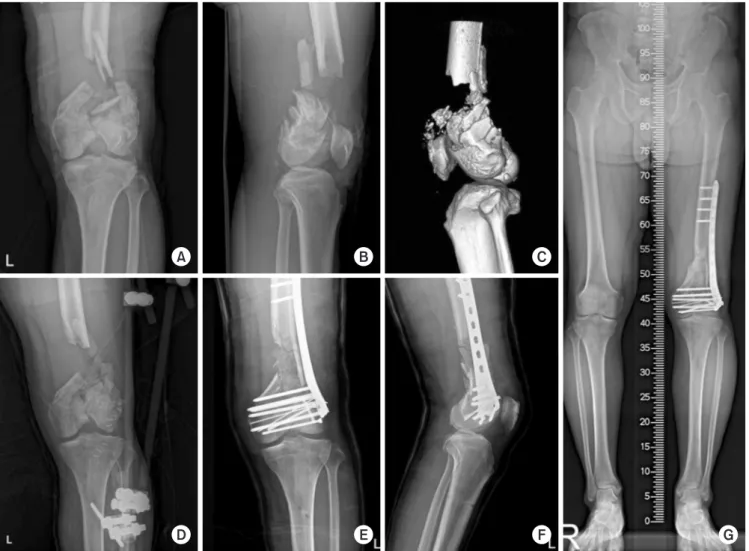
A B C
D E F G
Fig. 1. (A, B) A 58-year-old man with an open distal femoral fracture. He had a severe open comminuted distal femoral fracture. (C) The distal fem- oral segment was not large enough to allow rigid fixation. (D) Spanning temporary external fixation was used for severe soft tissue injury, which was maintained for 12 days. (E, F) Because fracture site instability was observed intraoperatively, cast immobilization was applied for 6 weeks. (G) Although bone union was observed after postoperative 9 months, the range of motion of the knee was not recovered completely.
other procedures. This case group was compared with the control group of patients with 12-month postoperative ROM of >90o and no MUA. Patient charts were reviewed to determine demographic data, injury mechanism, associ- ated injuries, and injury classification as defined by the AO/
OTA and Gustilo systems. The presence or absence of high- energy injury, comminuted fracture, intra-articular frac- ture, extensor mechanism injury, open fracture, temporary external fixation, severe osteoarthritis (Kellgren-Lawrence grade >4), and prolonged postoperative immobilization (>3 weeks) were compared between the case and control groups. In this study, high-energy injury included motor vehicle accidents and sports trauma. Lower-energy injury included low-energy fall fractures related to osteopenia in older patients. Comminuted fracture was defined as type 33A3, 33C2, or 33C3 and intra-articular fracture included type 33C1, 33C2, 33C3 fracture in the AO classification.
Extensor injury included patella fracture or open quadriceps injury after an open fracture. The use of spanning tempo- rary external fixation was used only for severe soft tissue injury, which was maintained only for 1 to 2 weeks before internal fixation. No patient who have external fixation over 2 weeks in all patients. The proportion of severe osteo- arthritis (Kellgren-Lawrence grade >4) was also compared.
Prolonged postoperative immobilization involved any pro- cedure that delay postoperative knee motion after internal fixation, such as cast immobilization. The proportion of prolonged postoperative immobilization (>3 weeks) was also compared between the 2 groups.
4. Clinical and radiologic assessments
In the clinical assessment, distal femoral fractures were evaluated using the postoperative ROM and Knee Society clinical rating system.19) The clinical score was evaluated at 1-year follow-up. Follow-up was performed every month with plain radiography until bone union. Callus bridging on 3/4 cortices and fading of fracture lines on the coronal and sagittal plane radiographs were considered signs of fracture union. Non-union was defined as the absence of bone union after 6 months. The diagnosis of nonunion is
generally made when there is no healing between two sets of x-ray after postoperative 6 months. The diagnosis was confirmed by persistent pain, abnormal movement at the fracture site, and presence of broken implant on x-ray. If there is doubt about the interpretation of nonunion on the x-ray, we performed bone scan or computed tomography scan for confirmation. After bone union, a whole-extremity scanogram was taken to determine the mechanical lateral distal femoral angle and anatomical lateral distal femoral angle.
5. Statistical analysis
A priori power analysis was performed to determine the sample size by use of the Fisher’s exact test at an α level of 0.05 and a power of 0.8. The results of a pilot study involv- ing each 10 cases indicated that 12 knees per group were required to detect a significant difference in risk factor of extensor mechanism injury between case group and con- trol group. This study finally involved 54 knees, indicating adequate power (0.886) for detecting a significant differ- ence between the groups. Statistical analysis was performed using IBM SPSS software ver. 20 (IBM Co., Armonk, NY, USA). Means and frequencies were compared with Mann- Whitney test and Fisher’s exact test for univariate analysis.
Backward conditional logistic regression was performed using the variables found to be significant in the univari- ate analysis, with p<0.05 as the criterion for entry into and p>0.10 as the criterion for removal from the equation.
Results
1. Patients and demographics
The mean patient age was 60±20 years (range, 19-95 years). The mean age, sex ratio, and follow-up period are listed in Table 1, and they were not significantly different between groups. Fourteen were open fractures (grade I, 4;
grade II, 8; grade III, 2), according to the Gustilo-Anderson criteria. The type of distal femoral fracture was 33-A in 24 and 33-C in 30. There were no 33-B type of fractures that
were treated with screw fixation only. The basic demo- graphic and diagnostic characteristics of the case and con- trol groups are summarized in Table 1.
2. Risk factor for knee stiffness
Among patients with knee stiffness, manipulation under general anesthesia was performed in 7 cases at a mean of 104 days (range, 72-146 days) after internal fixation of the distal femur. Among 7 cases of additional procedures, ar- throscopic lysis of adhesion was performed in 6 cases and open quadricepsplasty was done in 1 case. In the 7 cases with MUA, the mean ROM was improved from 82o to 100o. In the 7 cases with MUA, 3 cases still had a ROM of
<90o even after MUA.
The presence of high-energy injury, comminuted frac- ture, intra-articular fracture, extensor mechanism injury, open fracture, temporary external fixation, severe osteoar- thritis (Kellgren-Lawrence grade >4), and cast immobiliza- tion (>3 weeks) were more frequently observed in the case group than in the control group (Table 2). In the univariate analysis, all of the above-mentioned risk factors of knee stiffness had significantly higher incidence in the case group.
A backward conditional logistic regression model was con- ducted with the criterion for variable entry into the equation set at p<0.05 and that for variable exclusion from the equa- tion set at p>0.10. Backward conditional logistic regres-
sion was performed using variables identified as significant in univariate analysis. The model revealed that extensor mechanism injury was the only significant predictor among the risk factors of knee stiffness (p=0.001; odds ratio, 42.0;
95% confidence interval [CI], 5.0-350.7).
3. Clinical and radiologic outcomes
The mean ROM of all patients at 1 year was 115o±23o (70o-145o). The average flexion contracture of all patients was 1o±2o (0o-5o) and the average further flexion was 116o
±22o (70o-145o). The 3-month postoperative ROM of the knee joint was >120o in 24 patients (44.4%), between 90o and 120o in 16 patients (29.6%), and between 70o and 90o in 14 patients (26.0%). After 7 MUAs with quadricepsplasty or arthroscopic lysis of adhesion, the 12-month postopera- tive ROM was >120o in 24 patients (44.4%), between 90o and 120o in 20 patients (37.0%), and between 70o and 90o in 10 patients (18.6%). The mean Knee Society score of all patients was 86±9 (65-97). The functionality accord- ing to the 12-month postoperative Knee Society score was excellent (80-100) in 40 patients (74.1%), good (70-79) in 8 patients (14.8%), and fair (60-69) in 6 patients (11.1%).
The clinical and radiologic outcomes of the case and control groups are listed in Table 3. The ROM and Knee Society score were significantly lower, but the bone union time was significantly longer, in the case group. However, the coronal and sagittal alignment was not significantly different.
Table 1. Demographic Characteristics of the Case and Control Groups Characteristic Case (n=14) Control (n=40) p-value
Age (yr) 58±13 (37-74) 61±22 (19-95) 0.874
Sex (male:female) 4:10 16:24 0.678
Follow-up (mo) 19±8 (12-36) 18±8 (12-38) 0.957
Open fracture type 10 4
Grade I 2 2
Grade II 6 2
Grade III 2 0
AO classification
A 0 A1: 12, A2: 8, A3: 4
B 0 0
C C2: 2, C3: 12 C1: 4, C2: 6, C3: 6
Values are presented as mean±standard deviation (range) or number only.
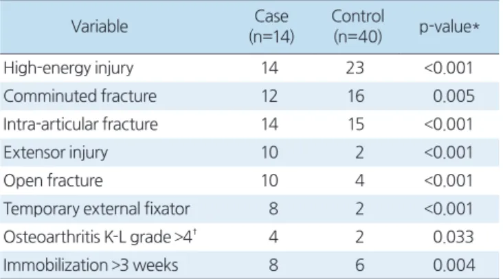
Table 2. Comparisons of the Frequency of Variables Known to be Risk Factors of Knee Joint Stiffness according to the Range of Motion
Variable Case
(n=14)
Control
(n=40) p-value*
High-energy injury 14 23 <0.001
Comminuted fracture 12 16 0.005
Intra-articular fracture 14 15 <0.001
Extensor injury 10 2 <0.001
Open fracture 10 4 <0.001
Temporary external fixator 8 2 <0.001
Osteoarthritis K-L grade >4† 4 2 0.033
Immobilization >3 weeks 8 6 0.004
Values are presented as number only. *Fisher’s exact test, †K-L grade:
Kellgren-Lawrence grade.
Discussion
Understanding the risk factors for knee stiffness after a distal femoral fracture may help surgeons in managing patients at a high risk for substantial knee stiffness. Supra- condylar and intra-articular fractures in the distal femur may lead to arthrofibrosis with adhesions in the parapatellar gutters, patellar tendon, and patellofemoral joint. There- fore, the surgical goals should include restoring the anatomy while providing stable fixation that allows early motion and aggressive postoperative mobilization to minimize the potential for debilitating loss of motion. The present study showed that the presence of comminuted fracture, intra- articular fracture, extensor mechanism injury, open frac- ture, temporary external fixation, severe osteoarthritis, and prolonged immobilization place patients at an increased risk for knee stiffness (ROM <90o) in the univariate analysis.
Extensor mechanism injury was the only significant pre- dictor in the logistic regression model. All of the above- described risk factors should be considered to avoid the possibility of knee stiffness in patients with distal femoral fractures. Especially in the case of extensor mechanism in- jury, firm fixation of the fracture and early joint motion are essential to prevent knee stiffness.
The pathogenesis is not clear how extensor mechanism injury causes knee stiffness. However, possible theory is that accelerated fibrosis around quadriceps muscle and patella fracture could bring about knee stiffness. Another possible theory is pain. Extensor mechanism injury usu- ally accompanied by high-energy injury, open fracture, and comminuted fracture. Therefore, patient could have severe pain to perform aggressive knee exercises. In the previ-
ous study, extensor mechanism injury was also risk factor for knee stiffness after periarticular fracture. Bishop et al.13) reported that backward conditional logistic regression test revealed two significant risk factors for knee stiffness, which were extensor mechanism injury and the need for ongoing wound management. The wound management was defined as the need for dressing changes, serial debridement, delayed closure, or eventual flap coverage by Bishop et al.13) In the present study, the wound management was not considered as a risk factor for knee stiffness. Because this study was confined to distal femoral fractures, there were rare cases that needed wound management like open tibial injury.
The characteristics of risk factors for knee stiffness have similarities and differences among arthroplasty, ligament reconstruction, and plate fixation of periarticular fractures.
In total knee arthroplasty, knee stiffness is associated with decreased preoperative knee motion, depression, poor pain tolerance, younger age, genetic predisposition, diabetes mellitus, low socioeconomic status, previous knee sur- gery, prosthesis malpositioning, inadequate resection, non- compliance of the patient to rehabilitation programs, and complex regional pain syndrome have all been proposed to influence this condition.20-23) However, there is no consen- sus on what factors might be associated with stiffness after total knee arthroplasty. As total knee replacement is typi- cally performed in elderly patients without acute injuries, the characteristics of patients with stiffness after fracture are rather different. In sports medicine, however, ligamen- tous reconstructions in the knee are often performed to treat acute traumatic injuries. Therefore, the characteristics of these patients are similar to our series of patients. After a ligament surgery, the risk factors for stiffness include ex-
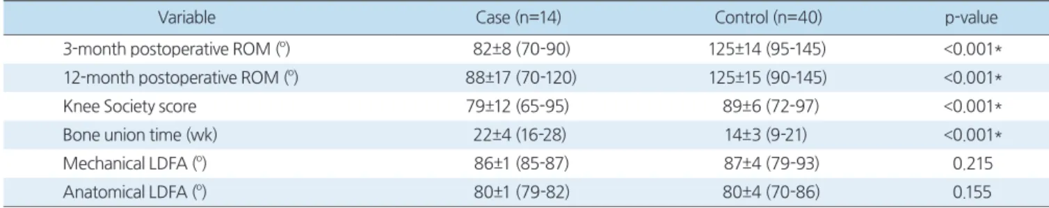
Table 3. Clinical and radiologic outcomes of the case and control groups
Variable Case (n=14) Control (n=40) p-value
3-month postoperative ROM (o) 82±8 (70-90) 125±14 (95-145) <0.001*
12-month postoperative ROM (o) 88±17 (70-120) 125±15 (90-145) <0.001*
Knee Society score 79±12 (65-95) 89±6 (72-97) <0.001*
Bone union time (wk) 22±4 (16-28) 14±3 (9-21) <0.001*
Mechanical LDFA (o) 86±1 (85-87) 87±4 (79-93) 0.215
Anatomical LDFA (o) 80±1 (79-82) 80±4 (70-86) 0.155
Values are presented as mean±standard deviation (range) only. *Significant difference. ROM: range of motion, LDFA: lateral distal femoral angle.
tensive soft tissue injury, acute reconstructive surgery, and lack of early knee motion.15,24,25) Extensive soft tissue injury could also be a risk factor for stiffness in distal femoral fractures. In the present study, extensive soft tissue injury is relevant to high-energy injury, extensor mechanism injury, open fracture, and comminuted fracture. In these cases, it is important to apply early exercise and firm fixation of fracture; however, these are sometimes difficult owing to pain and unstable fixation. Nonetheless, the best approach is to encourage patients to perform aggressive knee exercises after rigid fixation. In a 23-year-old female patient, early exercise and firm fixation resulted in good ROM although the patient had severe extensor mechanism injury and com- minuted fracture (Fig. 2).
Various daily activities require a different knee ROM.
Functional knee ROM requires 63o of flexion for walk- ing and up to 93o of flexion for standing up from a chair.26) More motion is required for activities such as getting out of a bathtub (138o), ascending stairs (98.5o), and standing up from a low chair (105o).27) According to gait analyses and biomechanical studies, patients require at least 83o of flexion to ascend stairs, 90o to 100o to descend stairs, 93o to 105o to rise from a standard or short chair, and >115o to squat or kneel.20,26) Although the goal is to maximize the knee ROM after fractures, the functional ROM should be considered in order to provide the best alternative results. If postoperative knee stiffness does not respond to physical therapy, it can be treated with MUA with other adjunctive procedures.14,28,29) At our institution, all patients with ROM ≤90o at 3 months after open reduction and internal fixation with no recent
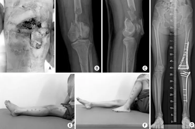
A B C
E F D
Fig. 2. (A) A 26-year-old woman with open distal femoral fracture. She had an open fracture and quadriceps muscle injury. A bony fragment penetrated through the skin and the wound was contaminated. (B, C) Preoperative radiographs demonstrating comminuted and intra-articular fractures in the distal femur. After complete debridement and a weak spanning external fixation, the patient underwent auto- and allo-bone graft and firm-plate fixation. Two days later, she started knee exercise. (D) Follow-up radiograph after 3 months demonstrating fracture union. (E, F) The range of motion of the knee was recovered completely through early exercise and firm internal fixation.
gains after physical therapy were offered MUA with ar- throlysis of knee adhesions. Some patients with ROM ≤ 90o did not have MUA and arthrolysis because they did not want additional surgery. Therefore, the case group includes both patients who underwent MUA and those with ROM
≤90o at postoperative 12 months in the present study.
Various studies have reported decreased knee ROM in patients with fractures around the knee. Ehlinger et al.16) reported that patients with supracondylar femoral fractures who undergo surgical fixation typically lose between 30o and 40o of knee flexion compared with the normal knee.
Kregor et al.17) reported that after submuscular plating of the distal femur, the average ROM of the knee was from 1o loss of extension to 109o of flexion. Kolb et al.18) reported that the ROM of the knee joint was >120o in 15 patients (48.3%), between 90o and 120o in 12 patients (38.7%), and between 70o and 90o in 4 patients (13.0%) after submuscular plating of the distal femur. Previous studies reported that the ROM decreases especially in flexion, and not frequently in exten- sion, after distal femoral fractures. In the present study, the mean loss of extension was 1o and further flexion was 116o. Loss of motion is usually observed in terminal flexion like in previous studies.
The present study has several limitations. Because of its retrospective design, some patients were excluded because they underwent a different fixation method, such as in- tramedullary nail fixation. This may have created a selec- tion bias. Second, the case and control groups were divided based on 90o of knee flexion. Although the functional knee ROM of daily activities was considered, there is no absolute standard for knee stiffness. Third, the numbers of patients in the case and control groups were small. Additionally, there was no guideline for the immobilization period. In cases of severe comminuted fracture in the distal femur, the period of immobilization was decided by the surgeon.
Conclusion
The present study showed that the presence of injuries that delay postoperative knee motion place patients at an increased risk for knee stiffness. Understanding these factors
may help surgeons to better identify patients at risk for sub- stantial stiffness. Especially, extensor mechanism injury such as patella fracture or open quadriceps injury, was an inde- pendent predictable factor associated with the knee stiffness (ROM <90o). Extensor mechanism injury related to exten- sile soft tissue injury place trauma patients at an increased risk for knee stiffness, and that the group of patients with knee stiffness have worse clinical and radiologic outcomes than the control group.
요 약
목적:
원위 대퇴골 골절을 근육 아래 잠김 금속판을 이용하여 치료한 환자에서 발생하는 슬관절 강직의 위험 인자에 대 해 조사하고 임상적 방사선적 결과에 대해 분석하고자 한다.
대상 및 방법:
본 연구는 후향적 환자 대조군 연구로 슬관절 강직의 환자군은 수술 후 12개월의 관절 운동 범위가 90도 이하 혹은 마취하 관절 수동술을 시행 받은 환자이고 대조군 은 정상 관절 운동 환자였다. 발생 가능한 위험인자의 빈도를 단변량 및 로지스틱 회귀 분석을 이용하여 분석하였다.결과:
환자군 14예, 대조군 40예로 54예의 환자가 연구에 포 함되었다. 단변량 분석상 분쇄골절, 관절내 골절, 개방성 골 절, 일시적 외고정 장치, 심한 퇴행성 관절염 및 장기간의 부 목 고정이 슬관절 강직의 위험 인자였다. 하지만, 다중 회 귀 분석상에서는 신전기전의 손상이 유일한 위험 인자였다 (p=0.001; odds ratio, 42.0; 95% confidence interval, 5.0- 350.7).결론:
원위 대퇴골 골절에서 슬관절 강직을 발생시킬 수 있는위험 인자들을 정확히 알고 있다면 슬관절 강직 예방에 도움 이 될 것으로 생각된다. 특히 슬개골 골절이나 대퇴 사두근의 개방성 손상과 같은 신전 기전의 손상은 슬관절 강직을 독립 적으로 예측할 수 있는 주요 위험 인자이다.
색인 단어:
대퇴골 골절, 골고정 금속판, 관절 운동 범위, 위험 인자ORCID
손동욱, https://orcid.org/0000-0001-8222-2843 김형수, https://orcid.org/0000-0002-2279-4222 최우영, http://orcid.org/0000-0002-4037-4969
References
1. Court-Brown CM, Caesar B: Epidemiology of adult fractures: a review. Injury, 37: 691-697, 2006.
2. Martinet O, Cordey J, Harder Y, Maier A, Bühler M, Barraud GE: The epidemiology of fractures of the distal femur. Injury, 31 Suppl 3: C62-63, 2000.
3. Arneson TJ, Melton LJ 3rd, Lewallen DG, O’Fallon WM: Epi- demiology of diaphyseal and distal femoral fractures in Roches- ter, Minnesota, 1965-1984. Clin Orthop Relat Res, (234): 188- 194, 1988.
4. Sanders R, Swiontkowski M, Rosen H, Helfet D: Double- plating of comminuted, unstable fractures of the distal part of the femur. J Bone Joint Surg Am, 73: 341-346, 1991.
5. Seinsheimer F 3rd: Fractures of the distal femur. Clin Orthop Relat Res, (153): 169-179, 1980.
6. Prevosti LG, Subramainian VA, Rothaus KO, Dineen P: A com- parison of the open and closed methods in the initial treatment of sternal wound infections. J Cardiovasc Surg (Torino), 30:
757-763, 1989.
7. Halpenny J, Rorabeck CH: Supracondylar fractures of the fe- mur: results of treatment of 61 patients. Can J Surg, 27: 606- 609, 1984.
8. Schandelmaier P, Partenheimer A, Koenemann B, Grün OA, Krettek C: Distal femoral fractures and LISS stabilization. Injury, 32 Suppl 3: SC55-63, 2001.
9. Bolhofner BR, Carmen B, Clifford P: The results of open reduc- tion and internal fixation of distal femur fractures using a bio- logic (indirect) reduction technique. J Orthop Trauma, 10: 372- 377, 1996.
10. Schütz M, Müller M, Krettek C, et al: Minimally invasive frac- ture stabilization of distal femoral fractures with the LISS: a prospective multicenter study. Results of a clinical study with special emphasis on difficult cases. Injury, 32 Suppl 3: SC48-54, 2001.
11. Ostrum RF, Geel C: Indirect reduction and internal fixation of supracondylar femur fractures without bone graft. J Orthop Trauma, 9: 278-284, 1995.
12. Haller JM, Holt DC, McFadden ML, Higgins TF, Kubiak EN:
Arthrofibrosis of the knee following a fracture of the tibial pla- teau. Bone Joint J, 97: 109-114, 2015.
13. Bishop J, Agel J, Dunbar R: Predictive factors for knee stiffness after periarticular fracture: a case-control study. J Bone Joint Surg Am, 94: 1833-1838, 2012.
14. Schiavone Panni A, Cerciello S, Vasso M, Tartarone M: Stiffness in total knee arthroplasty. J Orthop Traumatol, 10: 111-118, 2009.
15. Robertson GA, Coleman SG, Keating JF: Knee stiffness follow-
ing anterior cruciate ligament reconstruction: the incidence and associated factors of knee stiffness following anterior cruciate ligament reconstruction. Knee, 16: 245-247, 2009.
16. Ehlinger M, Dujardin F, Pidhorz L, Bonnevialle P, Pietu G, Vandenbussche E; SoFCOT: Locked plating for internal fixa- tion of the adult distal femur: influence of the type of construct and hardware on the clinical and radiological outcomes. Orthop Traumatol Surg Res, 100: 549-554, 2014.
17. Kregor PJ, Stannard JA, Zlowodzki M, Cole PA: Treatment of distal femur fractures using the less invasive stabilization system:
surgical experience and early clinical results in 103 fractures. J Orthop Trauma, 18: 509-520, 2004.
18. Kolb W, Guhlmann H, Windisch C, Marx F, Kolb K, Koller H:
Fixation of distal femoral fractures with the less invasive stabili- zation system: a minimally invasive treatment with locked fixed- angle screws. J Trauma, 65: 1425-1434, 2008.
19. Insall JN, Dorr LD, Scott RD, Scott WN: Rationale of the Knee Society clinical rating system. Clin Orthop Relat Res, (248): 13- 14, 1989.
20. Bong MR, Di Cesare PE: Stiffness after total knee arthroplasty. J Am Acad Orthop Surg, 12: 164-171, 2004.
21. Desai AS, Karmegam A, Dramis A, Board TN, Raut V: Ma- nipulation for stiffness following total knee arthroplasty: when and how often to do it? Eur J Orthop Surg Traumatol, 24: 1291- 1295, 2014.
22. Parvizi J, Tarity TD, Steinbeck MJ, et al: Management of stiff- ness following total knee arthroplasty. J Bone Joint Surg Am, 88 Suppl 4: 175-181, 2006.
23. Pivec R, Issa K, Kester M, Harwin SF, Mont MA: Long-term outcomes of MUA for stiffness in primary TKA. J Knee Surg, 26: 405-410, 2013.
24. Shelbourne KD, Patel DV: Treatment of limited motion after an- terior cruciate ligament reconstruction. Knee Surg Sports Trau- matol Arthrosc, 7: 85-92, 1999.
25. Magit D, Wolff A, Sutton K, Medvecky MJ: Arthrofibrosis of the knee. J Am Acad Orthop Surg, 15: 682-694, 2007.
26. Laubenthal KN, Smidt GL, Kettelkamp DB: A quantitative analysis of knee motion during activities of daily living. Phys Ther, 52: 34-43, 1972.
27. Rowe PJ, Myles CM, Walker C, Nutton R: Knee joint kinemat- ics in gait and other functional activities measured using flexible electrogoniometry: how much knee motion is sufficient for nor- mal daily life? Gait Posture, 12: 143-155, 2000.
28. Massè A, Biasibetti A, Demangos J, Dutto E, Pazzano S, Gal- linaro P: The judet quadricepsplasty: long-term outcome of 21 cases. J Trauma, 61: 358-362, 2006.
29. Lee DH, Kim TH, Jung SJ, Cha EJ, Bin SI: Modified judet quadricepsplasty and Ilizarov frame application for stiff knee af- ter femur fractures. J Orthop Trauma, 24: 709-715, 2010.