Pediatr Gastroenterol Hepatol Nutr 2018 October 21(4):219-233
PGHN
Review Article
Diagnosis of Helicobacter pylori Infection in Children and Adolescents in Korea
Ji-Hyun Seo, Ji-Sook Park, Kwang-Ho Rhee*, and Hee-Shang Youn
Departments of Pediatrics and *Microbiology, Gyeongsang National Institute of Health Sciences, Gyeongsang National University College of Medicine, Jinju, Korea
Helicobacter pylori plays an important role in the pathogenesis of chronic gastritis, peptic ulcer disease, gastric can- cer, and gastric mucosa-associated lymphoid tissue lymphoma. In Korea, the guidelines for the diagnosis and treat- ment of H. pylori infection in adults were revised in 2013. The European Helicobacter and Microbiota Study Group and Consensus panel released the fifth edition of the Maastricht Consensus Report for the management of H. pylori infection in 2015, and the European Society of Paediatric Gastroenterology, Hepatology and Nutrition and the North American Society of Paediatric Gastroenterology, Hepatology and Nutrition released the updated joint guidelines for children and adolescents in 2016. Considering these recommendations and recent progress in our research and that of other research teams, this study aimed to discuss the diagnostic strategies for H. pylori infection in children and adolescents.
Key Words: Helicobacter pylori, Diagnosis, Guideline, Child
Received:June 18, 2018, Accepted:June 21, 2018
Corresponding author: Hee-Shang Youn, Department of Pediatrics, Gyeongsang National Institute of Health Sciences, Gyeongsang National University College of Medicine, 15 Jinju-daero, 816-beon-gil, Jinju 52727, Korea. Tel: +82-55-750-8158, Fax: +82-55-752-9339, E-mail:
Copyright ⓒ 2018 by The Korean Society of Pediatric Gastroenterology, Hepatology and Nutrition
This is an openaccess article distributed under the terms of the Creative Commons Attribution NonCommercial License (http://creativecommons.org/licenses/by-nc/4.0/) which permits unrestricted noncommercial use, distribution, and reproduction in any medium, provided the original work is properly cited.
INTRODUCTION
Helicobacter pylori plays an important role in the pathogenesis of chronic gastritis, peptic ulcer dis- ease, gastric cancer, and gastric mucosa-associated lymphoid tissue (MALT) lymphoma. The seropreva- lence rates of anti-CagA antibodies among Koreans in 1988 to 1989 were as follows: 25% at 2 to 4 years of age, 50% at 5 years of age, and 80% to 90% after 7 years of age [1]; the same kind of study conducted in 2014 to 2015 revealed 15% at 5 to 14 years of age, 20%
at 15 to 19 years of age, 30% at 20 to 29 years of age and over 80% after 30 years of age (unpublished ob- servation). H. pylori infection begins at infancy and is established before 5 years of age, and non-infected persons have a very limited chance of acquiring the infection [2]. H. pylori, once acquired during child- hood, is rarely eliminated without specific treatment and the infected individuals live with it all their lives [3].
In Korea, the guidelines for the diagnosis and treatment of H. pylori infection in adults were revised
in 2013 (Korea Adult Guidelines) [4] and have been used in routine clinical practice up until today.
Recently, the European Helicobacter and Microbiota Study Group and Consensus panel released the fifth edition of the Maastricht Consensus Report for the management of H. pylori infection in 2015 (Maastricht Consensus V) [5], while the European Society of Paediatric Gastroenterology, Hepatology and Nutrition (ESPGHAN) and the North American Society of Paediatric Gastroenterology, Hepatology and Nutrition (NASPGHAN) released the updated joint ESPGHAN/
NASPGHAN guidelines for children and adolescents in 2016 (ESPGHAN/NASPGHAN guidelines) [6].
However, not only is it inappropriate to apply the di- agnosis and treatment strategies for H. pylori in- fection established in adults to children and adoles- cents, but it is also inappropriate to directly apply the guidelines developed in Europe and North America for children and adolescents to those living in Korea where the epidemiology of H. pylori is quite different.
To date, there is no established Korean consensus on the diagnosis and treatment of H. pylori infection in pediatric patients.
Considering these recommendations and recent progress in our research and other research teams, this study aimed to discuss the diagnostic strategies for H. pylori infection in children and adolescents.
WHO NEEDS A DIAGNOSTIC APPROACH?
The indications for the endoscopic diagnosis and treatment of H. pylori infection in the Korean Adult Guidelines are (i) peptic ulcer disease including scar, (ii) low-grade gastric MALT lymphoma, (iii) re- section of early gastric cancer, (iv) chronic atrophic gastritis or intestinal metaplasia, (v) a family history of gastric cancer, (vi) functional dyspepsia, (vii) long-term use of nonsteroidal anti-inflammatory drugs/aspirin medication, and (viii) previous long-term use of proton pump inhibitor (PPI); H. pylori erad- ication is also indicated in patients with idiopathic thrombocytopenic purpura who are infected with H.
pylori [4].
In the Maastricht Consensus V, H. pylori gastritis is defined as an infectious disease irrespective of symp- toms and complications, and all adult patients with signs of active H. pylori infection should be diagnosed and treated. Even a test and treat strategy is consid- ered appropriate for relatively young adult patients with uninvestigated dyspepsia in high prevalence H.
pylori populations. However, an endoscopy-based strategy should be considered in patients with dys- peptic symptoms to rule out significant esophageal pathologies, particularly in low prevalence H. pylori populations. Unlike the Korea Adult Guidelines, the Maastricht Consensus V includes unexplained iron deficiency anemia despite appropriate evaluation and vitamin B12 deficiency in the indication and ex- cludes asymptomatic individuals with family history of gastric cancer from the indication [5].
Unlike the Korean and European adult guidelines, the ESPGHAN/NASPGHAN guidelines for children and adolescents stated that the indications for endo- scopic diagnosis and treatment of H. pylori infection only include gastric or duodenal ulcers and re- fractory iron deficiency anemia in which other etiol- ogies have been ruled out, and noninvasive diag- nostic testing for H. pylori infection may be consid- ered when investigating the underlying causes of chronic immune thrombocytopenic purpura. It does not warrant a test and treat strategy for H. pylori in- fection in children. The ESPGHAN/NASPGHAN guidelines do not recommend invasive diagnostic testing for H. pylori infection in children if peptic ul- cers are not clinically suspected or identified by en- doscopy, in children with functional abdominal pain, in children with iron deficiency anemia as part of the initial investigation, and when investigating the causes of short stature because based on current evidence, the treatment provided to eliminate H. py- lori infection is not expected to improve the symp- toms and signs in children, except in cases of peptic ulcer disease. The ESPGHAN/NASPGHAN guide- lines generally do not recommend an aggressive di- agnostic approach to H. pylori infection in children.
When H. pylori infection is confirmed by a diagnostic test, the decision to treat or not to treat should be de-
termined after a careful discussion with parents and older patients about the advantages and dis- advantages of anti-H. pylori therapy. This strategy might be helpful in the prevention of peptic ulcer and gastric cancer later in life. However, it would be ac- companied with the risk of reinfection after erad- ication, the theoretical increased risk of allergic dis- eases in young children, non-relief of symptoms, treatment failure, cramps, diarrhea, and undesirable alteration of the gut microbiome after the treatment [6].
Although the H. pylori infection rate in Korea is de- creasing, the seroprevalence of anti-CagA im- munoglobulin (Ig) G antibodies in adolescents re- mains at 20%. Many Korean adolescents experience stress from academic problems and experience much more academic burnouts, such as emotional ex- haustion, cynicism, and academic inefficacy, than those of developed countries [7]. Individuals with high levels of perceived everyday life stress are at in- creased risk of developing peptic ulcer disease [8], and stress may aggravate the severity of H. pylori in- fection [9]. H. pylori-infected Korean adolescents are at higher risk of progression to severe gastritis, peptic ulcer disease, atrophic gastritis, and intestinal meta- plasia throughout the adolescent period than those in developed countries. Therefore, Korean adoles- cents with upper gastrointestinal symptoms includ- ing dyspepsia could be managed in accordance with the Korean Adult Guidelines by referring to the Maastricht Consensus V. The purpose of diagnosing the H. pylori infection in patients with gastro- duodenal disorders is to basically eradicate the H. py- lori and help patients recover from the disease.
Frequently prescribed anti-H. pylori therapeutic agents could be administered at adult doses in ado- lescents aged over 10 to 12 years with body weight over 35 kg [6]. Bismuth and tetracycline, the compo- nents of the bismuth-based quadruple therapy, could be administered at adult doses in adolescents aged over 10 and 12 years, respectively. However, it remains uncertain whether sufficient anti-microbial concentrations could be maintained in the gastric mucosa in young children to eradicate H. pylori living
on it when anti-H. pylori therapeutic agents are ad- ministered according to body weight-based dosing.
The treatment provided to eliminate H. pylori in- fection is not also expected to improve symptoms in young children in developed countries. Therefore, Korean children under 10 years of age should be managed in accordance with the ESPGHAN/
NASPGHAN guidelines. However, if the organic cause of abdominal pain or dyspepsia is suspected, upper gastrointestinal endoscopy should be ar- ranged even in young children.
Diagnostic tests are usually categorized into in- vasive (biopsy-based) and noninvasive methods.
Invasive diagnostic tests include endoscopy, culture, histology, rapid urease test (RUT), and polymerase chain reaction (PCR). Noninvasive diagnostic tests include stool antigen test (SAT), 13C-urea breath test (UBT), and serology. Each diagnostic method has its own advantages, disadvantages, and limitations (Table 1).
RECOMMENDED STANDARD INITIAL DIAGNOSTIC APPROACH
The ESPGHAN/NASPGHAN guidelines recom- mended that H. pylori infection in children should be diagnosed either by positive culture or by finding H.
pylori gastritis on histopathology plus one more pos- itive test such as RUT. If the result of histopatho- logical examination and RUT do not match, diag- nosis is determined by carrying additional non- invasive tests such as the UBT or the H. pylori SAT [6].
The Korean Adult Guidelines do not recommend a test and treat strategy for uninvestigated dyspepsia as part of the initial investigation because of the cheaper endoscopy costs than other countries [4]. As part of the initial investigation, the Maastricht Consensus V recommends a test and treat strategy for uninvestigated dyspepsia in high prevalence H.
pylori populations (≥20%) and an endoscopy-based strategy in low prevalence H. pylori populations to rule out significant esophageal pathologies. However, an endoscope and treat strategy is preferred in older patients or patients with alarm symptoms or signs of
Table 1. Diagnostic Tests for the Detection of Helicobacter pylori Infection Joint ESPGHAN/NASPGHAN Guidelines (update 2016)Maastricht V/Florence Consensus ReportAdvantagesDisadvantages Invasive Culture •At least 1 biopsy from the antrum and 1 from the corpus for culture (if available) as part of the initial investigation • Antimicrobial sensitivity be obtained for the infecting Helicobacter pylori strain(s), and eradication therapy tailored accordingly
•After a first failure, if an endoscopy is carried out, culture and standard anti- microbial susceptibility testing are recommended. •After failure of second-line treatment, culturewith susceptibility testing or molecular determination of genotype resistance is recommended in order to guide treatment.
•The gold standard and the most specific method •Used for determining antibiotic susce- ptibility
•Variable sensitivity; microbiologist’s experience, transport media, and speci- men quality •Relatively difficult to perform, expensive, time-consuming, and needs special media and equipment Histology • Two biopsies from the antrum and 2 biopsies from the corpus for the histopathological evaluation
•A minimum standard biopsy setting is two biopsies from the antrum (greater and lesser curvature 3 cm proximal to the pyloric region) and two biopsies from the middle of the body. Additional biopsy from the incisura is considered for detection of precancerous lesions. • Most cases of H. pylori infection can be diagnosed from gastric biopsies using histochemical staining alone. •IHC could be useful in patients with chronic gastritis (active or inactive), atrophic gastritis (extensive intestinal metaplasia) or infollow-up biopsies after eradication treatment for H. pylori, when no organisms are identified by using histochemical stains.
•H&E stain detects H. pylori •Specificity can be improved by special stains such as modified Giemsa, Warthin- Starry silver, Genta, and IHC stains •Direct visualization of pathologic changes related to H. pylori infection including severity of inflammation, grade of atrophy, the development of intestinal metaplasia, and malignancy
•Accuracy affected by antibiotics, bismuth, and PPIs •Expensive (endoscopy and special stains), •Need skilled personnel, interobserver variability Rapid urease test (RUT) • At least 1 biopsy for any additional diagnostic tests from the antrum (rapid urease, or molecular-based assays)
•In clinical practice RUT is recommended as a first-line diagnostic test. In the case of a positive test, it allows immediate treatment. •One biopsy should be taken from the corpus and one from the antrum. • RUT is not recommended as a test for H. pylori eradication assessment after treatment.
•Rapid, inexpensive, commonly available, and highly specific •Minimum of 10,000 organisms are usually required for a positive RUT result.
•Density of bacteria affects the reaction time. •Varying results according to the location which the biopsy is obtained. •False negative: bismuth, antibiotics, PPIs
Table 1. Continued Joint ESPGHAN/NASPGHAN Guidelines (update 2016)Maastricht V/Florence Consensus ReportAdvantagesDisadvantages Polymerase chain reaction • At least 1 biopsy for any additional diagnostic tests from the antrum (rapid urease, or molecular-based assays)
•Perform clarithromycin susceptibility testing when a standard clarithromycin- based treatment is considered as the first-line therapy, except in populations or regions with well documented low clarithromycin resistance (<15%). This test can beperformed either bya standard method (antibiogram) after culture or by a molecular test directly on the gastric biopsy specimen. •After failure of second-line treatment, culture withsusceptibility testing or molecular determination of genotype resistance is recommended to guide treatment.
•Faster, more accurate and sensitive detection •Useful for expected false negative in biopsy-based test, antibiotic resistance, virulence determinants, bacterial quan- tification •Tissue, stool, dental plaque, saliva, environmental contamination Noninvasive Stool antigen test (SAT) • Useful for post-treatment evaluation•Two-step monoclonal SAT can be used in the a test and treat strategy.•No age restriction •Useful in both pre- and post-treatment diagnosis
•False negative: unformed or watery stool samples, bleeding ulcer, atrophic gastritis, intestinal metaplasia, gastric cancer, MALT lymphoma, partial gast- rectomy •Interpretation of weakly positive results in one-step SAT 13 C-urea breath test (UBT) • Useful for post-treatment evaluation•UBT is the most investigated and best recommended noninvasive test in the context of a test and treat strategy.
•Useful in both pre- and post-treatment diagnosis•False positive: <6 years of age,acid-free stomach (atrophic gastritis, a long term use of PPIs) •False negative: bleeding ulcer,atrophic gastritis, intestinal metaplasia, gastric cancer, MALT lymphoma, partial gastr- ectomy
gastric cancer [5]. The ESPGHAN/NASPGHAN guidelines for children do not recommend a test and treat strategy in all pediatric patients and state that the primary goal of the clinical investigation of gas- trointestinal symptoms should be to determine the underlying etiology of the symptoms and not solely the presence of H. pylori infection. The guidelines do not recommend performing invasive diagnostic test- ing for mere detection of H. pylori infection if peptic lesions are not clinically suspected or identified by endoscopy. The diagnostic investigation for H. pylori in children is justified only in cases where the ex- pected benefits exceed the costs and risks of testing and subsequent treatment [6].
A test and treat strategy could not be recom- mended in Korean children and adolescents in view of the Korean Adult Guidelines and the ESPGHAN/
NASPGHAN guidelines. As part of the initial inves- tigation, the restricted use of diagnostic approach to H. pylori detection might be applied to Korean chil- dren aged under 10 to 12 years in accordance with the ESPGHAN/NASPGHAN guidelines, and an ac- tive endoscopy-based diagnostic strategy should be applied to Korean adolescents aged over 10 to 12 years as stated in the Korea Adult Guidelines.
However, biopsy-based diagnostic evaluation should be performed if the diagnostic endoscopies are car- ried out for any reason.
The recommended noninvasive tests useful for the test and treat strategy as part of the initial inves- tigation in the Maastricht Consensus V are UBT, two-step monoclonal SAT, and locally validated se- rological tests [5].
During the initial diagnostic endoscopic examina- tion, the Maastricht Consensus V recommends ob- taining two biopsies from the antrum (greater and lesser curvature, 3 cm proximal to the pyloric re- gion), two biopsies from the middle of the corpus, and additional biopsy from the incisura (5) for his- tology; one biopsy from the corpus and one from the antrum (2) for RUT; and one gastric biopsy specimen (1) for culture followed by MIC determination or molecular test to verify the clarithromycin suscepti- bility if clarithromycin resistance is >15% [5]. The Table 1. Continued Joint ESPGHAN/NASPGHAN Guidelines (update 2016)Maastricht V/Florence Consensus ReportAdvantagesDisadvantages Serology •Do not recommend using antibody- based tests (IgG, IgA) for H. pylori in serum, whole blood, urine, and saliva in the clinical setting
•Serological tests can be used only after validation. •Rapid (‘office’) serology tests using whole blood should be avoided. •Useful in the clinical situations such as GI bleeding, atrophic gastritis, gastric MALT lymphoma, and gastric carcinoma in which a low bacterial load in the stomach and a decreased sensitivity of other diagnostic methods are expected. •H. pylori serology kits should ideally be developed using local H. pylori strains and local titers should be established.
•Useful in screening and epidemiological studies •Do not produce false negative results in patients receiving treatment (PPIs and antibiotics) or presenting acute bleeding • Inexpensive and noninvasive •A quantitative ELISA test; IgG titers of paired sera from the acute and conva- lescent (6 to 12 months) phase and can confirm eradication of the infection.
•Serologic tests require validation at the local level using regional strains •Serology does not reliably distinguish between active and past infection. ESPGHAN: European Society of Paediatric Gastroenterology, Hepatology and Nutrition, NASPGHAN: North American Society of Paediatric Gastroenterology, Hepatology and Nutrition,IHC: immunohistochemistry,PPI:proton pumpinhibitor, MALT: mucosa-associatedlymphoid tissue,Ig: immunoglobulin, GI: gastrointesinal, ELISA: enzyme-linked immunosorbent assay.
ESPGHAN/NASPGHAN guidelines recommended obtaining two biopsies from the antrum and two bi- opsies from the corpus (4) for histological evalua- tion, at least one biopsy from the antrum and one from the corpus (2) for culture (if available), and at least one biopsy (1) for any additional diagnostic tests from the antrum for RUT or molecular-based assays [6].
Endoscopy-based diagnostic methods such as cul- ture, histology, and RUT and noninvasive methods such as SAT and UBT should be performed to avoid false-negative test results at least 2 weeks after stop- ping PPI and 4 weeks after stopping antibiotics.
INVASIVE TESTS
Endoscopy
Conventional endoscopy in adults is usually per- formed to diagnose H. pylori-related diseases, such as peptic ulcer diseases, atrophic gastritis, MALT lym- phoma, and gastric cancer. Although several at- tempts including a careful close-up observation of the gastric mucosal findings, such as redness, mu- cosal swelling, or nodular change, with standard en- doscopy have been made for real-time diagnosis of H.
pylori infection during endoscopic examination, those attempts may be time consuming and do not provide better results than other biopsy-based tests.
In addition to conventional endoscopy, chromoendo- scopy with phenol red, magnifying endoscopy with or without indigo carmine staining, confocal laser endomicroscopy, magnifying endoscopy with nar- row band imaging, and magnifying endoscopy with I-scan have been used to detect H. pylori infection in adults. However, highly skilled operators are re- quired to operate these advanced technologies, and a careful examination using magnifying with or with- out image-enhanced technique is also time consum- ing and may make a patient more uncomfortable compare with other biopsy-based tests. Those factors usually limit the clinical use of magnifying endos- copy to detect H. pylori infection in the clinical prac- tice [10]. In adults, it is recommended to collect biop- sies of even normal-appearing gastric mucosa for H.
pylori detection during endoscopy in patients with dyspeptic symptoms [11].
The recent advancement in medical technology had made endoscopic procedures feasible not only in young children but also in newborns. The endoscopic findings of an H. pylori-infected gastric mucosa in children are distinct from those seen in adults. If there are nodular changes in the antrum or erosions and ulcers in the stomach or duodenum, H. pylori in- fection should be suspected in children. A distinct nodular gastritis, rarely identified during adult en- doscopic examinations, is a common endoscopic manifestation and may be a pathognomonic finding of H. pylori infection in children [12]. In a conven- tional or magnifying endoscopy, the absence of regu- lar arrangement of collecting venules on the corpus mucosa in children could be associated with H. pylori infection as in adults [13].
Culture
Culturing of H. pylori from gastric biopsy specimen is the gold standard and the most specific method but the sensitivity of culture shows significant variations. Detection of H. pylori through cultural iso- lation is expensive, relatively difficult to perform, and time-consuming, and represents a procedure that requires equipment and microbiological ex- pertise, and it is significantly influenced by the con- ditions of transport of the biopsy specimen from the endoscopy unit to the microbiology laboratory.
To obtain proper biopsy specimen for the high- yield H. pylori isolation, the endoscopic instrument including the biopsy channel must be vigorously washed with anti-septic solutions with normal sal- ine and biopsy should be performed before the biop- sy forceps are contaminated with formalin. The ob- tained biopsy specimens for culture should be placed quickly into a container with a few drops of saline and transferred to the microbiology laboratory by ex- perienced microbiologists.
The Maastricht Consensus V recommended that H. pylori culture and antibiotic susceptibility testing should be performed with one gastric biopsy speci- men if primary resistance to clarithromycin is higher
than 15% in a given geographical area, after the first failure to tailor the treatment, except if a bis- muth-based quadruple therapy is considered, or af- ter failure of second-line treatments as a treatment guide [5]. The ESPGHAN/NASPGHAN guidelines recommend obtaining one biopsy from the antrum and one from the corpus for culture, obtaining in- formation about the antimicrobial sensitivity of the infecting H. pylori strain(s), and ensuring that erad- ication therapy should be tailored accordingly as an initial approach for diagnosing H. pylori infection in children [6]. The guidelines for children recommend obtaining biopsy specimens from two places because several strains with different antibiotic sensitivity profiles can exist within the same child [6]. However, compared with the Maastricht Consensus V and the ESPGHAN/NASPGHAN guidelines, the Korean Adult Guidelines do not recommend H. pylori culture for antimicrobial sensitivity as the initial diagnostic approach. It was not also recommended for Korean children and adolescents because the manpower and laboratory facilities with expertise in H. pylori culture are only available in few research laboratories in Korea. However, considering the increasing failure rate of standard therapies due to increased antibiotic resistance in Korea, bacterial culture will serve as an important method to prevent treatment failures.
Therefore, several regional microbiology laboratories with a good quality assurance and quality control program shall be established so that the transfer of the biopsy specimens for H. pylori culture and MIC determination at least after failure of second-line treatment could be possible.
Biopsy specimens could be transferred to a distant microbiology laboratory for successful H. pylori iso- lation if kept in a conventional transport medium, such as Portagerm pylori, Stuart’s transport medium, serum-free transport medium with cyanobacterial extract, brain heart infusion broth plus vancomycin or amphotericin B, and nalidixic acid for up to 24 hours at 4oC, and a newly developed semisolid GESA transport medium for up to 10 days at 4oC [14].
Histology
Standard hematoxylin and eosin staining of the gastric tissue is accepted as one of the good sensitive methods to detect H. pylori infection. It provides ad- ditional information about the presence and severity of inflammation, atrophy, and intestinal metaplasia based on the updated Sydney System in which gas- tric biopsy specimens are collected from the follow- ing five sites: the lesser curvature of the corpus ap- proximately 4 cm proximal to the angulus (i), from the lesser (ii) or greater curvature of the antrum (iii), both within 2 to 3 cm of the pylorus, from the middle portion of the greater curvature of the corpus, ap- proximately 8 cm from the cardia (iv), and from the incisura angularis (v). The ESPGHAN/NASPGHAN guidelines for children, in whom intestinal meta- plasia is very rare, recommended obtaining two biop- sies from the antrum and two biopsies from the cor- pus for histological evaluation. Obtaining at least two biopsy specimens from the antrum and the cor- pus, respectively, increases the sensitivity to detect H. pylori. The corpus biopsy is important for the diag- nosis of H. pylori in patients with atrophic gastritis.
The sensitivity and specificity to detect H. pylori in- fection can be improved by using special stains such as modified Giemsa, Warthin-Starry silver, Genta, and immunohistochemical stains. It is known that the accuracy is affected by preceding antibiotics, bis- muth, and PPIs and that the histological methods need skilled personnel for specimen processing and intrinsically have an inter-observer variability in the diagnosis [15]. In a study comparing Giemsa and im- munohistochemistry (IHC) to diagnose H. pylori in- fection, Giemsa showed significantly lower sensi- tivity (83.3%) than IHC (98.8%). Moreover, the sen- sitivity of Giemsa markedly dropped to 33.6% in pa- tients without active inflammation. The sensitivity of Giemsa strongly depends on H. pylori density and, accordingly, on the presence of active inflammation [16]. IHC has been proved to increase the sensitivity for detection of H. pylori infection, and this method has a lower inter-observer variation when compared to histochemical stains. The use of IHC could be rec- ommendable in patients with chronic active or in-
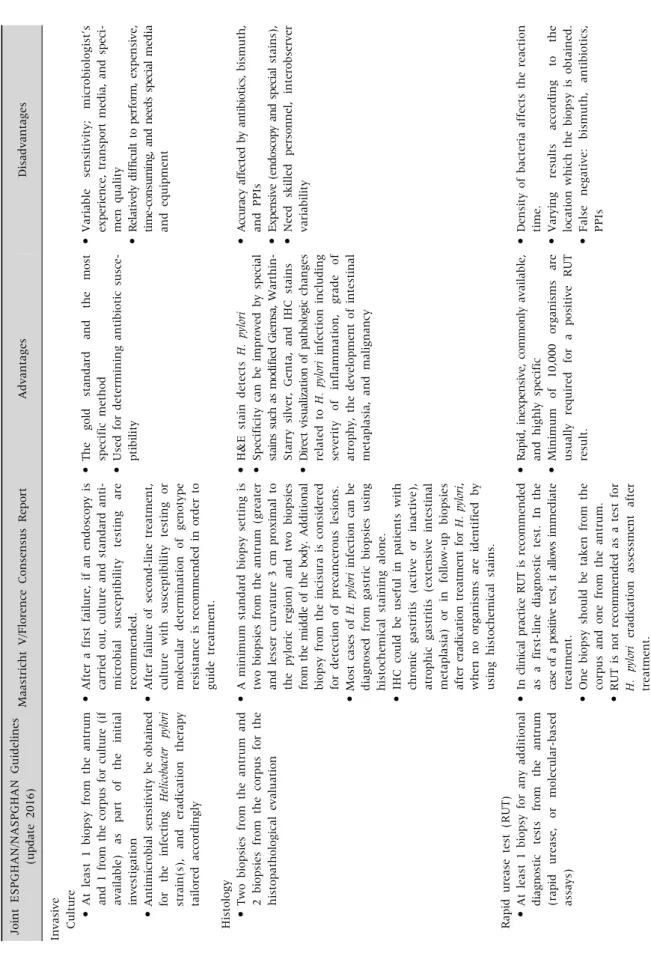
Fig. 1. A comparison of the results of multiple rapid urease tests (RUTs) between 25 initial RUT-positive (A) and 25 RUT-negative (B) pediatric patients from whom five or more gastric antral biopsy specimens were also available for 13C-urea breath tests revealed that RUTs using one biopsy specimen show a maximum of 62.5% false-negative results.
active gastritis, atrophic gastritis, intestinal meta- plasia, or in follow-up biopsies after the eradication of H. pylori, when no organisms are identified on his- tochemical staining [5].
H. pylori is usually observed in the gastric mucosa.
However, the mucous layer, together with the bac- teria, is lost during conventional tissue processing in which formalin is used for fixation. When the num- ber of H. pylori in the gastric mucosa is limited as in pediatric patients, the bacteria may not be detected by conventional histologic methods. The preserva- tion of mucous layer using Carnoy’s solution as a fix- ative and the following immunohistochemical de- tection of H. pylori might aid in further increasing the diagnostic yield in pediatric patients [17].
Peptide nucleic acid fluorescent in situ hybrid- ization (PNA-FISH) is highly sensitive and specific when it comes to detecting H. pylori infection and can identify coccoid forms of H. pylori. Recently, PNA- FISH is increasingly used to detect H. pylori clari- thromycin resistance in gastric biopsy specimens [10].
Rapid urease test
In routine clinical practice, the RUT is the most useful invasive test for the diagnosis of H. pylori in- fection because it is a simple and inexpensive meth- od that requires no special technique to perform and read the result. The biopsy specimen is placed into a solution or gel-containing urea and a pH indicator. If H. pylori is present, the urea is broken down into CO2
and ammonia, which increases the pH of the sol- ution or gel and causes a subsequent color change in the pH indicator. The RUT produces a result in a range of minutes up to 24 hours, depending on the number of bacteria in the biopsy specimen. A pos- itive RUT may require approximately 103 to 105 H. py- lori in the biopsy sample to change the color depend- ing on the experimental conditions [18,19].
In the Maastricht Consensus V, the RUT is recom- mended as a first-line diagnostic test in the endos- copy-based strategy when there is no contra- indication for biopsy. In cases of positive tests, an im- mediate treatment against H. pylori infection is
guaranteed. One biopsy should be taken from the corpus and one from the antrum. RUT is not recom- mended for the evaluation of H. pylori eradication af- ter treatment [5]. In the ESPGHAN/NASPGHAN guidelines, at least one biopsy for RUT or molec- ular-based assay from the antrum is recommended [6].
Multiple RUTs were performed in 50 pediatric pa- tients, consisting of 25 initial RUT-positive and 25 RUT-negative patients, with more than 5 gastric an- tral biopsy specimens collected from October 1991 to April 1992 and stored frozen in the Biobank to ex- plore the nature of patchy distribution of H. pylori in- fection and to verify whether these are more accen- tuated in children. Of 50 patients, 30 showed con- sistent results on all RUTs whereas 20 showed incon- sistent results. The result of RUT was determined positive if at least one out of six or more RUTs yielded positive results, and 32 out of 50 patients were con-
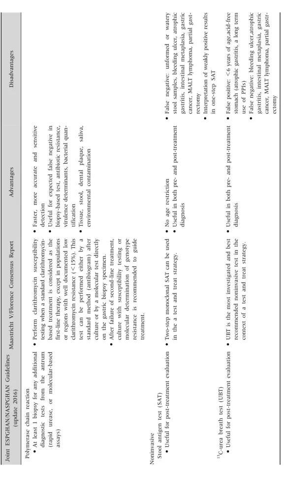
Fig. 2. The difference in the positivity rate of rapid urease tests (RUTs) between one and three biopsy specimens obtained from the gastric antrum in the same pediatric patient according to the age groups. The positivity rate of RUTs increased with age irrespective of the number of gastric biopsy specimens. The positivity rate of RUTs with three biopsy specimens was higher than RUTs with one biopsy specimen in patients aged below 10 years of age [20].
GU1: RUT using one biopsy specimen, GU3: RUT using three biopsy specimens.
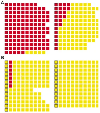
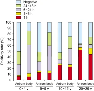
Fig. 3. The positivity rate and time to positivity of rapid urease tests (RUTs) in the antrum and body according to age.
The positivity rate of RUTs in the antrum was higher in the age group 20 to 29 years compared with that in other three age groups, and the positivity rate of RUTs in the body decreased with increasing age (p<0.0001). The most frequent time to positivity occurred within 1 hour in the age group 20 to 29 years and within 6 to 24 hours in children (p<0.0001).
The proportions of positive reactions within 1 hour were similar for the antrum and body in all groups [21].
sidered as RUT positive. Some RUTs revealed neg- ative results in 20 out of 32 RUT-positive cases.
Therefore, RUTs using one biopsy specimen might show up to 62.5% false-negative results (Fig. 1) (unpublished observation).
The RUT using one and three biopsy specimens from each of the 255 children were compared to check the correlation between the number of gastric biopsy specimens and the positivity rate. The pos- itivity rate of the RUTs with three biopsy specimens was higher than with RUTs with one biopsy specimen. The difference between one and three bi- opsy specimens was higher in the children aged 0 to 9 years. The RUT might be a more accurate diagnostic modality when it is performed with three or more bi- opsy specimens especially in children (Fig. 2) [20].
The differences in the results of RUTs according to age and sampling site were also evaluated. The pos- itive color change of the RUTs in adults primarily oc-
curred within 1 hour both in the antrum and in the corpus. The proportions of positive RUT reaction within 1 hour were similar between the antrum and the corpus regardless of age. The positive color change of the RUTs in children primarily occurred in the corpus within 6 to 24 hours. The discrepancy in the positivity rates between the antrum and the cor- pus in children was mainly due to the difference in the proportion of positive reactions in the RUTs with- in 6 to 24 hours (Fig. 3) [21]. The higher positivity rate of corpus RUTs than of antrum RUTs in children, although positive color change did not occur within 6 hours, might be related to the increased H. pylori urease activity at the corpus where parietal cells se- crete acid into the mucous layer. Therefore, the RUT may be more accurate when three or more specimens from both the antrum and corpus are used in children.
However, there is a high possibility of false-neg-
ative results with RUT due to decreased urease activ- ity, which could be caused by a recent intake of anti- biotics, bismuth compounds, or PPIs. Moreover, a false-negative urease test can be observed in patients with achlorhydria and bleeding. The risk of a false-positive result in RUTs may increase with in- creasing incubation time [15].
Polymerase chain reaction
PCR provides excellent sensitivity and specificity compared with other conventional tests and can ac- curately detect the presence of H. pylori even in pa- tients with bleeding. PCR also enables clinicians to make fast and more accurate decisions on patient’s treatment. The target genes such as ureA, glmM, ureC, 16S rRNA, 23S rRNA, hsp60, and vacA are frequently used in the diagnosis of H. pylori infection using PCR [10]. The conventional PCR might be helpful in the diagnosis of H. pylori infection even when few organ- isms exist in the gastric biopsy specimen and even if other biopsy-based tests are expected to show false-negative results. However, it has a high risk of false-negative and false-positive results, unless standard precautions are observed during specimen collection and PCR procedures. The presence of cross-reacting nucleotide sequences and the high ge- netic variability of the bacterial strains as well as the DNA segments of dead bacterium in the gastric mu- cosa of patients after treatment or if dead or live H.
pylori strains remain in the biopsy channel are among the limitations of PCR-based diagnosis [22].
Detection of virulence factors such as cagA, vacA, or dupA by PCR helps to evaluate the genetic variation within virulence factors of H. pylori and provides more information to understand the clinical discrep- ancies among H. pylori-infected patients. It is also useful in epidemiological studies [10].
Furthermore, it can be used to identify specific mutations associated with resistance to antimicrobial agents such as clarithromycin (23S rRNA), quino- lones (gyrA), tetracycline (16S rRNA), rifabutin (rpoB) and amoxicillin (pbp-1a) [10]. This antibiotic resistance profile may provide an important in- formation for the clinicians to determine the anti-H.
pylori therapeutic strategy.
The recent progress in the PCR procedures and the invention of real-time PCR made the molecular method useful in the quantitative detection of H. py- lori in biopsy specimens following anti-H. pylori ther- apy as well as in stool, dental plaque, saliva, and en- vironmental samples with high sensitivity and high specificity [10]. Many research papers using PCR have been published, and many of them used stools instead of biopsies to diagnose H. pylori infection.
NONINVASIVE TESTS
Stool antigen test
SAT can detect bacterial antigens of H. pylori and therefore can be used in the initial and post-treat- ment diagnosis of H. pylori infection. There are two types of SATs used for H. pylori detection. One is an enzyme immunoassay (EIA), the so-called two-step SAT, and the other is an immunochromatography assay (ICA), the so-called one-step SAT. Both types use either polyclonal or monoclonal antibodies. In general, monoclonal antibody-based tests are more accurate than polyclonal antibody-based tests, and EIA-based tests provide more reliable results than ICA-based tests. The rapid monoclonal ICA-based SAT has a high specificity but has a low sensitivity [10]. The ICA-based tests may have problems in in- terpreting weakly positive results [23]. The sensi- tivity and specificity of the laboratory-based mono- clonal EIA are comparable to those of UBT.
Monoclonal SAT is the recommended noninvasive test in the context of a test and treats strategy in adults [5].
In addition to assessment of eradication therapy, monoclonal SAT is a convenient and useful test for the initial diagnosis of H. pylori infection in pediatric patients. The use of SAT in the diagnosis of H. pylori has theoretically no age restriction and needs no starvation before the diagnosis. Stool samples could be stored for 24 hours at room temperature or 72 hours at 4oC before SAT.
The accuracy of SATs is decreased when the stool samples are unformed or watery, because H. pylo-