http://dx.doi.org/10.5625/lar.2013.29.1.27
Usefulness of a Helicobacter pylori stool antigen test for diagnosing H. pylori infected C57BL/6 mice
Dae-In Moon
1,2, Eun-Hye Shin
2, Hong-Geun Oh
2, Jin-Sik Oh
3, Sunhwa Hong
1, Yungho Chung
4, Okjin Kim
1*
1
Center for Animal Resources Development, Wonkwang University, Iksan, Korea
2
Huvet. Inc., Iksan, Korea
3
BioNote Inc., Hwaseong, Korea
4
Department of Companion Animal and Animal Recourses Science, Joongbu University, Kummsan, Korea
Among several diagnostic tests, a Helicobacter pylori stool antigen (HpSA) test may offer a useful non- invasive method for diagnosing infection without sacrificing animals. In this study, male C57BL/6 mice (n=6) were infected with H. pylori ATCC 49503 (1×10
8CFU/mouse) by intragastric inoculation three times at 2-day intervals, and H. pylori infected stool specimens were collected 1, 3, 5, 7, 14, 21 days after infection to assess reliability of the HpSA test. Five of six specimens were positive at 5-21 days after infection, and the sensitivity of the HpSA test was 83.33%. The presence of H. pylori infection was confirmed by the rapid urease test and genomic DNA polymerase chain reaction (PCR), and showed the same results as the HpSA. However, the rapid urease test and genomic DNA PCR are invasive tests and require animal sacrifice to detect H. pylori in gastric biopsy samples. We suggest that an HpSA test kit would be useful and effective for monitoring H. pylori in various laboratory animals, as H. pylori can be easily monitored without sacrificing animals.
Key words: Helicobacter pylori, H. pylori stool antigen KIT, non-invasive, CLO, HpSA
Received 1 November 2012; Revised version received 1 March 2013; Accepted 2 March 2013
Helicobacter pylori is a Gram-negative, microaerobic, spiral bacterium with polar flagella that lives near the surface of the human gastric mucosa. H. pylori has been found in the stomachs of >50% of humans [1,2]. In 1983, Marshall and Warren discovered an unidentified curved bacterium that was present in almost all patients with active chronic gastritis, duodenal ulcers, or gastric ulcers [3]. Spicy food, acid, stress, and lifestyle were considered the major causes of ulcers before 1982, when this bacterium was discovered. Since H. pylori was first identified, a complete paradigm shift has occurred in the clinical approach to gastric diseases, and efforts have been invested in the development of noninvasive diagnostic tests for H. pylori infection [4].
Modern humans were infected with H. pylori before
their migration from Africa, and H. pylori seems to have spread from east Africa around 58,000 yr ago [5]. Korea is an area where the H. pylori infection rate is high, as these bacteria are found in 60-90% of Korean adult stomachs [6]. Since the discovery of H. pylori in 1983, it has become one of the most common bacterial pathogens in humans causing chronic gastritis and peptic ulcer disease and is associated with gastric adenocarcinoma [7,8]. In 1994, the International Agency for Research on Cancer classified H. pylori as a definite carcinogenic factor (Group I) in humans [9]. Thus, testing and treatment of patients with H. pylori infection has become a familiar part of general practice. Although many researchers have tried to identify the mechanism associating H. pylori with gastric carcinogenesis, it is
*Corresponding author: Okjin Kim, Center for Animal Resources Development, Wonkwang University, 460 Iksandaero, Iksan, Jeonbuk 570-749, Korea
Tel: +82-63-850-6668; Fax: +82-63-850-7308; E-mail: [email protected]
This is an Open Access article distributed under the terms of the Creative Commons Attribution Non-Commercial License (http://creativecommons.org/licenses/
by-nc/3.0) which permits unrestricted non-commercial use, distribution, and reproduction in any medium, provided the original work is properly cited.
poorly understood [10,11].
H. pylori produces large amounts of urease to catalyze urea hydrolysis. Urease neutralizes stomach acid by generating ammonia from urea, which is essential for survival of H. pylori in the host [12,13]. Thus, an H.
pylori diagnosis is based on detecting urease. Several methods have been proposed and used to diagnose H.
pylori infection. Increasing interest has been directed toward non-invasive tests, compared to endoscopy- based invasive methods (histology and rapid urease test), as non-invasive methods do not require an endoscopic assessment [14].
The
13C-urea breath test (UBT) is the most recommended non-invasive test for detecting H. pylori infection and has high sensitivity and specificity [15]. However, the UBT cannot be applied to experimental animals due to its high cost and the requirement for expensive analytical instruments [16]. Thus, many researchers have used polymerase chain reaction (PCR) assays to monitor the infection in feces without biopsy or sacrifice of animals and confirmed the infection with the
13C-Urea breath test [17,18].
Mongolian gerbils are an optimal laboratory animal model to study H. pylori infection [19]. However, gerbils have several limits such as the limited number of littermates and experimental reagents. Many investigators have recently chosen to use mouse models because of their widespread availability, short breeding cycles, and accessibility to appropriate experimental reagents. The C57BL/6 mice strain has been extensively studied for investigating H. pylori [20]. Although there is no single model that is the best for all applications, the mouse is undeniably the most convenient and appropriate model today based on cost and availability of immunological reagents and genome information [20]. Vaccine candidates have been studied in H. pylori infected mouse models [21-24]. New therapeutic strategies against H. pylori have been investigated in the mouse model [25,26].
The aim of the present study was to evaluate the sensitivity and specificity of a H. pylori stool antigen (HpSA) kit-based detection method for monitoring various kinds of laboratory animals without sacrifice.
We performed an experiment using the SD Bioline H.
pylori Ag Kit (SD, Inc., Yongin, Korea) to detect H.
pylori infection in C57BL/6 mice. We determined the detection limit of the kit in a murine model.
Materials and Methods
H. pylori culture and identification
H. pylori (ATCC 49503) was obtained from the Helicobacter pylori Korean Type Culture Collection (Jinju, Korea), and cultured on Brucella broth agar with 10% bovine serum in an anaerobic chamber with 10%
CO
2, 5% O
2, and 85% N
2at 37
oC with sufficient humidity. The bacteria were identified as H. pylori by positive Gram staining, the urease test, and PCR.
Animals and treatment
Male C57BL/6 mice (weight, 20-24 g) were obtained from Samtako Bio Korea (Osan, Korea), and housed in a room with constant environmental conditions (22±2
o
C; 40-70% relative humidity; 12-hour light-dark cycle;
150-300 lux brightness). Pelleted feed and purified water were available ad libitum. All animal experiments were conducted according to standard operating procedures and were approved by the Institutional Animal Care and Use Committee of Wonkwang University, Korea (Approval No. WKU-1201).
H. pylori inoculation
Six pathogen-free male C57BL/6 8-week-old mice were used. After a 24-hour fast, the mice were orally inoculated with H. pylori (1×10
9CFU/mouse) three times at 2-day intervals.
HpSA kit
After inoculation, stool specimens were gathered in days 1, 3, 5, 7, 14, and 21. The H. pylori antigen was evaluated using the commercially available SD Bioline H. pylori Ag kit (Standard Diagnostics, Inc) according to the manufacturer’s instructions. Specimens (250 mg) were incubated with a diluent solution at room temperature for 30 min and then 100 µL was placed on the H. pylori Ag examination device. The test results were checked about 15 min later. One red line indicated negative and a double red line indicated an H, pylori positive result.
Biopsy and rapid urease test
The mice were sacrificed 21 days after H. pylori
inoculation, and their gastric mucosa was biopsied to
detect H. pylori. Biopsy samples (3×3 cm) from the
gastric pylorus were minced and applied to confirm
infection using CLO Helicobacter-detection kits (Asan Pharm Co., Ltd., Seoul, Korea) by incubating the biopsy specimens at 35
oC for 24 hours to examine urease activity. The reaction (color change) was determined as bright yellow (negative), bright yellow false (partially) positive, or bright (dark) red for positive.
Genomic DNA extraction and PCR
Gastric tissue of H. pylori infected mice was homogenized, resuspended in PBS, and DNA was extracted from the homogenates. Briefly, genomic DNA was isolated using an AccuPrep Genomic DNA Extraction kit (Bioneer Corp., Daejeon, Korea), according to the manufacturer’s instructions. DNA was eluted in Tris- EDTA buffer (pH 8.0), and an aliquot was used for PCR amplification. All DNA samples were stored at −20
oC until the PCR assays were performed. Amplification of the CagA DNA was performed with the primers H- cagA-F (5'-ATA ATG CTA AAT TAG ACA ACT TGAA GCG A) and H-cagA-R (5'-TTA GAA TAA TCA ACA AAC ATC ACG CCA T): 298 bp. The template DNA (400 ng) and 20 pmol of each primer were added to a PCR mixture tube (AccuPower PCR PreMix; Bioneer) containing 2.5 U of Taq DNA polymerase, 250 µM of each deoxynucleoside triphosphate, 10 mM Tris-HCl (pH 8.3), 40 mM KCl, 1.5 mM MgCl
2, and gel loading dye. The final volume was adjusted to 20 µL with distilled water. The reaction mixture was subjected to denaturation at 94
oC for 5 min followed by 35 cycles of 95
oC for 1 min, 57
oC for 30 s, 72
oC for 30 s, and a final extension step of 72
oC for 10 min. Samples were maintained at 4
oC until analysis. Reactions were conducted using My Genie 32 Thermal Block PCR (Bioneer).
Eight microliters of each sample were mixed with 2 µL of loading buffer, and electrophoretically separated on 1.5% agarose gels stained with 0.5 µg/mL ethidium bromide. DNA bands were observed under ultraviolet light.
Statistical analysis
The statistical analysis was performed using analysis of variance and SPSS for Windows v.12.0 (Chicago, IL, USA). A P-value<0.05 was considered statistically significant. The positive ratio was measured using MINITAB software (Minitab Inc., State College, PA, USA) and 95% confidential intervals. A positive ratio indicated a significant difference.
Results
HpSA kit
We used a stool antigen kit to determine the H. pylori detection range. We serially diluted the H. pylori bacterial stock to 1×10
8CFU/100 µL and applied the stock solution to the HpSA kit. The detection range of the SD Bioline H. pylori Ag Kit was 1×10
6CFU/100 µL.
After the final inoculation of H. pylori into C57BL/6 mice, we collected stool specimens on days 1, 3, 5, 7, 14, and 21. The stools contained less water. We incubated the stool specimens in diluent solution for 30 min with vortexing every 10 min. Then, we placed 100 µL of sample on the SD Bioline H. pylori Ag kit detection device. On day 1, we observed no positive results. But, the positive ratio increased on days 3 (16.7% [1/6]), 5 (66.7% [4/6]), and 7-21 (83.3% [5/6]) (Table 1).
Rapid urease test
Repeated intragastric inoculation (1×10
9CFU, three times) of H. pylori into C57BL/6 mice revealed a positive reaction (red color) 21 days after the final inoculation, indicating a bacterial infection in the stomach.
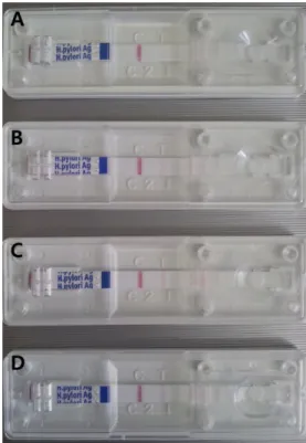
Figure 1. Results of Helicobacter pylori Stool Ag Kit based on
the number of H. pylori ATCC 49503 cells. (A) 1×10
5CFU/100
µL; (B) 1×10
6CFU/100 µL; (C) 1×10
7CFU/100 µL; (D) 1×10
8CFU/100 µL.
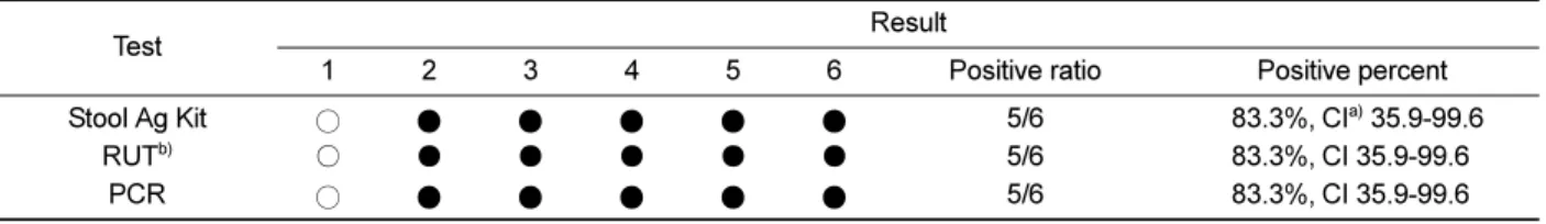
Five of six gastric biopsy specimens were positive by the rapid urease test on day 21. We did not detect a positive reaction from sample no. 1 (5/6, 83.3%) (Table 2).
Genomic DNA RT-PCR of gastric specimens
A RT-PCR analysis method using CagA DNA was employed to detect H. pylori infection. The CagA gene (298 bp) was specifically amplified by PCR with the H.
pylori CagA-specific primers (H-CagAF and H-CagAR).
The target nucleic acid fragments were specifically amplified by RT-PCR with CagA DNA primers. As a result, five of six gastric specimens were positive by the PCR reaction. However, we did not detect a positive reaction from sample no. 1 (5/6, 83.3%) (Table 2).
Discussion
H pylori infection is the main cause of chronic gastritis, which is the most frequent chronic bacterial disease [1,27]. H. pylori induces an inflammatory immune response in the gastric mucosa, and many studies have demonstrated a significant correlation between H. pylori density and acute and chronic inflammatory grade.
Treatments that suppress H. pylori density and virulence could become strategies to prevent H. pylori-associated disease in the future. The present study focused on the influence of some factors to eradicate H. pylori infection [28]. It has been widely reported over the last decade that the success of H. pylori eradication treatment has decreased [29,30].
Diagnosis of H pylori infection can be made with both invasive and noninvasive tests. Invasive tests include histology, culture, and the rapid urease test, which require endoscopy to obtain gastric mucosa biopsies.
Noninvasive tests for diagnosis of H pylori infection, which are based on analyzing breath, blood, or stool samples, have been developed [8]. However, serological tests are unable to distinguish active from past infections [31].
Noninvasive tests require demonstrating the microorganism in gastric biopsy samples; therefore, an endoscopy must be performed. The current gold standard for diagnosing H pylori infection is endoscopic biopsy of gastric tissue for the rapid urease test, histology, and culture. However, such an invasive procedure has major disadvantages of anesthesia, discomfort, and possible ethical problems [32]. But, noninvasive tests are easy to perform and do not produce significant discomfort and allow a patient to avoid the discomfort and risk of invasive endoscopy. These include serological antibody testing for H. pylori, the urea breath test, and the HpSA test [32].
Among several diagnostic tests, the HpSA test for diagnosing H. pylori infection may offer a useful non- invasive method without sacrificing animals during an in vivo study. Kazemi et al. recently performed a validity comparison of five diagnostic tests including HpSA and rapid urease test in patients [33]. In our study, we carries out experiments to evaluate the utility of the SD Bioline H. pylori stool Ag Kit (Standard Diagnostics, Inc., Korea) in a mouse model. As a result, the sensitivity of the HpSA test was 83.33%. We detected the presence of
Table 1. Helicobacter pylori stool Antigen (HpSA) test for diagnosing H. pylori infected C57BL/6 mice
Day H. pylori Inoculation
a)n Positive reaction
b)(Positive percent)
1 Yes 6 0 (0%, CI
c)0-39.3)
3 Yes 6 1 (16.7%, CI 0.42-64.1)
5 Yes 6 4 (66.7%, CI 0.22-0.96)
7 Yes 6 5 (83.3%, CI 35.9-99.6)
14 Yes 6 5 (83.3%, CI 35.9-99.6)
21 Yes 6 5 (83.3%, CI 35.9-99.6)
a)
H. pylori inoculation was three times every 2 days.
b)
A positive reaction revealed H. pylori colonization, which was observed as a red colored double line.
c)
Incidence percentage (95% confidential interval) was calculated with MiniTab statistical software.
Table 2. Comparison test results of mice infected with Helicobacter pylori
Test Result
1 2 3 4 5 6 Positive ratio Positive percent
Stool Ag Kit ○ ● ● ● ● ● 5/6 83.3%, CI
a)35.9-99.6
RUT
b)○ ● ● ● ● ● 5/6 83.3%, CI 35.9-99.6
PCR ○ ● ● ● ● ● 5/6 83.3%, CI 35.9-99.6
○, negative; ●, positive.
a)
Incidence percentage (95% confidential interval) was calculated with MiniTab statistical software.
b)