ABSTRACT
Background: The objective of this study was to examine changes in the prevalence of cytotoxic-associated gene A (CagA) positive Helicobacter pylori infection in Jinju, Korea, over the last 20 years.
Methods: Three cross-sectional analyses were conducted concurrently. A total of 1,305 serum samples were collected from 1994–1995, 2004–2005, and 2014–2015, respectively. The presence of immunoglobulin (Ig) G, IgA, and IgM antibodies against H. pylori CagA protein was examined by western blotting.
Results: Overall, seropositivity for anti-CagA IgG antibody was significantly decreased from 63.2% to 42.5% over the last 20 years (P < 0.001). Anti-CagA IgG seropositivities in children and young adults aged 10–29 years decreased from 1994 (60.0%–85.0%) to 2015 (12.5%–
28.9%). The age when plateau of increasing IgG seropositivity was reached in each study period shifted from the 15–19 year-old group in 1994–1995 (85.0%) to the 40–49 year-old group in 2014–2015 (82.5%). Overall seropositive rates of anti-CagA IgA and IgM antibodies did not change significantly either over the last 20 years.
Conclusion: H. pylori infection rate in children and young adults declined over 20 years in Jinju, probably due to improved sanitation, housing, or economy.
Keywords: Prevalence; Helicobacter pylori; CagA protein; Western Blotting
INTRODUCTION
Helicobacter pylori is a risk factor of peptic ulcer, atrophic gastritis, and gastric carcinoma.1 Environmental, host-related, and virulent factors associated with the bacterium, including cytotoxin-associated gene A (CagA), vacuolating cytotoxin A (VacA), and outer inflammatory protein (OipA) are involved in the development of H. pylori-related diseases.2 Of these factors,
Original Article
Ji Sook Park ,1,2* Jin-Su Jun ,1,2* Eo Young Ryu ,1,2 Jung Sook Yeom ,1,2 Eun Sil Park ,1,2 Ji-Hyun Seo ,1,2 Jae Young Lim ,1,2 Chan-Hoo Park ,1,2 Hyang-Ok Woo ,1,2 Seung-Chul Baik ,2,3 Woo-Kon Lee ,2,3 Myung-Je Cho ,2,3 Kwang-Ho Rhee ,2,3 and Hee-Shang Youn 1,2
1 Department of Pediatrics, Gyeongsang National University College of Medicine and Gyeongsang National University Hospital, Jinju, Korea
2Institute of Health Sciences, Gyeongsang National University, Jinju, Korea
3Department of Microbiology, Gyeongsang National University College of Medicine, Jinju, Korea
Changes in Seroprevalence of
Helicobacter pylori Infection over 20 Years in Jinju, Korea, from Newborns to the Elderly
Received: Feb 13, 2020 Accepted: Jun 18, 2020 Address for Correspondence:
Hee-Shang Youn, MD, PhD Department of Pediatrics, Gyeongsang National University College of Medicine and Gyeongsang National University Hospital, 15 Jinju-daero 816-beon-gil, Jinju 52727, Republic of Korea.
E-mail: [email protected]
*Ji Sook Park and Jin-Su Jun contributed equally to this work.
© 2020 The Korean Academy of Medical Sciences.
This is an Open Access article distributed under the terms of the Creative Commons Attribution Non-Commercial License (https://
creativecommons.org/licenses/by-nc/4.0/) which permits unrestricted non-commercial use, distribution, and reproduction in any medium, provided the original work is properly cited.
ORCID iDs Ji Sook Park
https://orcid.org/0000-0002-4704-2246 Jin-Su Jun
https://orcid.org/0000-0002-6382-6286 Eo Young Ryu
https://orcid.org/0000-0002-0265-4951 Jung Sook Yeom
https://orcid.org/0000-0003-0688-7493 Eun Sil Park
https://orcid.org/0000-0001-9344-7191 Ji-Hyun Seo
https://orcid.org/0000-0002-0691-3957
Gastroenterology &
Hepatology
Jae Young Lim
https://orcid.org/0000-0001-5205-202X Chan-Hoo Park
https://orcid.org/0000-0001-8979-1467 Hyang-Ok Woo
https://orcid.org/0000-0001-8849-9341 Seung-Chul Baik
https://orcid.org/0000-0001-6033-4078 Woo-Kon Lee
https://orcid.org/0000-0003-3913-2265 Myung-Je Cho
https://orcid.org/0000-0002-4958-9827 Kwang-Ho Rhee
https://orcid.org/0000-0002-4422-4992 Hee-Shang Youn
https://orcid.org/0000-0002-5498-838X Funding
This work was supported by grants (NRF-2013R1A1A2057513 and NRF- 2019R1G1A1100590) of the Basic Science Research Program through the National Research Foundation of Korea (NRF), funded by the Ministry of Education.
Disclosure
The authors have no potential conflicts of interest to disclose.
Author Contributions
Conceptualization: Ryu EY, Youn HS. Data curation: Park JS, Ryu EY, Yeom JS, Park ES, Seo JH, Lim JY, Park CH, Woo HO. Formal analysis: Baik SC, Lee WK, Cho MJ, Rhee KH.
Investigation: Jun JS. Methodology: Jun JS, Baik SC, Lee WK, Cho MJ, Rhee KH. Software:
Park JS. Validation: Park JS. Visualization: Youn HS. Writing - original draft: Park JS. Writing - review & editing: Youn HS.
CagA protein is associated with a severe clinical outcome. Most strains of H. pylori isolated in East Asia, including Korea, have been confirmed CagA-positive.3-5
It is thought that approximately half of the world's population harbors H. pylor
6 and H. pylori-related diseases represent a significant medical burden.7,8 Although the prevalence of H. pylori infection in developed countries has declined over recent decades, infection rate in the rest of the world remains high.6,9,10 In Korea, H. pylori infection of late adolescents and adults has decreased due to rapid socio-economic growth and improved housing.11-13 However, the previous studies had been designed to diagnose H. pylori infection using enzyme linked immunosorbent assay (ELISA), some of which used crude antigens of H.
pylori isolated outside Korea and had enrolled populations aged 16 years and older due to the limitations of its use in diagnosing H. pylori infection in children.11-15 H. pylori infection can take hold during infancy or early childhood and colonization generally persists for life if left untreated.16-18
H. pylori induces systemic and mucosal immune responses in most infected patients.
Serologic tests can detect H. pylori infection, and serum immunoglobulin (Ig) G, A, and M antibodies may indicate whether infection is acute or chronic.19,20 H. pylori CagA antigen might be one of the major antigens that produce the strongest antibody responses and might be related to high levels of anti-H. pylori antibodies in infected humans.21 The patients remain positive for anti-CagA antibodies longer than other anti-H. pylori antibodies even after eradication of H. pylori.3,22
Jinju is a city of around 340,000 inhabitants that lies in the southern part of Korea and is surrounded by rural areas. Compared with other cities in Korea, the inflow and outflow of citizens are relatively low (www.index.go.kr). The relatively low migration rate (0.1% during 2006–2016) makes Jinju suitable for evaluating the lifetime trend of H. pylori infection, together with birth cohort effects. Few reports have examined the prevalence of H. pylori infection in the general population, including infants and children, along with lifetime trends.23 Thus the aim of the present study was to investigate changes in the seroprevalence of CagA positive H. pylori infection over the last 20 years in the general population of Jinju, ranging from neonates to the elderly. In addition, the lifetime trend of H. pylori infection was estimated by western blot analysis of IgG, IgA, and IgM antibodies against H. pylori CagA.
METHODS
Study population
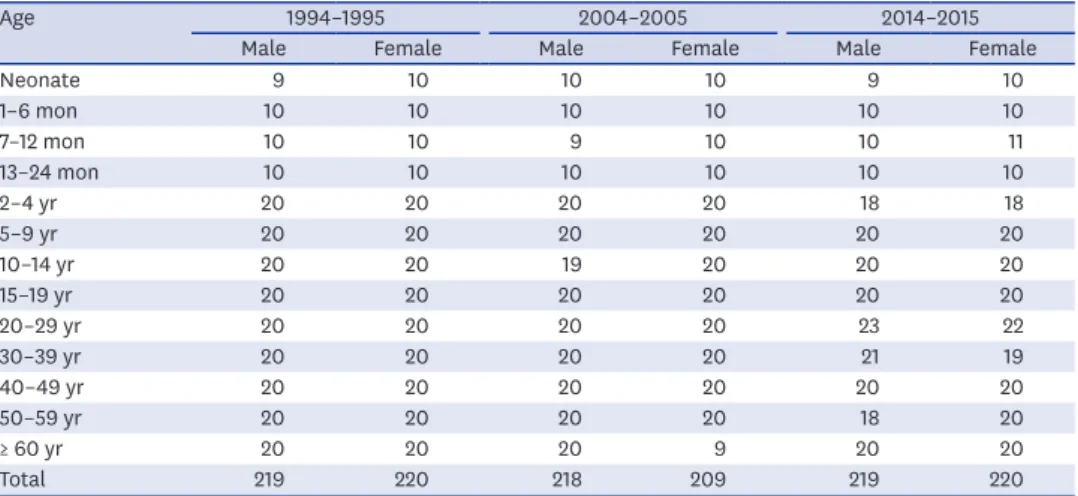
Three retrospective cross-sectional analyses using collected serum samples were conducted concurrently. These cross-sectional analyses covered 1994–1995, 2004–2005, and 2014–2015, respectively, spanning a total of 20 years. A total of 1,305 serum samples matched with age, sex and collection period were obtained from the Gyeongsang National University Hospital (GNUH) Biobank, a member of the Korea Biobank Network and analyzed. All of the serum samples were preserved in the deep freezer before analysis. To evaluate the age when acute or initial H. pylori infection occurs, and to differentiate serologic positivity due to infection from that due to transplacental transmission of anti-H. pylori CagA IgG antibodies during infancy and early childhood, samples were grouped according to age as follows: neonate, 1–6 months, 7–12 months, 13–24 months, 2–4 years, and 5–9 years (infants and children), and 5 to10-year intervals thereafter (Table 1).
Antigen preparation and western blot analysis
Whole cell extracts of CagA positive-H. pylori strain 51 (obtained from the Korean type Culture Collection; HpKTCC; http://hpktcc.knrrc.or.kr, NCBI Taxonomy ID: 290847) were prepared as described previously.24 H. pylori strain 51 was isolated from a patient with duodenal ulcer at GNUH in 1988 and has been studied extensively since then.14,15,18,22,24-27
Briefly, H. pylori strain 51 was cultured for 18 hours under the environment of 37°C, 5%–10%
CO2 and 100% humidity on Mueller-Hinton agar supplemented with 10% bovine serum.
Bacterial cells from each plate were harvested and pelleted by centrifugation at 4,000 × g for 15 minutes. These cells were then suspended in sterile phosphate buffer, broken by ultrasonic treatment using an Ultrasonic W380 (Sonics & Materials Inc., Danbury, CT, USA), and stored at −70°C. The sonicated H. pylori whole cell lysate was then used as an antigen. The presence of anti-CagA IgG, IgA, and IgM antibodies in the serum was then examined by western blot analysis. Briefly, cell lysates were run on 10%–20% sodium dodecyl sulfate-polyacrylamide gels overlaid with a 3% stacking gel, as described by Laemmli.28 These gels were loaded with samples containing 100 μg of antigen along with molecular mass markers (Bio-Rad Laboratories Inc., Philadelphia, PA, USA). Proteins were separated under a constant current of 15 mA for 60 minutes until the bromophenol blue dye migrated out of the gel. Proteins were then transferred onto pre-wetted nitrocellulose (NC) membranes (0.2 micron, Bio- Rad Laboratories Inc.). These membranes were then cut into strips. Strips were incubated with 1:20 diluted serum for 30 minutes at 37°C, rinsed three times with Tris buffered saline containing Tween-20 (TBST; 50 mM Tris-HCl, 150 mM NaCl, 0.05% Tween-20, pH 7.5), and incubated at 37°C for 30 minutes with alkaline phosphatase-conjugated goat anti-human IgG, IgA, or IgM antibodies (Bethyl Laboratories Inc., Montgomery, TX, USA). Three different NC membrane strips incubated with one serum sample were used for incubation with goat anti-human IgG, IgA and IgM antibodies, respectively. After washing with TBST buffer, these strips were incubated with 5-bromo-4-chloro-3-indolyl phosphate as a substrate and nitroblue tetrazolium as a chromogenic indicator. Reactions were stopped after 15 minutes by rinsing these strips several times with buffer (20 mM Tris-HCl, 50 mM EDTA, pH 8.0).
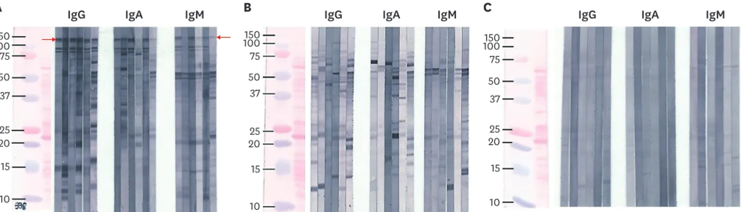
These strips were then dried before mounting. Samples showing a band at 116–120 kDa were considered positive for H. pylori CagA IgG, IgA, or IgM antibody (Fig. 1).14,15 Results were analyzed by two investigators who were blinded to information about the serum samples including age and sex of subjects, and collection time. The authors interpreted results of western blotting based on Fig. 1 to avoid inconsistency.
Table 1. Numbers and age distribution of subjects
Age 1994–1995 2004–2005 2014–2015
Male Female Male Female Male Female
Neonate 9 10 10 10 9 10
1–6 mon 10 10 10 10 10 10
7–12 mon 10 10 9 10 10 11
13–24 mon 10 10 10 10 10 10
2–4 yr 20 20 20 20 18 18
5–9 yr 20 20 20 20 20 20
10–14 yr 20 20 19 20 20 20
15–19 yr 20 20 20 20 20 20
20–29 yr 20 20 20 20 23 22
30–39 yr 20 20 20 20 21 19
40–49 yr 20 20 20 20 20 20
50–59 yr 20 20 20 20 18 20
≥ 60 yr 20 20 20 9 20 20
Total 219 220 218 209 219 220
Statistical analysis
The χ2 or Fisher's exact test was used to compare differences in seropositivities for anti-CagA IgG, IgA, and IgM antibodies according to age, sex, and study period. All statistical analyses were performed using SPSS ver 25.0 (IBM, Armonk, NY, USA). The level of significance was set at 0.05. GraphPad Prism 8 (Graph-Pad Software, San Diego, CA, USA) was used for graphics.
Ethics statement
The study protocol was approved by the Institutional Review Board at Gyeongsang National University Hospital (GNUH 2015-06-010). Informed consent was waived by the board.
RESULTS
A total of 1,305 serum samples were tested, of which 656 were from males and 713 were from subjects younger than 20 years old. Of these samples, 439, 427, and 439 were obtained from 1994 to 1995, 2004 to 2005, and 2014 to 2015, respectively (Table 1).
Anti-H. pylori CagA IgG antibody seropositivity according to age and study period
The overall seropositive rate, except for the anti-CagA IgG positive rate in infants aged 0–12 months, was 63.2% from 1994 to 1995, 54.9% from 2004 to 2005, and 42.5% from 2014 to 2015 (P < 0.001), respectively. There was no difference in overall anti-CagA IgG positive rate between males and females (male, 54.9%; female, 50.4%; P = 0.108).
Age-specific seropositive rates of anti- CagA IgG antibodies in each study period are plotted in Fig. 2. Anti-CagA IgG antibody seropositivities according to age in the three cross- sectional periods were decreased from newborn to 7–12 month-old group or 2–4 year-old group and then increased with age (Fig. 2). Seropositive rate of neonates was 73.7% in 1994–1995, 75.0% in 2004–2005, and 63.2% in 2014–2015 (P = 0.765). The lowest seropositive rate in each study period was 25.0% in those age 2–4 years in 1994–1995, 15.0% in those age 13–24 months in 2004–2005, and 0.0% in those age 7–12 months in 2014–2015. Significant reductions in age-specific anti-CagA IgG seropositivities, particularly in those aged between 7 and 12 months, 10 and 14 years, 15 and 19 years, and 20 and 29 years were presented over
A IgG B C
100150 75 50 37
2025
15 10
100150 75 50 37
2025
15
10
100150 75 50 37 2025
15
10
IgA IgM IgG IgA IgM IgG IgA IgM
Fig. 1. Three different western blot patterns (A, B, and C) were identified. Only (A) showed reactivity with a 116–120 kDa band (red arrow).Thus it was considered positive for IgG, IgA, and IgM anti-CagA-H. pylori antibodies. (B) showed several other bands after reacting with whole cell lysate of H. pylori 51. No definitive band was observed in (C). (C) Phosphate buffered saline was used as a negative control.
Ig = immunoglobulin, CagA = cytotoxic-associated gene A, H. pylori = Helicobacter pylori.
20 years (P < 0.05) (Fig. 2 and Table 2). The age when the seropositivity for anti-CagA IgG reached a plateau was delayed over the 20 years and occurred in the 15–19 year-old group in 1994–1995 (85.0%), the 20–29 year-old group in 2004–2005 (72.5%), and the 40–49 year-old group in 2014–2015 (82.5%), respectively. And then the highest rate in each study period occurred in the 15–19 year-old group in 1994–1995 (85.0%), and the 50–59 year-old group in 2004–2005 (92.5%) and 2014–2015 (89.5%), respectively. Seropositivities in early childhood (2–4 years old) during the three study periods decreased numerically from 1994 to 2015 (25%
in 1994–1995, 17.5% in 2004–2005 and 13.9% in 2014–2015). However, the decrease was not statistically significant (P = 0.484) (Table 2). According to the birth cohort of the 2–4 year-old group in 1994–1995, positive rates were not significantly changed until individuals were in their 20s in 2014–2015 (25.0% in 1994–1995, 35.9% in 2004–2005 and 28.9% in 2014–2015, P = 0.587) (Table 2).
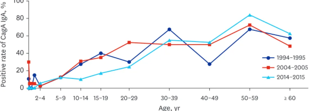
Anti-H. pylori CagA IgA seropositivity according to age and study period The overall rate of anti-CagA IgA seropositivity in each study period was 31.4% in 1994–1995, 34.0% in 2004–2005, and 29.4% in 2014–2015 (P = 0.354). The anti-CagA IgA seropositive
≥ 60 50–59
40–49 30–39
20–29
5–9 15–19
Positive rate of CagA IgG, %
Age, yr
2–4 10–14
0 20 60 100
40 80
1994–1995 2004–2005 2014–2015
Fig. 2. Changes in anti-CagA IgG antibody seropositivity according to age during a 20-year study in Jinju. Each line connects values for each study period: 1994–1995 (line with circle), 2004–2005 (line with square), and 2014–2015 (line with triangle). There were significant differences in age specific seropositivity with time (from 1994 to 2015) between those aged 7–12 months and 10–29 years (P < 0.05). Anti-CagA IgG seropositivity in early childhood (age, 2–4 years) remained low and stable over 20 years.
CagA = cytotoxic-associated gene A, Ig = immunoglobulin.
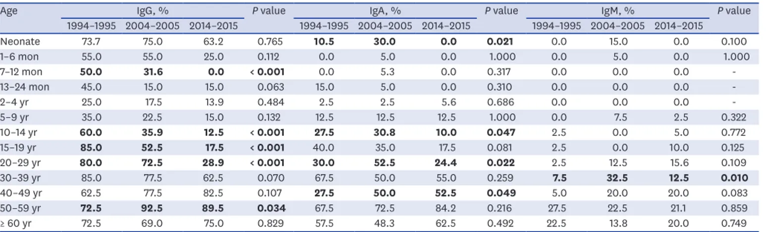
Table 2. Positive rate of anti-CagA IgG, IgA, and IgM according to age and year
Age IgG, % P value IgA, % P value IgM, % P value
1994–1995 2004–2005 2014–2015 1994–1995 2004–2005 2014–2015 1994–1995 2004–2005 2014–2015
Neonate 73.7 75.0 63.2 0.765 10.5 30.0 0.0 0.021 0.0 15.0 0.0 0.100
1–6 mon 55.0 55.0 25.0 0.112 0.0 5.0 0.0 1.000 0.0 5.0 0.0 1.000
7–12 mon 50.0 31.6 0.0 < 0.001 0.0 5.3 0.0 0.317 0.0 0.0 0.0 -
13–24 mon 45.0 15.0 15.0 0.063 15.0 5.0 0.0 0.310 0.0 0.0 0.0 -
2–4 yr 25.0 17.5 13.9 0.484 2.5 2.5 5.6 0.686 0.0 0.0 0.0 -
5–9 yr 35.0 22.5 15.0 0.132 12.5 12.5 12.5 1.000 0.0 7.5 2.5 0.322
10–14 yr 60.0 35.9 12.5 < 0.001 27.5 30.8 10.0 0.047 2.5 0.0 5.0 0.772
15–19 yr 85.0 52.5 17.5 < 0.001 40.0 35.0 17.5 0.081 2.5 0.0 10.0 0.125
20–29 yr 80.0 72.5 28.9 < 0.001 30.0 52.5 24.4 0.022 2.5 12.5 15.6 0.109
30–39 yr 85.0 77.5 62.5 0.070 67.5 50.0 55.0 0.259 7.5 32.5 12.5 0.010
40–49 yr 62.5 77.5 82.5 0.107 27.5 50.0 52.5 0.049 5.0 20.0 20.0 0.083
50–59 yr 72.5 92.5 89.5 0.034 67.5 72.5 84.2 0.216 27.5 22.5 21.1 0.859
≥ 60 yr 72.5 69.0 75.0 0.829 57.5 48.3 62.5 0.492 22.5 13.8 20.0 0.749
Statistically significant differences according to age and study period are shown in bold.
CagA = cytotoxic-associated gene A, Ig = immunoglobulin.
rate was relatively low in infancy and early childhood (2–4 years old group) in each study period.
After early childhood, anti-CagA IgA positive rate appeared to increase with age (Fig. 3).
Anti-H. pylori CagA IgM seropositivity according to age and study period Overall seropositivities for anti-CagA IgM antibodies in 1994–1995, 2004–2005, and 2014–
2015 were 6.4%, 10.8%, and 9.8%, respectively (P = 0.060). Overall seropositive rates of anti- CagA IgM over the last 20 years were generally low compared with those of anti-CagA IgG and anti-CagA IgA (Fig. 4). According to age, anti-CagA IgM seropositive rates were generally lower in individuals younger than 20 compared with those in adults ≥ 20 years old.
DISCUSSION
The present study showed that the overall seroprevalence of anti-H. pylori CagA IgG antibodies in Jinju, Korea, decreased from 63.2% to 42.5% between 1994 and 2015. Recent decline in the incidence of H. pylori infection with time was similar to a previous study.11 However, the rate of H. pylori in adolescents and adults aged 15 years and older was higher in Jinju (73.8% in 2004–2005) than other regions of Korea (59.6% in 2005).
≥ 60 50–59
40–49 30–39
20–29
5–9 15–19
Positive rate of CagA IgA, %
Age, yr
2–4 10–14
0 20 60 100
40 80
1994–1995 2004–2005 2014–2015
Fig. 3. Changes in anti-CagA IgA seropositivity according to age over the 20-year study period. Each line connects values for each study period: 1994–1995 (line with circle), 2004–2005 (line with square), and 2014–2015 (line with triangle). There was no significant reduction in seropositivity according to age over time.
CagA = cytotoxic-associated gene A, Ig = immunoglobulin.
≥ 60 50–59
40–49 30–39
20–29
5–9 15–19
Positive rate of CagA IgM, %
Age, yr
2–4 10–14
0 20 60 100
40 80
1994–1995 2004–2005 2014–2015
Fig. 4. Changes in anti-CagA IgM seropositivity according to age during the 20-year study period in Jinju. Each line connects values for each study period: 1994–1995 (line with circle), 2004–2005 (line with square), and 2014–2015 (line with triangle). There was no significant reduction in seropositivity according to age over time.
CagA = cytotoxic-associated gene A, Ig = immunoglobulin.
Anti-CagA IgG seropositivity increased with age from early childhood. Subjects older than 10 years of age in 1994–1995, 20 years of age in 2004–2005 and 30 years of age in 2014–2015 showed seropositive rates of over 60% (Table 2 and Fig. 2). Age-related increase of seropositivity observed in each study period in the present study may represent a cohort phenomenon23,29 rather than newly acquired H. pylori infections as individuals get older. Previous studies have also noted that birth cohort effects can lead to an increasing seroprevalence of H. pylori infection with age and decreasing seroprevalence observed in subsequent generations.11,30 In this study, the age to a plateau of anti-CagA IgG positive rate tended to be delayed by 10–20 years of age for each study period (Fig. 2). The shifts of age to a plateau over 20 years and the marked reduction of positive rates in the 10–20s age groups in the present study suggests that the prevalence of CagA positive H. pylori infection in Jinju will decline further in the coming decades. However, the relatively stable positive rate in the cohort of children aged 2–4 years in 1994–1995 (25%) over 20 years (35.9% of 10–14 year-old group in 2004–2005 and 28.9% of 20–29 year-old group in 2014–2015, P = 0.587) (Table 2 and Fig. 2) may contribute to persistently stable colonization of H. pylori and the incidence of H. pylori-associated disease in Jinju in the future, albeit low. Further studies on incidences of CagA positive H. pylori-associated diseases are warranted.
During the three study periods, neonates and infants showed high rates of anti-CagA IgG seropositivity (Table 2 and Fig. 2). However, such high rates could not be evidence of H. pylori infection in newborns and infants. Rather, they could represent positive rates in femalesof childbearing age. This was supported by the finding that rates of anti-CagA IgG seropositivity in those younger than 6 months of age was similar to those in females aged 15–49 years in each study period. The rate of anti-CagA IgG seropositivity in those younger than 6 months of age was 64.1% in 1994–1995, 65.0% in 2004–2005, and 43.6% in 2014–2015. Corresponding rates in females aged 15–49 years were 78.8%, 66.3%, and 46.9%, respectively. Transplacental transmission of anti-CagA IgG might cause high positivities in neonates and infants because IgG can transfer passively from a mother to a fetus. Therefore, the overall seropositive rates of anti-CagA IgG in this study were calculated excluding neonates and infants. Serum samples from babies and females aged 15–49 years were randomly assigned and the mother-infant relationship was unknown in the present study. Further studies with paired serum samples from babies and their mothers would be necessary.
Overall rates of anti-CagA IgA seropositivity were similar among the three study periods (P
= 0.354). Unlike those measured in a previous study,30 the authors found that the anti-CagA IgA seropositivity during each study period gradually increased with age, particularly after 2–4 years of age (Fig. 3). Anti-CagA IgG and IgA seropositivities around 1–4 years of age showed lower rates than others. And IgG and IgA seropositivities in those older than 5 years increased along with age for each period. Thus, the authors tentatively estimated the time of H. pylori infection for each period might have occurred in early childhood, especially of those younger than 4 years old in Jinju, Korea (Figs. 2 and 3).
Generally, IgM and IgA antibodies could be produced as a primary response. Greater quantity of IgG antibodies could be produced as a secondary response after antigen exposure. Specific IgG could persist several months or years. Positive anti-CagA IgA could represent mucosal immune response in H. pylori infection. In addition, mucosal immune response in a person infected with H. pylori might occur repeatedly.21 Based on humoral immune response sequences and our results of higher anti-CagA IgG positivities in the older birth cohort and anti-CagA IgA seropositivity patterns along with age, especially in those older than 5 years,
anti-CagA IgA antibodies might be repetitively produced by the active mucosal immune response of already existing bacteria rather than acute infection.19-21 Our findings could support that H. pylori infection is acquired during early childhood while new infections during adulthood are rare (Figs. 2 and 3).16,17,31
Humoral IgA response in newborns and infants is usually weak. The low rate of anti-CagA IgA seropositivity in neonates and infants younger than 2 years of age in the present study might represent sporadic acute H. pylori infection.21 Rates of anti-CagA IgA seropositivity in those aged 60 years and older were lower than those in subsequent generations during the three study periods (Fig. 3). Development of atrophic gastritis, intestinal metaplasia, or gastric cancer in the elderly may have contributed to the lower anti-CagA IgA seropositivity because H. pylori disappears from the gastric mucosa after these diseases develop.16,17 However, further studies are warranted to determine the relationship between anti-H. pylori CagA IgA seropositivity and mucosal inflammation or histologic status of the gastric mucosa.
In children and adolescents, anti-CagA IgA seropositivity might reflect inflammation of the gastric mucosa caused by H. pylori based on our unpublished research, which showed that 81.5% (202 of 247) of anti-CagA IgA-positive subjects aged less than 20 years in Jinju had active gastritis.
The present study showed that age-specific rates of anti-CagA IgM seropositivity during the three study periods were very low compared with those of anti-CagA IgG and IgA seropositivity (Fig. 4). Anti-CagA IgM antibody has not been well established as a meaningful marker of H. pylori infection yet.20 However, a small number of acute H. pylori infections could be acquired sporadically at any age including the newborn period. In addition, H. pylori might invade the gastric mucosa or re-infect an already infected person during their lifetime, although this is likely to be uncommon.32
This study has several limitations. First, any information regarding housing or economic circumstance was not provided. Neither medical information such as H. pylori eradication nor clinical outcome of subjects associated with CagA-positive H. pylori was provided. Second, this was a retrospective cross sectional cohort study based on limited geography. Regional limitation may account for our finding of a higher seroprevalence of H. pylori infection than that of a previous nationwide survey.11 Third, the authors used western blotting for qualitative analyses of IgG, IgA and IgM antibodies. However, western blotting has been extensively validated as a method for the detection of several antibodies at the same time.
And it is, in fact, more sensitive and specific for anti-H. pylori antibodies in younger children including infants than an ELISA.15,33 Finally, presence of CagA negative H. pylori among the study population and change in antigenicity of CagA over 20 years might be possible.
However, since CagA positive H. pylori comprises a large proportion of strains identified in Korea so far,4 CagA negative H. pylori might account for a small portion in the present study. Nevertheless, the present study has a strength; it examined temporal changes in CagA positive H. pylori infection in a relatively constant population from birth to old age, over a period of 20 years.
Results of this study suggest that Jinju may experience a lower prevalence of CagA positive H. pylori infection and associated diseases in the future. However, the low and stable seroprevalence of CagA positive H. pylori infection in early childhood over 20 years suggests that low, albeit constant, colonization by the bacterium may continue. Transmission of H. pylori could be highly affected by intrafamilial and environmental exposure,24,31,34-36
and infection could occur in early childhood and persist without treatment.16-18 Therefore, education regarding personal hygiene such as hand washing and eating habits in family,37 and aggressive diagnosis and eradication could help reduce colonization.38,39 Establishment of a diagnostic and therapeutic strategy in children with H. pylroi infection in Korea is necessary.
ACKNOWLEGMENTS
Serum samples used for the present study were provided by Gyeongsang National University Hospital, a member of the Korea Biobank Network supported by the Ministry of Health, Welfare and Family Affairs. All samples from the Korea Biobank Network were obtained with informed consent under institutional review board-approved protocols. H. pylori strain 51 was provided by Helicobacter pylori Korean type Culture Collection (HpKTCC), a member of the Korean National Research Resource Center supported by the Ministry of Science, ICT and Future Planning.
REFERENCES
1. Correa P, Piazuelo MB. Natural history of Helicobacter pylori infection. Dig Liver Dis 2008;40(7):490-6.
PUBMED | CROSSREF
2. Yamaoka Y. Mechanisms of disease: Helicobacter pylori virulence factors. Nat Rev Gastroenterol Hepatol 2010;7(11):629-41.
PUBMED | CROSSREF
3. Shiota S, Suzuki R, Yamaoka Y. The significance of virulence factors in Helicobacter pylori. J Dig Dis 2013;14(7):341-9.
PUBMED | CROSSREF
4. Kim JY, Kim N, Nam RH, Suh JH, Chang H, Lee JW, et al. Association of polymorphisms in virulence factor of Helicobacter pylori and gastroduodenal diseases in South Korea. J Gastroenterol Hepatol 2014;29(5):984-91.
PUBMED | CROSSREF
5. Hatakeyama M. Oncogenic mechanisms of the Helicobacter pylori CagA protein. Nat Rev Cancer 2004;4(9):688-94.
PUBMED | CROSSREF
6. Eusebi LH, Zagari RM, Bazzoli F. Epidemiology of Helicobacter pylori infection. Helicobacter 2014;19 Suppl 1:1-5.
PUBMED | CROSSREF
7. Dorer MS, Talarico S, Salama NR. Helicobacter pylori's unconventional role in health and disease. PLoS Pathog 2009;5(10):e1000544.
PUBMED | CROSSREF
8. Parkin DM. The global health burden of infection-associated cancers in the year 2002. Int J Cancer 2006;118(12):3030-44.
PUBMED | CROSSREF
9. Wex T, Venerito M, Kreutzer J, Götze T, Kandulski A, Malfertheiner P. Serological prevalence of Helicobacter pylori infection in Saxony-Anhalt, Germany, in 2010. Clin Vaccine Immunol 2011;18(12):2109-12.
PUBMED | CROSSREF
10. den Hoed CM, Vila AJ, Holster IL, Perez-Perez GI, Blaser MJ, de Jongste JC, et al. Helicobacter pylori and the birth cohort effect: evidence for stabilized colonization rates in childhood. Helicobacter 2011;16(5):405-9.
PUBMED | CROSSREF
11. Lim SH, Kwon JW, Kim N, Kim GH, Kang JM, Park MJ, et al. Prevalence and risk factors of Helicobacter pylori infection in Korea: nationwide multicenter study over 13 years. BMC Gastroenterol 2013;13(1):104.
PUBMED | CROSSREF
12. Lee SY, Moon HW, Hur M, Yun YM. Validation of western Helicobacter pylori IgG antibody assays in Korean adults. J Med Microbiol 2015;64(Pt 5):513-8.
PUBMED | CROSSREF
13. Lim SH, Kim N, Kwon JW, Kim SE, Baik GH, Lee JY, et al. Trends in the seroprevalence of Helicobacter pylori infection and its putative eradication rate over 18 years in Korea: a cross-sectional nationwide multicenter study. PLoS One 2018;13(10):e0204762.
PUBMED | CROSSREF
14. Youn HS, Baik S, Lee WK, Cho MJ, Ryou HH, Choi HJ, et al. Serodiagnosis of Helicobacter pylori infection.
J Korean Soc Microbiol 1990;25(6):463-74.
15. Jeong HL, Jung YS, Jun JS, Yeom JS, Park JS, Seo JH, et al. Comparison of four commercial ELISA kits and in-house immunoblotting for diagnosis of Helicobacter pylori infection. Pediatr Gastroenterol Hepatol Nutr 2012;15(2):85-90.
CROSSREF
16. Rhee KH, Youn HS, Baik SC, Lee WK, Cho MJ, Choi HJ, et al. Prevalence of Helicobacter pylori infection in Korea. J Korean Soc Microbiol 1990;25(6):475-90.
17. Gologan A, Graham DY, Sepulveda AR. Molecular markers in Helicobacter pylori-associated gastric carcinogenesis. Clin Lab Med 2005;25(1):197-222.
PUBMED | CROSSREF
18. Seo JH, Youn JH, Kim EA, Jun JS, Park JS, Yeom JS, et al. Helicobacter pylori antigens inducing early immune response in infants. J Korean Med Sci 2017;32(7):1139-46.
PUBMED | CROSSREF
19. Malaty HM, Kim JG, Kim SD, Graham DY. Prevalence of Helicobacter pylori infection in Korean children:
inverse relation to socioeconomic status despite a uniformly high prevalence in adults. Am J Epidemiol 1996;143(3):257-62.
PUBMED | CROSSREF
20. Patel SK, Pratap CB, Jain AK, Gulati AK, Nath G. Diagnosis of Helicobacter pylori: what should be the gold standard? World J Gastroenterol 2014;20(36):12847-59.
PUBMED | CROSSREF
21. Sörberg M, Engstrand L, Ström M, Jönsson KA, Jörbeck H, Granström M. The diagnostic value of enzyme immunoassay and immunoblot in monitoring eradication of Helicobacter pylori. Scand J Infect Dis 1997;29(2):147-51.
PUBMED | CROSSREF
22. Seo JH, Lim CW, Park JS, Yeom JS, Lim JY, Jun JS, et al. Correlations between the CagA antigen and serum levels of anti-Helicobacter pylori IgG and IgA in children. J Korean Med Sci 2016;31(3):417-22.
PUBMED | CROSSREF
23. Banatvala N, Mayo K, Megraud F, Jennings R, Deeks JJ, Feldman RA. The cohort effect and Helicobacter pylori. J Infect Dis 1993;168(1):219-21.
PUBMED | CROSSREF
24. Youn HS, Baik SC, Cho YK, Woo HO, Ahn YO, Kim K, et al. Comparison of Helicobacter pylori infection between Fukuoka, Japan and Chinju, Korea. Helicobacter 1998;3(1):9-14.
PUBMED | CROSSREF
25. Rhee KH, Cho MJ, Kim JB, Choi SK, Park CK, Kim YC, et al. A prospective study on the Campylopbacter pylori isolated from patients of gastroduodenal inflammatory conditions. J Korean Soc Microbiol 1988;23(1):9-16.
26. Song JY, Choi SH, Byun EY, Lee SG, Park YH, Park SG, et al. Characterization of a small cryptic plasmid, pHP51, from a Korean isolate of strain 51 of Helicobacter pylori. Plasmid 2003;50(2):145-51.
PUBMED | CROSSREF
27. Seo JH, Jun JS, Youn HS, Yeom JS, Park JS, Park CH, et al. Development of an ELISA for quantitative detection of immunoglobulin G (IgG) and IgA antibodies to Helicobacter pylori for use in Korean patients with H. pylori-associated diseases. Gut Liver 2013;7(4):437-42.
PUBMED | CROSSREF
28. Laemmli UK. Cleavage of structural proteins during the assembly of the head of bacteriophage T4. Nature 1970;227(5259):680-5.
PUBMED | CROSSREF
29. den Hollander WJ, Holster IL, den Hoed CM, van Deurzen F, van Vuuren AJ, Jaddoe VW, et al. Ethnicity is a strong predictor for Helicobacter pylori infection in young women in a multi-ethnic European city. J Gastroenterol Hepatol 2013;28(11):1705-11.
PUBMED | CROSSREF
30. Mourad-Baars PE, Verspaget HW, Mertens BJ, Mearin ML. Low prevalence of Helicobacter pylori infection in young children in the Netherlands. Eur J Gastroenterol Hepatol 2007;19(3):213-6.
PUBMED | CROSSREF
31. Zabala Torrres B, Lucero Y, Lagomarcino AJ, Orellana-Manzano A, George S, Torres JP, et al. Review:
prevalence and dynamics of Helicobacter pylori infection during childhood. Helicobacter 2017;22(5):e12399.
PUBMED | CROSSREF
32. Andersen LP, Rosenstock SJ, Bonnevie O, Jørgensen T. Seroprevalence of immunoglobulin G, M, and A antibodies to Helicobacter pylori in an unselected Danish population. Am J Epidemiol 1996;143(11):1157-64.
PUBMED | CROSSREF
33. Raymond J, Sauvestre C, Kalach N, Bergeret M, Dupont C. Immunoblotting and serology for diagnosis of Helicobacter pylori infection in children. Pediatr Infect Dis J 2000;19(2):118-21.
PUBMED | CROSSREF
34. Cho MJ, Lee WK, Jeon YS, Kim KH, Kim SH, Baik SC, et al. Intrafamilial transmission of Helicobacter pylori detected by random amplified polymorphic DNA fingerprinting. Mol Cells 1995;5(5):508-13.
35. Kivi M, Tindberg Y, Sörberg M, Casswall TH, Befrits R, Hellström PM, et al. Concordance of Helicobacter pylori strains within families. J Clin Microbiol 2003;41(12):5604-8.
PUBMED | CROSSREF
36. Krueger WS, Hilborn ED, Converse RR, Wade TJ. Environmental risk factors associated with Helicobacter pylori seroprevalence in the United States: a cross-sectional analysis of NHANES data. Epidemiol Infect 2015;143(12):2520-31.
PUBMED | CROSSREF
37. O'Ryan ML, Rabello M, Cortés H, Lucero Y, Peña A, Torres JP. Dynamics of Helicobacter pylori detection in stools during the first 5 years of life in Chile, a rapidly developing country. Pediatr Infect Dis J 2013;32(2):99-103.
PUBMED | CROSSREF
38. Seo JH, Park JS, Rhee KH, Youn HS. Diagnosis of Helicobacter pylori infection in children and adolescents in korea. Pediatr Gastroenterol Hepatol Nutr 2018;21(4):219-33.
PUBMED | CROSSREF
39. Jun JS, Seo JH, Park JS, Rhee KH, Youn HS. Changes in the treatment strategies for Helicobacter pylori infection in children and adolescents in Korea. Pediatr Gastroenterol Hepatol Nutr 2019;22(5):417-30.
PUBMED | CROSSREF