ISSN 2234-3806 • eISSN 2234-3814
https://doi.org/10.3343/alm.2019.39.3.317
Laboratory Diagnosis of Clostridium difficile Infection in Korea: The First National Survey
Hae-Sun Chung, M.D., Ph.D.1, Jeong Su Park, M.D., Ph.D.2, and Bo-Moon Shin , M.D., Ph.D.3
1Department of Laboratory Medicine, Ewha Womans University College of Medicine, Seoul, Korea; 2Department of Laboratory Medicine, Seoul National University Bundang Hospital, Seongnam, Korea; 3Department of Laboratory Medicine, Sanggye Paik Hospital, School of Medicine, Inje University, Seoul, Korea
In May 2015, we conducted a voluntary online survey on laboratory diagnostic assays for Clostridium difficile infection (CDI) across clinical microbiology laboratories in Korea. Re- sponses were obtained from 66 laboratories, including 61 hospitals and five commercial laboratories. Among them, nine laboratories reported having not conducted CDI assays.
The toxin AB enzyme immunoassay (toxin AB EIA), nucleic acid amplification test (NAAT), and C. difficile culture, alone or in combination with other assays, were used in 51 (89.5%), 37 (64.9%), and 37 (64.9%) of the remaining 57 laboratories, respectively, and 23 (40.4%) of the laboratories performed all three assays. Only one laboratory used the glutamate de- hydrogenase assay. Nine laboratories used the toxin AB EIA as a stand-alone assay. The median (range) of examined specimens in one month for the toxin AB EIA, NAAT, and C.
difficile culture was 160 (50–2,060), 70 (7–720), and 130 (9–750), respectively. These findings serve as valuable basic data regarding the current status of laboratory diagnosis of CDI in Korea, offering guidance for improved implementation.
Key Words: Clostridium difficile infection, Laboratory diagnosis, Toxin AB enzyme immu- noassay, Nucleic acid amplification test, Culture, Survey, Korea
Received: August 2, 2018
Revision received: October 11, 2018 Accepted: December 11, 2018
Corresponding author: Bo-Moon Shin, M.D.
https://orcid.org/0000-0001-8432-9556 Department of Laboratory Medicine, Sanggye Paik Hospital, School of Medicine, Inje University, 1342 Dongil-ro, Nowon-gu, Seoul 01757, Korea
Tel: +82-2-950-1227 Fax: +82-2-950-1244
E-mail: [email protected]
© Korean Society for Laboratory Medicine This is an Open Access article distributed under the terms of the Creative Commons Attribution Non-Commercial License (http://creativecom- mons.org/licenses/by-nc/4.0) which permits unrestricted non-commercial use, distribution, and reproduction in any medium, provided the original work is properly cited.
Clostridium difficile infection (CDI) has become the most com- mon cause of healthcare-associated diarrhea, with an increas- ing prevalence in high-income countries [1-4]. In the United States, C. difficile is the most frequently reported nosocomial pathogen. The incidence of CDI has increased from 4.5 per 1,000 adult discharges in 2001 to 8.2 per 1,000 adult discharges in 2010. Patients with CDI have higher health care costs: annual attributable costs exceed $1.5 billion in the United States [2]. In Korea, a nationwide study revealed that total incidence of CDI has increased significantly from 1.7 per 1,000 adult admissions in 2004 to 2.7 per 1,000 adult admissions in 2008 [5].
Rapid and accurate diagnosis of CDI is crucial for patient care, infection control, and surveillance. Various assays are currently available for diagnosing CDI, including the cell cytotoxicity neu- tralization assay (CCNA), toxigenic culture (TC), toxin AB en-
zyme immunoassay (toxin AB EIA), glutamate dehydrogenase (GDH) assay, and nucleic acid amplification tests (NAATs). Al- gorithmic approaches have also been developed to improve the diagnostic performance, and several guidelines for CDI diagno- sis have been established [5-8]. However, this wide variation in approaches has hindered universal application of these guide- lines. Moreover, there is currently no consensus for the best CDI diagnostic assay or strategy to adopt in Korea. As a first step to- ward standardization of CDI diagnosis in Korea, we conducted a national survey to investigate the diagnostic assays for CDI used in clinical laboratories.
In May 2015, we administered a voluntary online survey on laboratory diagnosis for CDI to health professionals in 120 clini- cal microbiology laboratories (https://docs.google.com/forms/
u/0/). Questions covered the current assays used for CDI diag-
2017-03-16 https://crossmark-cdn.crossref.org/widget/v2.0/logos/CROSSMARK_Color_square.svg
nosis, including the toxin AB EIA, NAAT, C. difficile culture, GDH assay, and CCNA, and the number of examined specimens. This study was approved by the Institutional Review Board of Inje Uni- versity Sanggye Paik Hospital (IRB No. SGPAIK-2018-10-010), which waived the requirement for informed consent. The data was organized and analyzed using Microsoft Excel 2016 (Micro- soft, Redmond, WA, USA). Statistical analysis was performed using MedCalc Version 10.0 (MedCalc Software bvba, Ostend, Belgium). The Mann-Whitney test was used to compare the number of examined specimens between assays. P <0.05 was considered statistically significant.
Responses were obtained from 66 laboratories, including 61 hospitals (number of beds≥1,000, N=11; 500–1,000, N=42;
300–500, N=3; <300, N=5) and five commercial laboratories (CL). The 61 hospitals were located in 6 metropolitan cities (Seoul, N =26; Incheon, N =4; Daegu, N =4; Busan, N =3, Gwangju, N=3; Daejeon, N=1; Ulsan, N=1) and 5 provinces (Gyeonggi, N =12; Chungbuk, N =2; Gyeongnam, N =2; Jeonbuk, N =2;
Jeonnam, N=1). Among them, nine laboratories reported hav- ing not conducted any CDI assay. All hospitals with ≥1,000 beds
performed CDI assays, whereas 88.1% (37/42) of hospitals with 500–1,000 beds and 50.0% (4/8) of hospitals with <500 beds (including the 300–500 and <300 beds categories) performed CDI assays.
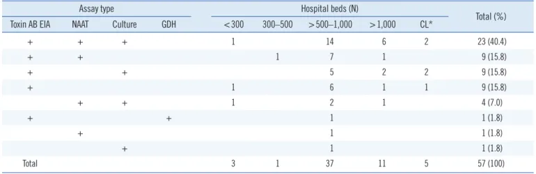
The various assay methods used in the participating laborato- ries are summarized in Table 1. The toxin AB EIA was the most popular assay. Among the 57 laboratories that reported perform- ing CDI assays, 51 (89.5%) used the toxin AB EIA, either alone or in combination with other assays. NAATs and C. difficile cul- ture, alone or in combination with other assays, were used in 37 (64.9%) laboratories. Only one laboratory used the GDH assay, which was conducted in combination with the toxin AB EIA. Forty- five (78.9%) laboratories used more than one assay. However, no laboratory reported performing the CCNA. Table 2 shows combinations of assay types for diagnosis of CDI according to the size of hospitals. Assay type (single or combination) did not significantly differ by hospital size.
Table 3 shows the median (range) of examined specimens in one month for the toxin AB EIA, NAATs, and C. difficile culture.
More specimens were examined with the toxin AB EIA than with
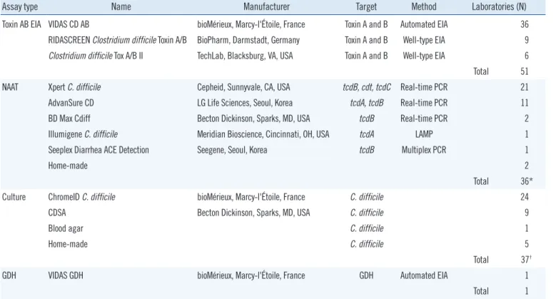
Table 1. Clostridium difficile assay methods and the numbers of laboratories that participated in the survey
Assay type Name Manufacturer Target Method Laboratories (N)
Toxin AB EIA VIDAS CD AB bioMérieux, Marcy-l'Étoile, France Toxin A and B Automated EIA 36
RIDASCREEN Clostridium difficile Toxin A/B BioPharm, Darmstadt, Germany Toxin A and B Well-type EIA 9
Clostridium difficile Tox A/B II TechLab, Blacksburg, VA, USA Toxin A and B Well-type EIA 6
Total 51
NAAT Xpert C. difficile Cepheid, Sunnyvale, CA, USA tcdB, cdt, tcdC Real-time PCR 21
AdvanSure CD LG Life Sciences, Seoul, Korea tcdA, tcdB Real-time PCR 11
BD Max Cdiff Becton Dickinson, Sparks, MD, USA tcdB Real-time PCR 2
Illumigene C. difficile Meridian Bioscience, Cincinnati, OH, USA tcdA LAMP 1
Seeplex Diarrhea ACE Detection Seegene, Seoul, Korea tcdB Multiplex PCR 1
Home-made 2
Total 36*
Culture ChromeID C. difficile bioMérieux, Marcy-l'Étoile, France C. difficile 24
CDSA Becton Dickinson, Sparks, MD, USA C. difficile 9
Blood agar C. difficile 1
Home-made C. difficile 5
Total 37†
GDH VIDAS GDH bioMérieux, Marcy-l'Étoile, France GDH Automated EIA 1
Total 1
*Two laboratories used two NAAT methods (Xpert C. difficile and AdvanSure CD, Xpert C. difficile and home-made). One laboratory did not specify the NAAT method; †Two laboratories used two culture methods (ChromeID and CDSA, CDSA and home-made).
Abbreviations: EIA, enzyme immunoassay; GDH, glutamate dehydrogenase assay; LAMP, loop-mediated isothermal amplification; NAAT, nucleic acid ampli- fication test; CDSA, C. difficile selective agar.
NAATs (P =0.021). In addition, although the same number of laboratories reported performing NAATs and C. difficile culture (N=37), there were more specimens examined with the latter method, though this difference was not significant. Moreover, the number of specimens examined using C. difficile culture was higher for hospitals with ≥1,000 beds than those with 500–1,000 beds (P =0.008). The number of examined specimens might reflect the disease burden of CDI in the hospital and/or the in- fection control policy, including the screening strategy for CDI, number of laboratory personnel, and reimbursement of medical insurance. The assays covered by medical insurance were per- formed more frequently.
Toxin AB EIA is more frequently used possibly because of its advantages of short turnaround time and cost-efficiency. How- ever, this assay is often criticized for its poor sensitivity and should therefore no longer be considered as a stand-alone assay for the
diagnosis of CDI [1, 2, 6, 7, 9-12]. Therefore, the nine (15.8%) laboratories that use only the toxin AB EIA for CDI diagnosis should reconsider their diagnostic strategy.
Since the clinical guidelines for CDI provided by the Infectious Diseases Society of America (IDSA) and Society for Healthcare Epidemiology of America (SHEA) were updated in 2010 [13], many hospitals in the United States have switched the toxin AB EIA to NAATs for CDI diagnosis. Wong et al. [10] reported that 84.5% of the hospitals surveyed in Ohio, USA, used NAATs as a stand-alone assay in 2014. However, the proportion of laborato- ries using NAATs as a stand-alone assay was lower in other coun- tries: only 3% and 6% of small (<500 beds) and large (>500 beds) hospitals in Italy in 2012–2013, respectively [11], 0.9%
of participating laboratories in Spain in 2013 [12], and 11.1%
(2/18) of hospitals in Israel in 2012 [14]. In general, NAATs are more commonly used in combination with other assays, as ob-
Table 2. Combinations of assays types for diagnosis of Clostridium difficile infection according to hospital size
Assay type Hospital beds (N)
Total (%) Toxin AB EIA NAAT Culture GDH <300 300–500 >500–1,000 >1,000 CL*
+ + + 1 14 6 2 23 (40.4)
+ + 1 7 1 9 (15.8)
+ + 5 2 2 9 (15.8)
+ 1 6 1 1 9 (15.8)
+ + 1 2 1 4 (7.0)
+ + 1 1 (1.8)
+ 1 1 (1.8)
+ 1 1 (1.8)
Total 3 1 37 11 5 57 (100)
*CLs were not classified according to size.
Abbreviations: CL, commercial laboratory; EIA, enzyme immunoassay; GDH, glutamate dehydrogenase; NAAT, nucleic acid amplification test.
Table 3. Numbers of specimens examined for CDI diagnosis according to assay types and hospital size per month in 2015 Size of hospital
(N of beds)
Toxin AB EIA NAAT Culture
Hospitals
(N) Specimens, median
(range) Hospitals
(N) Specimens, median
(range) Hospitals
(N) Specimens, median (range)
< 300 2 140 (80–200) 2 25 (10–40) 2 42 (10–74)
300–500 1 90 1 7 0
< 500–1,000 33 155 (50–489) 23 70 (10–373) 22 100 (9–200)
> 1,000 10 200 (80–750) 7 80 (35–300) 9 300 (80–750)
Subtotal 46 160 (50–750) 33 70 (7–370) 33 120 (9–750)
CL 5 568 (65–2,060) 2 365 (10–720) 4 140 (95–340)
Total 51 160 (50–2,060) 35* 70 (7–720) 37 130 (9–750)
*Two laboratories did not specify the number of specimens examined by the NAAT.
Abbreviations: see Table 2.
served in 16% and 34% of small and large hospitals in Italy in 2012–2013, respectively [11], 38.2% of participating laborato- ries in Spain in 2013 [12], and 38.9% (7/18) of hospitals in Is- rael in 2012 [14]. In our study, 36 of 57 (63.2%) of the labora- tories conducting CDI assays also used NAATs in combination with other assays, except for one laboratory (1.8%) that reported using NAATs as a stand-alone assay.
Approximately 60% (34/57) of the laboratories reported per- forming C. difficile culture, and the majority used chromogenic media for culture (Table 2), which has been reported to be more sensitive than conventional culture media [15-17]. The C. diffi- cile cultures performed in many laboratories are not TC, and thus, an additional toxin assay might be needed because not all C. difficile strains produce toxins [8]. The extent to which C. dif- ficile culture is used differs by region: for example, in a 2006 study, only six of the 25 (24%) participating laboratories in Ire- land performed C. difficile culture [18], whereas 19 of 24 (79%) Finnish laboratories performed it [19]. In 2012–2013, 25%
(38/151) and 37% (24/65) of small and large hospitals in Italy performed C. difficile culture, respectively, either alone or in combination with other assays [11]. Given the gap in time be- tween these aforementioned studies, the extent to which NAATs and C. difficile culture are used for laboratory diagnosis of CDI varies noticeably across countries. However, in our study, more than 80% of the participating hospitals (excluding CL) used NAATs and/or C. difficile culture with or without the toxin AB EIA. This finding may reflect the greater concern about CDI in Korean hospitals in recent years, which has resulted in the need for more rapid and sensitive diagnosis.
The GDH assay was only recently introduced in the last five years and approved for reimbursement in Korea since 2016.
Thus, this assay was not popular at the time of conducting the sur- vey, with only one laboratory reporting its use in combination with the toxin AB EIA. GDH has been reported as a sensitive marker for the detection of C. difficile and is recommended as a screening assay for CDI diagnosis [6, 7]; however, GDH-positive results should be followed by an assay to confirm toxin production [1, 2, 20].
The recently updated clinical guidelines for CDI by IDSA and SHEA recommend using a stool toxin assay as part of a multistep algorithm (i.e., GDH plus toxin; GDH plus toxin, arbitrated by NAAT; or NAAT plus toxin) rather than an NAAT alone for all specimens received in the clinical laboratory when there are no pre-agreed institutional criteria for patient stool submission. When there are pre-agreed institutional criteria for patient stool submis- sion, it is recommended to use an NAAT alone or a multistep al- gorithm for testing [6]. The European Society of Clinical Microbiol-
ogy and Infectious Diseases (ESCMID) strongly recommends us- ing a two-step algorithm instead of a single assay as a stand-alone assay. The algorithm should start with either the NAAT or GDH assay, and specimens with a positive first assay result should be tested further with the toxin AB EIA. An alternative algorithm is to screen specimens with both the GDH assay and toxin AB EIA [7].
Although approximately 80% of the laboratories in our study used more than one assay, we did not enquire about the sequen- ces and/or detailed processes used for multiple assays. The di- agnostic algorithms applied in Korean hospitals or laboratories are currently not clear; thus, further investigation is necessary to clarify this aspect.
In a survey conducted in Europe in 2014, 24 of the 35 re- sponding countries reported one or more changes in the na- tional/subnational laboratory diagnostics for CDI since 2011 [9].
The main changes included the availability of commercial diag- nostic assays, new or revised guidelines for CDI diagnostics, rel- evant legislation, and reimbursement policies for diagnostic as- says. The main barriers to applying appropriate assays accord- ing to the guidelines were financial restrictions, along with insuf- ficient reimbursement and trained staff [9]. Although this was not explicitly explored in our survey, a similar situation is expected to be occurring in Korea.
There were several limitations in this study. The number and area of participating laboratories were restricted. As mentioned above, the sequences and/or detailed processes used for multi- ple assays were not investigated, which are the important issues that need to be addressed in order to develop multistep algorith- mic approaches for diagnosis of CDI in Korea.
Despite these limitations, this study represents the first survey on the laboratory diagnosis for CDI conducted in Korea. We found considerable variation in the assays used for CDI diagnosis among laboratories in Korea, and some laboratories were still using in- appropriate methods such as the toxin AB EIA as a stand-alone assay. NAATs were more rapidly introduced than expected, uti- lized in approximately 65% of participating laboratories. These findings suggest the need for establishing optimized guidelines for CDI diagnosis in Korea. Thus, our study can provide valuable basic data on the current situation, as a first step towards stan- dardizing laboratory diagnosis of CDI in Korea.
Authors’ Disclosures of Potential Conflicts of Interest
No potential conflicts of interest relevant to this article were re- ported.
Ohio acute care hospitals. Am J Infect Control 2017;45:306-7.
11. Spigaglia P, Barbanti F, Morandi M, Moro ML, Mastrantonio P. Diagnos- tic testing for Clostridium difficile in Italian microbiological laboratories.
Anaerobe 2016;37:29-33.
12. Alcalá L, Reigadas E, Marín M, Martín A, Catalán P, Bouza E, et al. Im- pact of clinical awareness and diagnostic tests on the underdiagnosis of Clostridium difficile infection. Eur J Clin Microbiol Infect Dis 2015;34:
1515-25.
13. Cohen SH, Gerding DN, Johnson S, Kelly CP, Loo VG, McDonald LC, et al. Clinical practice guidelines for Clostridium difficile infection in adults:
2010 update by the society for healthcare epidemiology of America (SHEA) and the infectious diseases society of America (IDSA). Infect Control Hosp Epidemiol 2010;31:431-55.
14. Adler A, Schwartzberg Y, Samra Z, Schwartz O, Carmeli Y, Schwaber MJ, et al. Trends and changes in Clostridium difficile diagnostic policies and their impact on the proportion of positive samples: a national sur- vey. Clin Microbiol Infect 2014;20:O904-10.
15. Shin BM and Lee EJ. Comparison of chromID agar and Clostridium dif- ficile selective agar for effective isolation of C. difficile from stool speci- mens. Ann Lab Med 2014;34:15-9.
16. Yim JS, Hwang SM, Kim M, Lim HJ, Shin S, Chung HS, et al. Evaluation of a chromID C. difficile agar for the isolation of Clostridium difficile. Ko- rean. J Clin Microbiol 2012;15:88-91.
17. Han SB, Chang J, Shin SH, Park KG, Lee GD, Park YG, et al. Performance of chromID Clostridium difficile agar compared with BBL C. difficile se- lective agar for detection of C. difficile in stool specimens. Ann Lab Med 2014;34:376-9.
18. Fitzpatrick F, Oza A, Gilleece A, O’Byrne AM, Drudy D, C. difficile sub- committee of the Health Protection Surveillance Centre. Laboratory di- agnosis of Clostridium difficile-associated disease in the Republic of Ire- land: a survey of Irish microbiology laboratories. J Hosp Infect 2008;68:
315-21.
19. Könönen E, Rasinperä M, Virolainen A, Mentula S, Lyytikäinen O. Diag- nostic trends in Clostridium difficile detection in Finnish microbiology laboratories. Anaerobe 2009;15:261-5.
20. Shin BM, Lee EJ, Moon JW, Lee SY. Evaluation of the VIDAS glutamate dehydrogenase assay for the detection of Clostridium difficile. Anaerobe 2016;40:68-72.
Acknowledgments
We thank all the health professionals in clinical microbiology laboratories who responded to the survey.
REFERENCES
1. Burnham CA and Carroll KC. Diagnosis of Clostridium difficile infection:
an ongoing conundrum for clinicians and for clinical laboratories. Clin Microbiol Rev 2013;26:604-30.
2. Bagdasarian N, Rao K, Malani PN. Diagnosis and treatment of Clostrid- ium difficile in adults: a systematic review. JAMA 2015;313:398-408.
3. Leffler DA and Lamont JT. Clostridium difficile infection. N Engl J Med 2015;372:1539-48.
4. Lytvyn L, Mertz D, Sadeghirad B, Alaklobi F, Selva A, Alonso-Coello P, et al. Prevention of Clostridium difficile infection: a systematic survey of clini- cal practice guidelines. Infect Control Hosp Epidemiol 2016;37:901-8.
5. Kim YS, Han DS, Kim YH, Kim WH, Kim JS, Kim HS, et al. Incidence and clinical features of Clostridium difficile infection in Korea: a nation- wide study. Epidemiol Infect 2013;141:189-94.
6. McDonald LC, Gerding DN, Johnson S, Bakken JS, Carroll KC, Coffin SE, et al. Clinical practice guidelines for Clostridium difficile infection in adults and children: 2017 update by the Infectious Diseases Society of America (IDSA) and Society for Healthcare Epidemiology of America (SHEA). Clin Infect Dis 2018;66:e1-48.
7. Crobach MJ, Planche T, Eckert C, Barbut F, Terveer EM, Dekkers OM, et al. European Society of Clinical Microbiology and Infectious Diseases:
update of the diagnostic guidance document for Clostridium difficile in- fection. Clin Microbiol Infect 2016;22(S4):S63-81.
8. Surawicz CM, Brandt LJ, Binion DG, Ananthakrishnan AN, Curry SR, Gilligan PH, et al. Guidelines for diagnosis, treatment, and prevention of Clostridium difficile infections. Am J Gastroenterol 2013;108:478-98.
9. van Dorp SM, Notermans DW, Alblas J, Gastmeier P, Mentula S, Nagy E, et al. Survey of diagnostic and typing capacity for Clostridium difficile infection in Europe, 2011 and 2014. Euro Surveill 2016;21.
10. Wong KK, Choi B, Fraser TG, Donskey CJ, Deshpande A. Diagnostic testing methods for Clostridium difficile infection: a statewide survey of