Introduction
Recently, digital radiography has superseded film-based radiography because of its advantages. The lower diagnos- tic dose, no need for chemical development and fixation, the possibility of processing and analysis, image enhance- ment capacity, and image transfer are the main advantages of digital radiography.1-3Several studies have shown that digital radiography has equal or better diagnostic accuracy compared to film-based radiography;4-12therefore, digital radiography is widely used in dentistry, especially in endo- dontics.
Manipulation of images is one of the most important
properties of digital radiography.13,14 Several digital pro- cessing algorithms are available that enhance the display quality of digital images.14,15Since digital sensors produce a large amount of noise, different image processing schemes have been developed to improve image quality.16 Noise reduction with the use of different techniques might result in higher signal-to-noise ratios. For medical imaging, a high signal-to-noise ratio is considered essential for clini- cal diagnosis.16Noise reduction, sharpening-smoothening, and edge enhancement are examples of these algorithms.
It has been accepted that reduction of the noise value has a significant effect on the improvement of digital image quality. However, this process can lead to loss of some small structures on images.
Several studies have evaluated the use of various digital enhancement algorithms and their effects on diagnostic accuracy.17-25However, there has been only limited research on the task-specific use of noise reduction algorithms.20,26-30
Effect of digital noise reduction on the accuracy of endodontic file length determination
Mojdeh Mehdizadeh1, Abbas Ali Khademi2, Ali Shokraneh2, Nastaran Farhadi1,*
1Department of Oral and Maxillofacial Radiology, School of Dentistry, Isfahan University of Medical Sciences, Isfahan, Iran
2Department of Endodontics, School of Dentistry, Isfahan University of Medical Sciences, Isfahan, Iran
ABSTRACT
Purpose: The aim of the present study was to evaluate the measurement accuracy of endodontic file length on peri- apical digital radiography after application of noise reduction digital enhancement.
Materials and Methods: Thirty-five human single-rooted permanent teeth with canals measuring 20-24 mm in length were selected. ISO #08 endodontic files were placed in the root canals of the teeth. The file lengths were measured with a digital caliper as the standard value. Standard periapical digital images were obtained using the Digora digital radiographic system and a dental X-ray unit. In order to produce the enhanced images, the noise reduction option was applied. Two blinded radiologists measured the file lengths on the original and enhanced images. The measurements were compared by repeated measures ANOVA and the Bonferroni test (α==0.05).
Results: Both the original and enhanced digital images provided significantly longer measurements compared with the standard value (P⁄0.05). There were no significant differences between the measurement accuracy of the origi- nal and enhanced images (P¤0.05).
Conclusion: Noise reduction digital enhancement did not influence the measurement accuracy of the length of the thin endodontic files on the digital periapical radiographs despite the fact that noise reduction could result in the elimination of fine details of the images. (Imaging Sci Dent 2013; 43: 185-90)
KEY WORDS: Radiography, Dental, Digital; Endodontics; Noise; Radiographic Image Enhancement
Received February 11, 2013; Revised February 21, 2013; Accepted March 9, 2013
*Correspondence to : Dr. Nastaran Farhadi
Torabinejad Dental Research Center and Department of Oral and Maxillofacial Radiology, School of Dentistry, Isfahan University of Medical Sciences, Hezar Jirib Street, Isfahan 81746-73461, Iran
Tel) 98-913-210-0152, Fax) 98-311-668-7080, E-mail) [email protected]
Copyright ⓒ 2013 by Korean Academy of Oral and Maxillofacial Radiology
This is an Open Access article distributed under the terms of the Creative Commons Attribution Non-Commercial License (http://creativecommons.org/licenses/by-nc/3.0) which permits unrestricted non-commercial use, distribution, and reproduction in any medium, provided the original work is properly cited.
Imaging Science in Dentistry∙pISSN 2233-7822 eISSN 2233-7830
Some of these studies have shown that this type of enhance- ment does not change the diagnostic accuracy;20,29on the other hand, others have shown the opposite.26-28,30
The effect of this enhancement on the measurement accuracy is particularly important in endodontics. The accuracy of measurement can influence the final verifica- tion of the outcome of root canal treatment by affecting the precise working length, and consequently, the poten- tially successful treatment. Therefore, the aim of the pre- sent study was to evaluate whether the length measure- ment of small endodontic files can be affected by applica- tion of noise reduction enhancement or not.
Materials and Methods
Thirty-five human-extracted single-rooted permanent
teeth with intact crowns and roots were used in this expe- rimental study. The teeth had only one root canal, mea- suring 20-24 mm in length. Periodontal or prosthetic pro- blems were the reasons for extraction of the teeth. The samples were cleaned and disinfected by scaling and soak- ing in 0.5% sodium hypochlorite for 12 h and stored in distilled water at 4�C during the study. Conventional peri- apical radiography was used to verify any abnormalities or pathologies, such as internal/external root resorption, root canal obliteration/calcification, or severe curvature.
An anatomic access cavity was prepared with #008 and
#010 fissure burs and an Endo-Z bur (Dentsply Maillefer, Ballaigues, Switzerland) in a high-speed handpiece. Gates- Glidden #3 and #4 drills (Dentsply Maillefer, Ballaigues, Switzerland) were used to enlarge the coronal and middle thirds of the root canals. ISO #08 K-files (Mani Inc., Utsu-
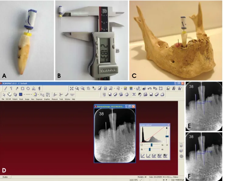
Fig. 1.A. Insertion of an endodontic file into the canal. B. File length measurement (standard value). C. Placement of tooth into cadaver socket. D. Scanora software. E. Original image. F. enhanced image.
A B C
D
E
F
nomiya, Japan) were inserted into the canals until the tips of the files were just visible at the apical foramen (Fig.
1A). The file length was measured with a digital caliper to the nearest 0.01 millimeter (Guilin Guanglu Measuring Instrument Co., Ltd., Guilin, China), and the file was short- ened by 0.5 mm, which was set as the standard value for the endodontic file length (Fig. 1B). Only teeth with a length of 20-24 mm were included. Then, the files were inserted into the canals again and fixed with flowable com- posite resin (Tetric®Flow, Ivoclar Vivadent AG, Schaan, Liechtenstein). Afterward, the samples were placed in suitable sockets of a dry human mandible to reproduce the bone density.
In order to eliminate the radiographic magnification, a 10-mm round orthodontic wire was placed in the adjacent dental socket and fixed with wax. A Rinn-Endo-Ray film holder (Dentsply/Rinn Corporation, Elgin, IL, USA) was used to ensure parallelism. The standard geometric confi- guration was fixed at a 30-cm source-to-object distance.
Radiographic images of each sample were obtained with Digora storage phosphor plates (Soredex Corp, Helsinki, Finland) and its special scanner, the Digora Optime (Sore- dex Corp, Helsinki, Finland), using a Prostyle dental X- ray unit (Planmeca OY, Helsinki, Finland) operating at 63 kVp, 8 mA, and 1.5-mm Al-equivalent filtration for 0.03 s.
The digital images were numbered by Scanora 5.0 soft- ware (Soredex Corp, Helsinki, Finland) (Fig. 1D) in a semi-dark room and saved in the DICOM format for fur- ther processing and analysis (Fig. 1E). The noise of each image was reduced by single clicking on the “noise reduc- tion” option on the diagnostic tools menu bar on the top of the operating window of the Scanora software, and the resultant image was saved again in the same format (Fig.
1F). Then, two blinded radiologists with 5 years or more experience in the interpretation of digital radiographs, identified the endodontic file tip and the most apical por- tion of the rubber stop of each file in the original and en- hanced digital images. They also determined the most coronal and apical point of the orthodontic wire in the adjacent dental socket. The endodontic file and orthodon-
tic wire lengths were measured by a third person using the measurement tool of the software to the nearest 0.1 mm.
The magnification coefficient of each image was deter- mined using real and radiographic orthodontic wire lengths.
To eliminate the magnification effect of the radiography, the obtained endodontic file length for each image was divided by the magnification coefficient. Then, the mean values of the radiologist measurements were taken as data.
The data were first verified with the Kolmogorov-Smir- nov test for the normality of data distribution. Repeated measures ANOVA and the Bonferroni test were used to compare the standard value and radiographic file length with and without enhancement. A SPSS software (ver. 11.0, SPSS Inc., Chicago, IL, USA) was used for analysis. Sta- tistical significance was set at a confidence level of 95%.
Also, Cohen’s kappa statistic was used to assess inter- observer reliability.
Results
Repeated measures ANOVA showed significant differ- ences between the standard, original, and enhanced images in the endodontic file length measurements (P⁄0.05). The
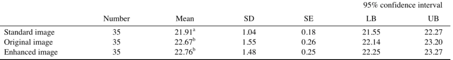
Table 1.Statistical characteristics of the three groups
95% confidence interval
Number Mean SD SE LB UB
Standard image 35 21.91a 1.04 0.18 21.55 22.27
Original image 35 22.67b 1.55 0.26 22.14 23.20
Enhanced image 35 22.76b 1.48 0.25 22.25 23.27
Means with different superscript letters are statistically different (P⁄0.05).
SD==standard deviation, SE==standard error, LB==lower bound, UB==upper bound
Fig. 2. Box plot of endodontic file length in the standard value and on original and enhanced digital radiographs.
26.00
25.00
24.00
23.00
22.00
21.00
20.00
Gold standard Original image Enhanced image Group
Endodontic file length
Bonferroni test showed no significant differences between the two sets of digital radiographs, suggesting no improve- ment or reduction in the linear measurement accuracy with noise reduction digital enhancement (P¤0.05); however, there was a tendency to overestimate the file length deter- mination by digital enhancement. Additionally, there were significant differences between the two sets of digital radio- graphs and the standard value (P⁄0.05). Both the original and enhanced digital images had a tendency to overesti- mate the file length measurements (P⁄0.05). The results are summarized in Table 1 and Figure 2.
In addition, inter-observer agreement was excellent for the assessment of digital images with and without enhance- ment, showing a kappa value of 0.86.
Discussion
The main aim of digital processing is to create images with sufficient detail. This is possible by displaying infor- mation that has been collected during imaging but is not visible.19Although processing should be able to improve diagnostic signals, some of the information might be lost during the process.18,19,23,31Therefore, digital enhancement should be used with caution, based on the diagnostic task.
This task-specific use of enhancements requires accurate investigation. The present study was the first one under- taken to evaluate the influence of noise reduction enhance- ment on the measurement accuracy of endodontic file length on images acquired using Digora storage phosphor plates. The accuracy was assessed by comparing the posi- tion of reproducible landmarks in the original and process- ed images.
The results of the present study showed that noise reduc- tion does not decrease the accuracy of file length measure- ment. From the clinical viewpoint, this option does not result in the loss of subtle information, including the posi- tion of the tip of a #08 endodontic file. This result is con- sistent with the results reported by Koob et al20and Haak and Wicht.29They showed that noise reduction does not change the diagnostic quality of images in identification of interproximal caries. In addition, Koob et al20reported that there were no significant differences between images with and images without noise reduction in determination of interproximal caries depth. In contrast, in three studies,26-28 Davies demonstrated that the use of noise elimination fil- ters resulted in size and shape distortions. Furthermore, Xu and Lai30reported that these distortions occurred in fine details such as the tip of endodontic files, particularly with
#06 and #08 files. In addition, Brullmann et al16,32evaluat-
ed noise reduction in two different situations. In the first study,16it was illustrated that noise filtering had no effect on the accuracy of length measurement of files greater than #10, but this enhancement resulted in underestima- tion of the length of #10 and smaller endodontic files. This is in contrast with the result of the present study showing overestimation in the length measurement. This contro- versy may be due to differences in the study design such as the use of charge-coupled device (CCD) digital radio- graphy rather than a PSP system, the use of digital soft- ware (Sidexis XG 2.4, Sirona, Bensheim, Germany) with a special filtering program (Borland C-Builder 6.0, Bor- land GmbH, Langen, Germany) rather than Scanora soft- ware, the presence of a marker in the apical portion of the root of the teeth that helps in the identification of the tip of the endodontic file, and the existence of the soft tissue of the mandible resulting in more scatter radiation and noise in that study. In the second study,32 it was shown that this filter was effective in the elimination of noise without the loss of diagnostic information but did not in- crease the odds of identification of dental root fracture.
Some studies have addressed subjective image quality with and without noise reduction based on observer per- formance.29,33,34 Their major limitation was reliance on subjective assessment of images. For example, Yalcin- kaya et al33compared the display quality of anatomical structures on conventional and digital periapical and pano- ramic radiographs. They reported that conventional radio- graphy was superior to unfiltered and noise-reduced filter- ed digital images, with no statistically significant differ- ences between filtered and unfiltered digital images. How- ever, it has been suggested that it may be more important to use a computable objective measure to predict diagnos- tic accuracy rather than subjective assessments. Näslund et al34 reported on both objective and subjective evalua- tions of image quality with and without post-processing noise reduction. The authors basically compared the ability of localization of anatomic landmarks on standard expos- ed, low-exposed, and a combination of low-exposed and noise-reduced digital cephalograms. They reported that the landmarks were identified more effectively on the low- exposed images than on the images with post-processing noise reduction, although the subjective evaluation of the image quality indicated the opposite. The standard-expos- ed digital cephalograms were the best in objective and subjective quality evaluations. The results of the objective evaluation contrasted with the result of the present study.
In addition, the results of the present study showed that both enhanced and unenhanced digital images tended to
overestimate the length of endodontic files, consistent with the results of studies by Mehdizadeh et al35and Williams et al.36In contrast, Schmitd et al37and Brito-Junior et al38 reported that the measurement accuracy of digital images was comparable to real measurements.
It is undisputed that the diagnostic impact of digital imag- ing depends on the task, the quality of source data, and the kind of image processing applied.19 Therefore, in a clinical situation, discrimination of small file tips may become more difficult. Selection of optimal exposure time is important because under-exposed images have a higher amount of noise.39In addition, the presence of soft tissues and the increase in the volume and density of hard tissues in the clinical situation results in a greater amount of scat- tered radiation, increasing the amount of noise. Therefore, clinical studies are suggested for further evaluation of the usefulness of noise reduction digital enhancement in prac- tice.
In addition, although a storage phosphor plate system was used in the present study, most endodontists benefit from the advantages of solid-state detectors to obtain peri- apical radiographs during root canal treatment. Therefore, the application of solid-state detectors is also recommend- ed for further investigation.
In conclusion, noise reduction enhancement did not change the measurement accuracy of endodontic file length on digital images. Therefore, it could be used, depending on the demands of the practitioner.
References
1. Vandenberghe B, Jacobs R, Bosmans H. Modern dental imag- ing: a review of the current technology and clinical applica- tions in dental practice. Eur Radiol 2010; 20: 2637-55.
2. Gormez O, Yilmaz HH. Image post-processing in dental prac- tice. Eur J Dent 2009; 3: 343-7.
3. Parks ET, Williamson GF. Digital radiography: an overview.
J Contemp Dent Pract 2002; 3: 23-39.
4. Almenar García A, Forner Navarro L, Ubet Castelló V, Mi~nana Laliga R. Evaluation of a digital radiography to estimate working length. J Endod 1997; 23: 363-5.
5. Kositbowornchai S, Nuansakul R, Sikram S, Sinahawattana S, Saengmontri S. Root fracture detection: a comparison of direct digital radiography with conventional radiography. Den- tomaxillofac Radiol 2001; 30: 106-9.
6. Kamburo˘glu K, Barenboim SF, Kaffe I. Comparison of con- ventional film with different digital and digitally filtered ima- ges in the detection of simulated internal resorption cavities - an ex vivo study in human cadaver jaws. Oral Surg Oral Med Oral Pathol Oral Radiol Endod 2008; 105: 790-7.
7. Borg E, Gröndahl K, Persson LG, Gröndahl HG. Marginal bone level around implants assessed in digital and film radio-
graphs: in vivo study in the dog. Clin Implant Dent Relat Res 2000; 2: 10-7.
8. Syriopoulos K, Sanderink GC, Velders XL, van der Stelt PF.
Radiographic detection of approximal caries: a comparison of dental films and digital imaging systems. Dentomaxillofac Radiol 2000; 29: 312-8.
9. Akdeniz B, So˘gur E. An ex vivo comparison of conventional and digital radiography for perceived image quality of root fillings. Int Endod J 2005; 38: 397-401.
10. Naoum HJ, Chandler NP, Love RM. Conventional versus stor- age phosphor-plate digital images to visualize the root canal system contrasted with a radiopaque medium. J Endod 2003;
29: 349-52.
11. Kavadella A, Karayiannis A, Nicopoulou-Karayianni K. De- tectability of experimental peri-implant cancellous bone lesions using conventional and direct digital radiography. Aust Dent J 2006; 51: 180-6.
12. Mohtavipour ST, Dalili Z, Azar NG. Direct digital radiography versus conventional radiography for estimation of canal length in curved canals. Imaging Sci Dent 2011; 41: 7-10.
13. Li G. Comparative investigation of subjective image quality of digital intraoral radiographs processed with 3 image-process- ing algorithms. Oral Surg Oral Med Oral Pathol Oral Radiol Endod 2004; 97: 762-7.
14. van der Stelt PF. Filmless imaging: the uses of digital radio- graphy in dental practice. J Am Dent Assoc 2005; 136: 1379- 87.
15. van der Stelt PF. Better imaging: the advantages of digital radiography. J Am Dent Assoc 2008; 139 Suppl: 7S-13S.
16. Brüllmann DD, Röhrig B, Sulayman SL, Schulze R. Length of endodontic files measured in digital radiographs with and without noise-suppression filters: an ex-vivo study. Dento- maxillofac Radiol 2011; 40: 170-6.
17. Castro V, Katz J, Hardman P, Glaros A, Spencer P. In vitro comparison of conventional film and direct digital imaging in the detection of approximal caries. Dentomaxillofac Radiol 2007; 36: 138-42.
18. Eickholz P, Riess T, Lenhard M, Hassfeld S, Staehle HJ. Digi- tal radiography of interproximal bone loss; validity of differ- ent filters. J Clin Periodontol 1999; 26: 294-300.
19. Kal BI, Baksi BG, Dündar N, Sen BH. Effect of various digi- tal processing algorithms on the measurement accuracy of endo- dontic file length. Oral Surg Oral Med Oral Pathol Oral Radiol Endod 2007; 103: 280-4.
20. Koob A, Sanden E, Hassfeld S, Staehle HJ, Eickholz P. Effect of digital filtering on the measurement of the depth of prox- imal caries under different exposure conditions. Am J Dent 2004; 17: 388-93.
21. Sund T, Møystad A. Sliding window adaptive histogram equa- lization of intraoral radiographs: effect on image quality. Dento- maxillofac Radiol 2006; 35: 133-8.
22. Møystad A, Svanaes DB, Risnes S, Larheim TA, Gröndahl HG. Detection of approximal caries with a storage phosphor system. A comparison of enhanced digital images with dental X-ray film. Dentomaxillofac Radiol 1996; 25: 202-6.
23. Raitz R, Assunção Junior JN, Fenyo-Pereira M, Correa L, de Lima LP. Assessment of using digital manipulation tools for diagnosing mandibular radiolucent lesions. Dentomaxillofac
Radiol 2012; 41: 203-10.
24. Shrout MK, Russell C, Potter B, Powell B, Hildebolt C. Dig- ital enhancement of radiographs: can it improve caries diag- nosis? J Am Dent Assoc 1996; 127: 469-73.
25. de Molon RS, Morais-Camillo JA, Sakakura CE, Ferreira MG, Loffredo LC, Scaf G. Measurements of simulated periodontal bone defects in inverted digital image and film-based radio- graph: an in vitro study. Imaging Sci Dent 2012; 42: 243-7.
26. Davies ER. Edge location shifts produced by median filters:
theoretical bounds and experimental results. Signal Processing 1989; 16: 83-96.
27. Davies ER. Median and mean filters produce similar shifts on curved boundaries. Electron Lett 1991; 27: 826-8.
28. Davies ER. Formulation of an accurate discrete theory of me- dian shifts. Signal Processing 2003; 83: 531-44.
29. Haak R, Wicht MJ. Grey-scale reversed radiographic display in the detection of approximal caries. J Dent 2005; 33: 65-71.
30. Xu Y, Lai EM. Restoration of images contaminated by mixed Gaussian and impulse noise using a recursive minimum-maxi- mum method. IEE Proc Vis Image Signal Process 1998; 145:
264-70.
31. Nair MK, Nair UP. Digital and advanced imaging in endo- dontics: a review. J Endod 2007; 33: 1-6.
32. Brüllmann D, Witzel V, Willershausen B, d’Hoedt B. Effect of digital noise filters on diagnostic radiographs for the diag- nosis of experimental root fractures. Int J Comput Dent 2008;
11: 107-14.
33. Yalcinkaya S, Künzel A, Willers R, Thoms M, Becker J. Sub- jective image quality of digitally filtered radiographs acquired by the Dürr Vistascan system compared with conventional radiographs. Oral Surg Oral Med Oral Pathol Oral Radiol Endod 2006; 101: 643-51.
34. Näslund EB, Møystad A, Larheim TA, Øgaard B, Kruger M.
Cephalometric analysis with digital storage phosphor images:
extreme low-exposure images with and without postprocess- ing noise reduction. Am J Orthod Dentofacial Orthop 2003;
124: 190-7.
35. Mehdizadeh M, Khademi AA, Nasr N. Canal length measure- ment by digital radiography and conventional parallel radio- graphy. Res J Biol Sci 2010; 5: 400-3.
36. Williams CB, Joyce AP, Roberts S. A comparison between in vivo radiographic working length determination and measure- ment after extraction. J Endod 2006; 32: 624-7.
37. Schmitd LB, Lima Tde C, Chinellato LE, Bramante CM, Gar- cia RB, de Moraes IG, et al. Comparison of radiographic mea- surements obtained with conventional and indirect digital imaging during endontic treatment. J Appl Oral Sci 2008; 16:
167-70.
38. Brito-Júnior M, Santos LA, Baleeiro EN, Pêgo MM, Eleutério NB, Camilo CC. Linear measurements to determine working length of curved canals with fine files: conventional versus digital radiography. J Oral Sci 2009; 51: 559-64.
39. Macdonald R. Digital imaging for dentists. Aust Dent J 2001;
46: 301-5.