*Corresponding Author. Tel: 82-31-219-5070; Fax: 82-31-219-5079; E-mail: sinsun@ajou.ac.kr
#These authors contributed equally to this work.
This work was supported by the Korea Science and Engineering Foundation (KOSEF) through the Chronic Inflammatory Disease Research Center (CIDRC), Ajou University, grant R13-2003-019.
Keywords: Interleukin 29 protein, monoclonal antibody
Seung-Ho Hong#, Jung-Sik Kim# and Sun Park*
Department of Microbiology and Immunology, Ajou University School of Medicine, Suwon, Korea
Background: Members belonging to the interferon-lambda (IFN-λ) family exert protective action against viral infection;
however, the mechanisms of their action have remained elusive. To study IFN-λ biology, such as endocytosis of IFN- λ, we produced monoclonal antibodies (Abs) against human IFN-λ and examined their usefulness. Methods: We purified recombinant human IFN-λ1 expressed in Escherichia coli by using affinity columns. Then, we generated hybridoma cells by fusing myeloma cells with splenocytes from IFN-λ1- immunized mice. For evaluating the neutralizing activity of the monoclonal Abs against IFN-λ1, we performed RT-PCR for the MxA transcript. In order to study the binding activity of IFN-λ and the monoclonal Ab complex on HepG2 cells, we labeled the monoclonal Ab with rhodamine and deter- mined the fluorescence intensity. Results: Four hybridoma clones secreting Abs specific to IFN-λ1 were generated and designated as HL1, HL2, HL3, and HL4. All the Abs reacted with IFN-λ1 in the denatured form as well as in the native form. Abs produced by HL1, HL3, and HL4 did not neutralize the induction of the MxA gene by IFN-λ1. We also de- monstrated the binding of the HL1 monoclonal anbitody and IFN-λ complex on HepG2 cells. Conclusion: Monoclonal Abs against IFN-λ1 were produced. These Abs can be used to study the cellular binding and internalization of IFN-λ.
[Immune Network 2008;8(1):7-12]
INTRODUCTION
Interferon-lambdas (IFN-λs) have recently been described as cytokines that bind to a distinct class II cytokine receptor complex composed of CRF2-12 and IL-10R2 (1–3). The sub-
types of the IFN-λ family are IFN-λ1, IFN-λ2, and IFN-λ3.
These 3 IFN-λ genes are located on chromosome 19 and en- code for 20-kDa monomeric proteins that are secreted (1).
IFN-λ1 has a potential site for N-linked glycosylation that is not present in IFN-λ2 or IFN-λ3. (1) IFN-λ1 shows 81%
identity with IFN-λ2 that is 96% homologous to IFN-λ3 (4).
Although the biological activities of IFN-λ have not been completely explored, its antiviral and immunomodulatory functions have been demonstrated. IFN-λs have suppressive activity against the encephalomyocarditis virus in in vitro as- says and against the herpes simplex type 2 virus in vivo (5).
Further, IFN-λ inhibits the replication of hepatitis B and C viruses (6,7). In relation to the antiviral activity of IFN-λ, the induction of several genes, including that of the 2′5′oligoa- denylation synthetase (2′5′-OAS) and MxA genes, has been demonstrated (8,9). Further, IFN-λs have been shown to generate tolerogenic dendritic cells (DCs) in vitro (10). IFN-λ -treated DCs expressing high levels of major histocompati- bility complex molecules with low levels of costimulatory molecules specifically induced the proliferation of the CD4+CD25+Foxp3+ T-cell subset and suppressed T-cell pro- liferation (10). IFN-λ1 also affects CD4 T cell functions. In the presence of IFN-λ1, Th cells stimulated with a mitogen or allogeneic DCs secrete markedly diminished levels of inter- leukin-13 (11). IFN-λ together with IFN-α mediates the sup- pression of CD4 T cells that are induced following infection with respiratory syncytial virus (12).
Several stimuli that activate IFN-λ genes and the mecha- nisms behind it have been studied but not completely elucidated. IFN-λ genes are expressed in cells along with other type I IFNs following viral infection (1,2). In addition
to viral stimulation, treatment with polyinosine-polycytidylic acid (poly(I·C)) or lipopolysaccharide induces IFN-λ ex- pression (13). In blood cells, IFN-λ expression is induced via Toll-like receptor (TLR)-7, TLR-8, and TLR-9 in an inter- leukin receptor-associated kinase (IRAK)-4-dependent manner but via TLR-3 and TLR-4 in an IRAK-4 independent manner (14). However, IFN-λ induction in fibroblasts by TLR-3 ago- nists and viruses is independent of IRAK-4 (14). Several regu- lators such as retinoic acid-inducible gene I (RIG-I), IFN pro- moter stimulator-1 (IPS-1), TANK-binding kinase 1 (TBK1), and interferon regulatory factor (IRF)-3 that are involved in the virus-induced activation of IFN-λ genes have been identi- fied (15). The IFN-λ1 gene is regulated by virus-activated IFN regulatory factor 3 (IRF3) and IRF7, whereas IFN-λ2 and IFN-λ3 gene expression is mainly controlled by IRF7 (16).
Although virtually any cell type following viral infection can express IFN-λ, DCs, particularly plasmacytoid DCs, appear to be the major producers of IFN-λ (13,17). Most of these studies analyzed the transcriptional levels of IFN-λ without analyzing the protein levels of IFN-λ.
In order to obtain monoclonal antibodies (Abs) that enable us to measure the production of IFN-λ protein and to study IFN-λ endocytosis, we generated hybridoma clones and ex- amined the reactivity of their monoclonal Abs against IFN-λ1.
MATERIALS AND METHODS Plasmids and protein purification
For the production of human recombinant IFN-λ1, we used 2 prokaryotic expression vectors: pMAL-IFN-λ1 (1) and pPROEX-IFN-λ1. pMAL-IFN-λ1 contains the cDNA of hu- man IFN-λ1 fused to the gene encoding the maltose binding protein (MBP). pPROEX-IFN-λ1 contains the cDNA of hu- man IFN-l1 tagged with histidine (His). Recombinant MBP- IFN-λ1 was purified using an amylose affinity column (New England Biolabs, Beverly, MA) and fast protein liquid chroma- tography (FPLC) (Amersham Biosciences Co., Piscataway, NJ, USA). Recombinant His-tagged IFN-λ1 was purified using a nickel-nitrilotriacetic acid (Ni-NTA) column (Qiagen, Valencia, CA, USA). Monoclonal Abs were purified using a protein A/G sepharose column (Amersham Biosciences Co.).
Generation of hybridoma cells
To develop B-cell hybridomas that secrete murine mono- clonal Abs against IFN-λ1, BALB/c mice (Daehan Biolink
Co., Ltd, Chungbuk, Korea) were subcutaneously injected with 50μg of His-tagged IFN-λ1 emulsified in complete Freund’s adjuvant (Sigma., St. Louis, MO, USA) and then sub- cutaneously injected with 25μg of His-tagged IFN-λ1 emul- sified in incomplete Freund’s adjuvant (Sigma) biweekly.
Three days prior to fusion, the mice were intraperitoneally injected with 50μg of MBP-IFN-λ1. Their splenocytes were fused with cultured F0 myeloma cells using 50% polyethylene glycol (Sigma). Hybridomas were screened for the production of Abs against IFN-λ1 by enzyme-linked immunosorbent as- say (ELISA) and cloned by limiting dilution.
ELISA
To screen hybridoma cells, each well of 96-well microtiter plates was coated with 100μl of 10μg/ml of His-tagged IFN- λ1, MBP-IFN-λ1, and MBP each and then blocked with 3%
BSA. The hybridoma supernatant (100μl) was added to each well, and the plate was incubated for 1 h at room temper- ature and washed 3 times with PBS containing 0.05%
Tween-20 (PBST). Alkaline phosphatase-conjugated goat an- ti-mouse IgG (100μl) (Pierce, Rockford, IL, USA) was added to the microtiter plate. One hour later, the plate was washed and ρ-nitrophenyl phosphate (Sigma) was added. Absor- bance at 415 nm was determined using the Microplate reader model 680 (Bio-Rad, Melville, NY, USA).
To determine the detection limit of ELISA for IFN-λ1, ELISA was performed as described above, using various con- centrations of IFN-λ1 (R&D systems Inc., Minneapolis, MN, USA) as a coating antigen and 10μg/ml of purified mono- clonal Abs as the primary Ab.
Western blot analysis
For western blot analysis, purified proteins were resolved on 12% sodiumdodecyl sulfate (SDS)-polyacrylamide gels and transferred to a polyvinylidene fluoride (PVDF) membrane (Millipore co. Bedford, MA, USA). The membrane was blocked in 10% powdered milk-PBS and incubated for 1 h at room temperature with the hybridoma culture supernatants or purified Abs. After washing the membrane with PBST, the membrane was incubated for 1 h at room temperature with horseradish peroxidase (HRP)-conjugated goat anti-mouse IgG. The membrane was washed 5 times with PBST and de- veloped using enhanced chemical luminescence (ECL) sol- ution (Amersham Biosciences Co.).
SDS-PAGE Western blot SDS-PAGE Western blot
Marker MBP
MBP-IFN-λ1 75kDa
50kDa
30kDa
MBP MBP-IFN-λ1
IFN-λ1-His Marker
(A) MBP-IFN-λ1 (B) His-IFN- λ1
IFN-λ1-His
20kDa 30kDa
SDS-PAGE Western blot SDS-PAGE Western blot
Marker MBP
MBP-IFN-λ1 75kDa
50kDa
30kDa
MBP MBP-IFN-λ1
IFN-λ1-His Marker
(A) MBP-IFN-λ1 (B) His-IFN- λ1
IFN-λ1-His
20kDa 30kDa
Figure 1. The purification of IFN-λ1 in E. coli. (A) INF-l1 fused with MBP was purified using amylose column chromatography and analyzed by SDS-PAGE (left) and western blotting (right). Western blotting was performed with rabbit anti-MBP Ab and HRP-conjugated anti-rabbit Ig Ab. (B) His-tagged IFN-λ1 was purified using a Ni-NTA column and analyzed by SDS-PAGE (left) and western blotting (right).
Western blotting was performed with mouse anti-His Ab and HRP- conjugated anti-mouse Ig Ab.
(A)
? 23kD
HL1 HL2 HL3 - +
Ab : (B)
HL4 (A)
? 23kD
HL1 HL2 HL3 - +
Ab : (B)
HL4
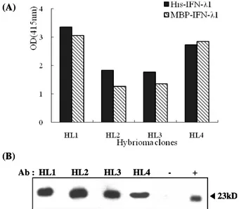
Figure 2. The binding activities of monoclonal Abs against IFN-λ1.
(A) The reactivity of the supernatant of the hybridoma clones to IFN-λ 1 was examined through indirect ELISA by using recombinant MBP-IFN-λ1 (1μg/well) and His-tagged IFN-λ1 (1μg/well) as Ags.
(B) The reactivity of the supernatant of the hybridoma clones to denatured His-tagged IFN-λ1 (50 ng/lane) was examined through- western blotting using HRP-conjugated anti-mouse Ig Ab. (–): mye- loma cell culture supernatant (+): rabbit polyclonal anti-IFN-λ1 Ab.
Assessment of IFN-λ1 neutralization of monoclonal Abs
Neutralization activity of monoclonal Abs against MxA gene induction by IFN-λ1 was evaluated using Huh-7 cells. The mixture of IFN-λ1 (5 ng/ml) and monoclonal Ab (800 ng/ml) was pre-incubated for 1 h at 4oC and then added to Huh-7 cells (5 × 10⁴/ml). After a 6-h incubation, total RNA was ex- tracted using RNA STAT-60 (Biogenesis Ltd, Poole, Dorset, UK) according to the manufacturer’s instructions. Reverse transcription of RNA was carried out at 42oC using the SuperscriptTM reverse transcription system (Invitrogen, Carsbade, CA, USA). PCR amplification of the MxA DNA frag- ment was performed by adding 2.5 mM dNTPs, Taq DNA polymerase (Bioneer, Daejeon, Korea), and 10μM of sense and antisense primers (9) under the following conditions: de- naturation for 5 minutes at 94oC, followed by 30 cycles with 30 s at 94oC, 30 s at 55oC, 60 s at 72oC, and a final extension for 10 min at 72oC. PCR products were analyzed in a 1% agar- ose gel.
Labeling of monoclonal Ab with rhodamine and immunofluorescence microscopy
HL1 Ab (230μg) was labeled with rhodamine using the FluoReporter Rhodamine Red-X Protein Labeling Kit (Molecu- lar Probes, Eugene, OR, USA). The HepG2 cell suspension (2 × 10⁵) was incubated in 1% BSA-1mM EDTA-PBS buffer
with or without 100 ng/ml of MBP-IFN-λ at 4oC for 30 min and then washed twice with 2% BSA PBS buffer. Then, the HepG2 cells were stained with rhodamine-conjugated HL1 Ab at 4oC for 30 min. After washing, the cells were fixed with 2% paraformaldehyde, and the fluorescence intensity was measured using a fluorescence reader (Molecular Device).
The cells were transferred to a 6-well plate and observed un- der a fluorescence microscope (Zeiss).
RESULTS AND DISCUSSION Purification of IFN-λ1
In order to immunize mice, we produced 2 forms of IFN-λ1.
The purified MBP-IFN-λ1 and His-tagged IFN-λ1 were sub- jected to SDS-polyacrylamide gel electrophoresis (PAGE) and western blotting (Fig. 1). In SDS-PAGE, 2 bands of approx- imately 65 kDa, which corresponds to MBP-IFN-λ1, were observed (Fig 1A). In western blotting, both the bands were detected with anti-MBP Ab. Thus, the smaller band might be a degradation product of MBP-IFN-λ1. Although no 43-kDa band, which corresponds to MBP, was detected in the lane of purified MBP-IFN-λ1 in the SDS-PAGE gel, such a band was observed on the western blot. Further, His-tagged IFN-λ1
◀
(A)
20kDa 50kDa
M HL1 HL2 HL3 HL4
30kDa
? Heavy chain
? Light chain
(B) (A)
20kDa 50kDa
M HL1 HL2 HL3 HL4
30kDa
? Heavy chain
? Light chain
(B)
Figure 3. The detection limit of ELISA for IFN-λ1 when monoclonal anti-IFN-λ1 Abs are used. (A) The purity of the monoclonal anti-IFN- λ1 Abs was examined by SDS-PAGE after protein A agarose column chromatography of the hybridoma culture supernatant. (B) Microtiter plates were coated with serially diluted IFN-λ1, and indirect ELISAs were performed using monoclonal anti-IFN-λ1 Abs (10μg/ml).
GAPDH (600bp) MxA (289bp) (A)
Ab - - HL1 HL3 HL4 R& D
IFN-λ - + + + + +
Ab - - HL1 HL3 HL4 R& D
IFN-λ - + + + + +
(B)
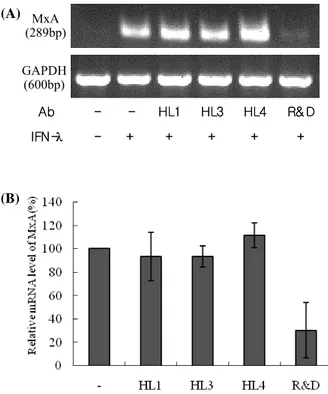
Figure 4. Neutralizing activity of monoclonal Abs. Huh7 cells were treated with MBP-IFN-λ1 alone or a mixture of MBP-IFN-λ1 and an indicated monoclonal Ab. Six hours later, the cells were harvested and the induction of MxA transcription was analyzed by semi-quantitative RT-PCR. (A) Agarose gel electrophoresis of the amplified PCR products. The data is the representative result of 3 independent experiments. (B) The levels of MxA transcription were normalized to GAPDH mRNA levels. Data represents the means ± SD of 3 independent experiments.
was observed as a single band of approximately 23 kDa by both SDS-PAGE and western blotting using anti-His Ab (Fig.
1B).
Generation of hybridoma cells producing mono- clonal Ab against IFN-λ1
From 3 mice immunized with MBP-IFN-λ1 and His-tagged IFN-λ1, 4 hybridoma clones showing reactivity against both MBP-IFN-λ1 and His-tagged IFN-λ1 were selected using ELISA and designated as HL1, HL2, HL3, and HL4 (Fig. 2A).
All 4 types of Abs showed binding activity to the denatured form of IFN-λ1 (Fig. 2B). These results indicate that all Abs may be used to detect both the native and denatured forms of IFN-λ1.
Detection limit of indirect ELISA using the HL mono- clonal Abs
The secretion of IFN-λ had not been determined until
Contoli et al. reported deficient induction of IFN-λs by rhi- novirus in asthmatic primary bronchial epithelial cells and al- veolar macrophages (18). Although the ELISA kit for IFN-λ is currently commercially available, it was not available when we performed this experiment. In order to establish an in- direct ELISA against IFN-λ1, Ab molecules were purified from the culture supernatant of each hybridoma clone. The purity of each Ab preparation was revealed to be similar based on SDS-PAGE analysis (Fig. 3A). We performed in- direct ELISA using IFN-λ1 expressed in eukaryotic cells. The detection limit of the indirect ELISA for IFN-λ1 was 20 ng/ml using HL1 Ab or HL3 Ab and 40 ng/ml using HL2 Ab or HL4 Ab (Fig. 3B). Compared to IFN-λ1, 4 ng/ml of MBP-IFN-λ1 was detected with indirect ELISA using HL3 Ab (data not shown). Considering that the molecular weight of IFN-λ1 is one-third that of MBP-IFN-λ1, the difference in detection limits between IFN-λ1 and MBP-IFN-λ1 might be more than
◀
◀
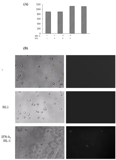
Figure 5. Binding of IFN-λ and HL1 complex on HepG2 cells. HepG2 cells were reacted with rhodamine-conjugated HL1 alone or sequentially with MBP-IFN-λ1 (100 ng/ml) and rhodamine-conjugated HL1 at 4oC. After fixation, the fluorescence intensity of the cells was measured (A) and fluorescence microscopy was performed (B).
10-fold, possibly due to the presence of carbohydrate moi- eties and the absence of the MBP fusion partner in IFN-λ1 molecules expressed by eukaryotic cells. In the first report adopting quantitative sandwich ELISA for IFN-λ using mono- clonal Abs and polyclonal Abs as the capture and secondary Ab, respectively, the detection limit was 10∼15 pg/ml (18).
Our HL monoclonal Abs may be utilized for sandwich ELISA to improve the sensitivity of IFN-λ detection.
Neutralization of HL Abs against IFN-λ1
Next, we examined the neutralizing activity of HL1, HL3, and
HL4 Abs. As the control, we included the commercially avail- able monoclonal Ab against IFN-λ1. Regardless of the pres- ence of HL1, HL3, or HL4 Ab, IFN-λ1 induced the MxA tran- script, while the presence of the control Ab suppressed the induction of MxA transcription by 50% as compared to when IFN-λ1 alone was used (Fig. 4). These results imply that HL1, HL3, and HL4 Abs do not neutralize IFN-λ1 activity.
Visualization of binding of IFN-λon HepG2 cells The finding that HL1 Ab does not possess neutralizing activity prompted us to examine if HL1 Ab could be used for visualiz-
ing the binding of IFN-λ on cells. Suspended HepG2 cells were incubated in the medium containing MBP-IFN-λ at 4oC for 30 min and then in the medium with HL1 Ab labeled with rhodamine. The fluorescence intensity of the HepG2 cells that reacted with HL1 Ab alone was the same as that of untreated HepG2 cells (Fig. 5A). However, the fluorescence intensity of HepG2 cells reacted with IFN-λ and HL1 Ab was higher than that of the control cells (Fig. 5A). In agreement with these findings, fluorescence microscopy of the control sam- ples (untreated and HL1 Ab-treated HepG2 cells) showed no signal, however, when HepG2 cells were reacted with both IFN- λ and HL1 Ab, it showed that these cells reacted with the IFN-λ and HL1 Ab complex to reveal rhodamine-labeled cells (Fig. 5B). These findings demonstrated that HL1 Ab did not interfere and instead visualized the binding of IFN-λ on HepG2 cells.
REFERENCES
1. Kotenko SV, Gallagher G, Baurin VV, Lewis-Antes A, Shen M, Shah NK, Langer JA, Sheikh F, Dickensheets H, Donnelly RP. IFN-lambdas mediate antiviral protection through a dis- tinct class II cytokine receptor complex. Nat Immunol 4;69- 77, 2003
2. Sheppard P, Kindsvogel W, Xu W, Henderson K, Schluts- meyer S, Whitmore TE, Kuestner R, Garrigues U, Birks C, Roraback J, Ostrander C, Dong D, Shin J, Presnell S, Fox B, Haldeman B, Cooper E, Taft D, Gilbert T, Grant FJ, Tackett M, Krivan W, McKnight G, Clegg C, Foster D, Klucher KM. IL-28, IL-29 and their class II cytokine receptor IL-28R. Nat Immunol 4;63-68, 2003
3. Dumoutier L, Lejeune D, Hor S, Fickenscher H, Renauld JC.
Cloning of a new type II cytokine receptor activating signal transducer and activator of transcription (STAT)1, STAT2 and STAT3. Biochem J 370;391-396, 2003
4. Uze G, Monneron D. IL-28 and IL-29: newcomers to the in- terferon family. Biochimie 89;729-734, 2007
5. Ank N, West H, Bartholdy C, Eriksson K, Thomsen AR, Paludan SR. Lambda interferon (IFN-lambda), a type III IFN, is induced by viruses and IFNs and displays potent antiviral activity against select virus infections in vivo. J Virol 80;
4501-4509, 2006
6. Marcello T, Grakoui A, Barba-Spaeth G, Machlin ES, Kotenko SV, MacDonald MR, Rice CM. Interferons alpha and lambda inhibit hepatitis C virus replication with distinct sig- nal transduction and gene regulation kinetics. Gastroentero- logy 131;1887-1888, 2006
7. Robek MD, Boyd BS, Chisari FV. Lambda interferon inhibits hepatitis B and C virus replication. J Virol 79;3851-3854, 2005
8. Holzinger D, Jorns C, Stertz S, Boisson-Dupuis S, Thimme R, Weidmann M, Casanova JL, Haller O, Kochs G. Induction of MxA gene expression by influenza A virus requires type I or type III interferon signaling. J Virol 81;7776-7785, 2007 9. Hong SH, Cho O, Kim K, Shin HJ, Kotenko SV, Park S. Effect
of interferon-lambda on replication of hepatitis B virus in human hepatoma cells. Virus Res 126;245-249, 2007 10. Mennechet FJ, Uze G. Interferon-lambda-treated dendritic
cells specifically induce proliferation of FOXP3-expressing suppressor T cells. Blood 107;4417-4423, 2006
11. Jordan WJ, Eskdale J, Srinivas S, Pekarek V, Kelner D, Rodia M, Gallagher G. Human interferon lambda-1 (IFN- lambda1/IL-29) modulates the Th1/Th2 response. Genes Immun 8;254-261, 2007
12. Chi B, Dickensheets HL, Spann KM, Alston MA, Luongo C, Dumoutier L, Huang J, Renauld JC, Kotenko SV, Roederer M, Beeler JA, Donnelly RP, Collins PL, Rabin RL. Alpha and lambda interferon together mediate suppression of CD4 T cells induced by respiratory syncytial virus. J Virol 80;5032- 5040, 2006
13. Coccia EM, Severa M, Giacomini E, Monneron D, Remoli ME, Julkunen I, Cella M, Lande R, Uze G. Viral infection and Toll-like receptor agonists induce a differential ex- pression of type I and lambda interferons in human plas- macytoid and monocyte-derived dendritic cells. Eur J Immunol 34;796-805, 2004
14. Yang K, Puel A, Zhang S, Eidenschenk C, Ku CL, Casrouge A, Picard C, von Bernuth H, Senechal B, Plancoulaine S, Al-Hajjar S, Al-Ghonaium A, Marodi L, Davidson D, Speert D, Roifman C, Garty BZ, Ozinsky A, Barrat FJ, Coffman RL, Miller RL, Li X, Lebon P, Rodriguez-Gallego C, Chapel H, Geissmann F, Jouanguy E, Casanova JL. Human TLR-7-, -8-, and -9-mediated induction of IFN-alpha/beta and -lambda Is IRAK-4 dependent and redundant for protective im- munity to viruses. Immunity 23;465-478, 2005
15. Onoguchi K, Yoneyama M, TakemuraA, Akira S, Taniguchi T, Namiki H, Fujita T. Viral infections activate types I and III interferon genes through a common mechanism. J Biol Chem 282;7576-7581, 2007
16. Osterlund PI, Pietila TE, Veckman V, Kotenko SV, Julkunen I. IFN regulatory factor family members differentially regu- late the expression of type III IFN (IFN-lambda) genes. J Immunol 179;3434-3442, 2007
17. Diebold SS, Montoya M, Unger H, Alexopoulou L, Roy P, Haswell LE, Al-Shamkhani A, Flavell R, Borrow P, Reis e Sousa C. Viral infection switches non-plasmacytoid den- dritic cells into high interferon producers. Nature 424;324- 328, 2003
18. Contoli M, Message SD, Laza-Stanca V, Edwards MR, Wark PA, Bartlett NW, Kebadze T, Mallia P, Stanciu LA, Parker HL, Slater L, Lewis-Antes A, Kon OM, Holgate ST, Davies DE, Kotenko SV, Papi A, Johnston SL. Role of deficient type III interferon-lambda production in asthma exacerbations.
Nat Med 12;1023-1026, 2006