90
Received on March 24, 2009. Revised on April 1, 2009. Accepted on April 15, 2009.
*Corresponding Author. Tel: 82-53-950-5388; Fax: 82-53-943-6925; E-mail: [email protected] Keywords: macrophage, atherosclerosis, inflammation, CD147, cyclophilin A
The Stimulation of CD147 Induces MMP-9 Expression through ERK and NF-κB in Macrophages: Implication for
Atherosclerosis
Ju-Young Kim1, Won-Jung Kim1, Ho Kim1, Kyoungho Suk2 and Won-Ha Lee1*
1The School of Life Sciences and Biotechnology, 2Department of Pharmacology, School of Medicine, Kyungpook National University, Daegu 702-701, Korea
Background: CD147, as a cellular receptor for cyclophilin A (CypA), is a multifunctional protein involved in tumor in- vasion, inflammation, tissue remodeling, neural function, and reproduction. Recent observations showing the expression of CD147 in leukocytes indicate that this molecule may have roles in inflammation. Methods: In order to investigate the role of CD147 and its ligand in the pathogenesis of athero- sclerosis, human atherosclerotic plaques were analyzed for the expression pattern of CD147 and CypA. The cellular re- sponses and signaling molecules activated by the stimulation of CD147 were then investigated in the human macrophage cell line, THP-1, which expresses high basal level of CD147 on the cell surface. Results: Staining of both CD147 and CypA was detected in endothelial cell layers facing the lu- men and macrophage-rich areas. Stimulation of CD147 with its specific monoclonal antibody induced the expression of matrix metalloproteinase (MMP)-9 in THP-1 cells and it was suppressed by inhibitors of both ERK and NF-κB.
Accordingly, the stimulation of CD147 was observed to in- duce phosphorylation of ERK, phosphorylation-associated degradation of IκB, and nuclear translocation of NF-κB p65 and p50 subunits. Conclusion: These results suggest that CD147 mediates the inflammatory activation of macro- phages that leads to the induction of MMP-9 expression, which could play a role in the pathogenesis of inflammatory diseases such as atherosclerosis.
[Immune Network 2009;9(3):90-97]
INTRODUCTION
CD147 (EMMPRIN/basigin/HAb18G/neurothelin/M6/TCSF), which has two immunoglobulin-like extracellular domains, is a mul- tifunctional transmembrane glycoprotein with short (39 amino acids long) intracellular domain (1). CD147 plays a critical role in many pathological and physiological processes involv- ing a variety of cell types such as various cancer cells, leuko- cytes, fibroblasts, and endothelial cells (2-7). As a tu- mor-derived MMP inducer, CD147 stimulates fibroblast and endothelial cells to facilitate tumor invasion, metastasis, and angiogenesis (7). In addition, CD147 enhances angiogenesis through stimulation of the production of vascular endothelial growth factor (VEGF) (8). The expression of CD147 has been shown to be induced in activated leukocytes such as gran- ulocytes, lymphocytes, and macrophages (4). Stimulation of CD147 in leukocytes is believed to be involved in in- flammatory processes associated with lung injury, rheumatoid arthritis (RA), chronic liver disease, heart failure, and athero- sclerosis (9-13).
The ligands for CD147 were identified to be the two cyclo- sporin A binding proteins: cyclophilin A and B (CypA and CypB) (14,15). A secreted form of CypA, which are expressed by smooth muscle cells (SMCs) and macrophages during in- flammatory conditions (16-18), has been shown to have cyto- kine-like functions (17,19). The expression of CypA and CD147 was detected in synovial macrophages of RA patients and stimulation of CD147 induced NF-κB-mediated ex- pression of MMP-9 and proinflammatory cytokines and en-
hanced cell migration in macrophages (20,21). Accordingly, blocking the interaction between CD147 and CypA in a colla- gen-induced arthritis model resulted in a significant reduction in arthritic symptoms (22). Furthermore, CypA has been shown to have chemoattractant activity toward CD4+ T cells, which up-regulate the expression of CD147 after activation (23).
Although CD147 has been shown to be expressed by mac- rophages in atherosclerotic plaques (11) and in patients with acute myocardial infarction (24), the expression pattern and role of CD147 in relation to CypA has not been investigated simultaneously in the context of atherosclerosis. In this manu- script, the expression patterns of CD147 and CypA were com- pared in human atherosclerotic plaques and the role of CD147, in relation to CypA, was investigated in macrophage activation and cell signaling.
MATERIALS AND METHODS
Monoclonal antibodies, cell lines, and reagents Monoclonal antibodies (mAbs) to CD68 (KP1) and rabbit pol- yclonal antibody to the von Willebrand factor (vWF) were purchased from DAKO (Glostrup, Denmark); rabbit poly- clonal antibody to CypA was from BIOMOL International (Plymouth Meeting, PA, USA); mAb for CD147 (clone MEM-M6/1) was from Abcam (Cambridge, MA, USA); rabbit polyclonal antibody to MMP-9 was from Chemicon (Temecula, CA, USA); mAb for TFIIB (clone 24/TFIIB) was from BD-Pharmingen (San Jose, CA, USA); rabbit polyclonal antibody to IκB, mAb to phospho-IκB (Ser32/36) (5A5), PD08059, U0126, and polyclonal antibodies for ERK, phos- phospho-ERK, p38, phospho-p38, AKT, and phospho-AKT (Ser473) originated from Cell Signaling (Danvers, MA, USA);
SB203580, LY294002, JNK inhibitor I (JNK-I1), a cell-per- meable fusion protein containing 20 AA of the JNK-binding domain of islet-brain and HIV-TAT48-57 (25), and its negative control containing only HIV-TAT were obtained from Calbiochem International Inc. (La Jolla, CA, USA); TPCK, eth- yl pyruvate, and sulfasalazine were purchased from Sigma (St. Louis, MO, USA); and mAb for NF-κB p65 subunit (F-6) and rabbit polyclonal antibodies for p50 and goat polyclonal antibody for actin were purchased from Santa Cruz (Santa Cruz, CA, USA). Human monocytic leukemia cell line THP-1 (26) was obtained from the American Type Culture Collection (Rockville, MD, USA).
Histological analysis
Carotid endoarterectomy specimens, generously provided by Dr. Jeong-Euy Park, Sungkyunkwan University, School of Medicine, were obtained from patients, aged between 63 to 81, who had undergone surgery at the Samsung Seoul Hospital. The current study was approved by the internal re- view board. Atherosclerotic plaque specimens were washed with saline and embedded to produce frozen sections. For the immunohistochemical analysis, standard 5-μm sections were stained using an LSAB kit (DAKO, Glostrup, Denmark) according to the manual provided by the manufacturer. The sections were then counterstained with Hematoxylin which stains the nucleus in blue. Finally, the slides were mounted in a 1:1 mixture of Xylene and Malinol (Muto Pure Chemicals, Tokyo, Japan).
Cell stimulation, Western blot analysis and gelatin zymogram
For the activation utilizing immobilized mAbs, 100μl/well of PBS containing 1 or 10μg/ml of antibody was incubated overnight on a 96-well plate. The wells were washed twice with PBS, after which THP-1 cells (1×10⁵/well) in 100μl of RPMI1640 medium supplemented with 0.1% serum were added. Cell lysates were prepared at appropriate times after activation in 100μl of triple-detergent lysis buffer. For the de- tection of nuclear proteins, cell lysate were prepared in 200 μl of NP-40 lysis buffer (0.1% NP-40, 25 mM KCl, 5 mM MgCl2, 10 mM Tris (pH 8.0), 1 mM PMSF, 1 mM Na3VO4, and 1 mM NaF). Cell debris containing nucleus was collected and nuclear extracts were isolated in 100μl of high salt lysis buf- fer (0.1% NP-40, 500 mM NaCl, 5 mM EDTA (pH 8.0), 10 mM Tris (pH 8.0), 1 mM PMSF, 1 mM Na3VO4, and 1 mM NaF). For the analysis of MMP-9, culture supernatants were concentrated 10-fold using a speedvac. Western blot analysis was performed as described previously (27). For the de- tection of MMP-9 using gelatin zymogram, culture super- natants were collected 24 hours after activation. The MMP-9 activity in the culture supernatant was determined by sub- strate gel electrophoresis as described previously (28).
RT-PCR
Five micrograms of total RNAs isolated from cells were treat- ed with RNase free DNase (BD-Pharmingen), and then used to generate first-strand cDNAs using a RevertAidTM first strand cDNA synthesis kit with 500 ng oligo (dT)12-18 primers. PCR
92
Figure 1. Endothelial cells and macrophages express CD147 and CypA in atherosclerotic plaques. Human carotid atherosclerotic plaques were sequentially sectioned and stained against CD68 (a macrophage marker), vWF (an endothelial cell marker), CD147, CypA. Mouse IgG (mIgG) was used for the staining as a negative control. Upper panel shows the plaques area facing the vessel lumen (L). Lower panel shows the shoulder area of a plaque which have macrophages and smooth muscle cells (SM). Note the low level staining of CypA in areas rich in SMCs.
primers were designed with ABI PRISM Primer Express 2.0 (Applied Biosystems, Foster City, CA, USA) and made by Geno Tech Corp (Daejeon, Korea). Primer sequences are 5’
GGCCAGAAAACGGAGTTCAA 3’ (forward) and 5’ GCGC- TTCTCGTAGATGAAGA 3’ (reverse) for CD147, 5’ ATCA- CTGCCACCCAGAAGAC 3’ (forward) and 5’ TGAGCTTGA CAAAGTGGTCG 3’(reverse) for GAPDH. After the PCR re- action, the PCR products were run on 2% agarose gel to con- firm the size and purity of the PCR products.
Immunofluorescence assay
The detection of intracellular localization of NF-κB p50 sub- unit was performed as described previously (20). Briefly, THP-1 cells were stimulated and fixed with 10μl of 4% form- aldehyde in PBS at appropriate time after stimulation. The fixed cells were then permeabilized with 1% Triton X-100 in PBS for 10 minutes at room temperature and the per- meabilized cells were then stained with 0.5μg/ml Hoechst staining solution (Sigma, St. Louis, MO, USA) for 30 minutes at 37oC and then washed. The cells were then sequentially treated with 10μg/ml anti-p50 polyclonal antibody for 45 mi- nutes at 37oC and with a 1:50 dilution of Alexa Fluor 594-la- beled goat anti-rabbit antibody (Invitrogen, Carlsbad, CA, USA) for 45 minutes at 37oC in a humid chamber. Finally, the cells were dried at room temperature and mounted in a 1:1 mixture of xylene and malinol.
Flow cytometry analysis
For the flow cytometric analysis of cell surface antigens, cells (5×10⁵) were sequentially incubated with 0.3μg of an- ti-CD147 mAb and FITC-labeled goat anti-mouse IgG in 30 μl of FACS solution (a PBS containing 0.5% BSA and 0.1%
Sodium Azide) for 20 minutes on ice. For background fluo- rescence, the cells were stained with an isotype-matching control antibody. The fluorescence profiles of 2×10⁴ cells were collected and analyzed using FACS-calibur (Becton- Dickinson, Mountain View, CA, USA).
RESULTS
In order to analyze the role of CD147 and its ligand (CypA) in atherogenic processes, human carotid atherosclerotic pla- ques were analyzed using immunohistochemical analysis (Fig.
1). The innermost layer of atherosclerotic plaque facing the lumen was lined with endothelial cells which are specifically stained with mAb against the von Willebrand factor (vWF).
vWF is a multimeric glycoprotein essential for thrombus for- mation and the plasma level of it has been shown to be ele- vated in patients with atherosclerosis (29,30). Since the ex- pression of vWF is restricted to platelets and endothelial cells, its presence has been used as a endothelial cell marker in a number of studies employing immunohistochemistry
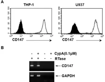
Figure 2. Human monocyte/macrophage cell lines express high levels of CD147. (A) THP-1 and U937 cells were stained with anti-CD147 mAb (empty area) or isotype-matching mouse IgG (filled area). (B) THP-1 cells were stimulated with or without indicated amounts of CypA for 20 hr. Total cellular RNAs were collected and the expression levels of CD147 or GAPDH mRNA were measured using RT-PCR analysis.
Figure 3. The stimulation of CD147 induced the expression of MMP-9 in THP-1 cells. (A) THP-1 cells were stimulated with anti-CD147 mAb or isotype-matching mouse IgG (mIgG) that had been immobilized at indicated concentrations. LPS was used as a positive control. Some of the cells were also stimulated with antibodies that had been heat inactivated at 95oC for 2 hr. Culture supernatants were collected 24 hr after activation and subjected to gelatin zymogram. (B) THP-1 cells were stimulated as in (A) and culture supernatants were concentrated (x10) and subjected to Western blot analysis using MMP-9 specific antibody. As loading control, the picture of the membrane used for Western blot analysis that had been stained with Coomassie Brilliant Blue is shown at the lower panel.
(31,32). Macrophages, stained with anti-CD68 mAb, were found in large numbers at the shoulder region of plaques in between thick layers of SMCs (Fig. 1, lower panel). CD147 expression was detected in the innermost layers facing the lumen, which corresponds to endothelial cells (Fig. 1, upper panel), and in the area corresponding to macrophage-rich re- gions in the shoulder region (Fig. 1, lower panel). The ex- pression of CD147 in SMCs was not detected. Interestingly, the expression pattern of CypA, the ligand for CD147, was similar to that of CD147: both endothelial cell- and macro- phage-rich areas.
Since macrophages in atherosclerotic plaques express CD147, monocyte/macrophage cell lines were used to test whether they express CD147. As shown in Fig. 2A, both THP-1 and U937 cells expressed high levels of CD147.
Stimulation of THP-1 cells with CypA did not affect the ex- pression levels of CD147, probably because the basal ex- pression level of CD147 was already high (data not shown).
The expression of CD147 in THP-1 was also confirmed using RT-PCR (Fig. 2B). THP-1 cells were then used to study the signaling pathway initiated from CD147. Since stimulation of THP-1 cells with CypA induced the expression of MMP-9 (20), CD147 on the surface of THP-1 cells were stimulated with anti-CD147 mAb and the cellular responses were analyzed.
Anti-CD147 mAb was used instead of CypA, to exclude the possibility that CypA may stimulate other yet unknown cel- lular receptors. Stimulation of the cells with immobilized an- ti-CD147 mAb induced the secretion of MMP-9 (Fig. 3A). The expression levels of MMP-2, which is known to be unaffected by cellular activation status, are shown as the internal control.
Isotype-matching mouse IgG failed to induce the expression of MMP-9 indicating that the induction of MMP-9 requires specific interaction between CD147 and the antibody.
Furthermore, heat inactivation of anti-CD147 mAb abolished the effect indicating that the activation was not induced by endotoxins that are heat resistant. The induction of MMP-9 expression was also confirmed in the protein level using Western blot analysis (Fig. 3B).
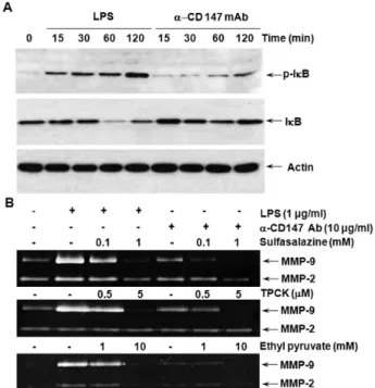
The expression of MMP-9 in macrophage requires the acti- vation of NF-κB in macrophages. NF-κB, a heterodimer of p65 and p50, stays in cytoplasm in its inactive status in associ- ation with IκB. When the activation signal(s) is transmitted, IκB become phosphorylated and, as a result, degraded by proteasome. The free NF-κB heterodimer then translocates into the nucleus. In order to analyze the requirement of NF- κB activation in the CD147-induced expression of MMP-9,
94
Figure 4. The stimulation of CD147 induces nuclear translocation of NF-κB p65 and p50 subunits. (A) THP-1 cells were stimulated with 1μg/ml of LPS or anti-CD147 mAb that had been immobilized at a concentration of 10μg/ml. Nuclear extracts were collected after indicated times and the Western blot analysis was performed using p65- or TFIIB-specific antibodies. TFIIB was used as a loading control for nuclear extracts. (B, C) THP-1 cells were stimulated with LPS, anti-CD147 mAb, or isotype-matching mouse IgG (mIgG) for 2 hr and analyzed with immunofluorescence analysis using an antibody specific for NF-κB p50 subunit. (B) shows representative pictures of cells in each samples and (C) shows the percentage of cells that had NF-κB p50 subunit at their nucleus.
Figure 5. CD147-induced expression of MMP-9 requires activation of NF-κB. (A) THP-1 cells were stimulated with 1μg/ml of LPS or immobilized anti-CD147 mAb (10μg/ml). Cell lysates were collected at indicated times and subjected to Western blot analysis using antibodies specific for phospho-IκB, IκB, and actin. (B) THP-1 cells were stimulated with indicated amounts of LPS or immobilized anti-CD147 mAb in the presence of indicated amounts of NF-κB inhibitors (sulfasalazine, TPCK, or ethyl pyruvate). Culture supern- atants were collected 24 hr after activation and subjected to gelatin zymogram.
immunohistochemistry and Western blot analysis was per- formed using p65 or p50 specific antibodies. As shown in Fig.
4A, the level of nuclear p65 was increased 30 to 60 min after activation with anti-CD147 mAb. In accordance with this data, nuclear translocation of NF-κB p50 subunit was also detected in cells stimulated with anti-CD147 mAb (Fig. 4B and 4C).
The activation of NF-κB requires the phosphorylation and degradation IκB in advance. When IκB levels were analyzed after the stimulation of CD147 (Fig. 5A), phosphorylation of IκB was observed as early as 15 min after stimulation, which continued up to two hours. Accordingly, degradation of IκB was observed 30 and 60 min after stimu- lation. IκB level started to increase two hours after stimulation due to accumulation of newly synthesized IκB.
Cells stimulated with LPS was used as a positive control. The requirement of NF-κB in CD147-induced MMP-9 secretion was also confirmed using NF-κB-specific inhibitors such as ethyl pyruvate, sulpasalazine, and N-tosyl-L-phenylalanine chloromethyl ketone (TPCK). These inhibitors blocked CD147- induced expression of MMP-9 in a dose-dependent manner (Fig. 5B). These data are in agreement with previous data showing the requirement of NF-κB activation for the ex-
pression of MMP-9 in CypA-treated THP-1 cells (20).
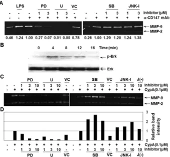
CypA has been reported to induce the activation of ERK1/2 in various cell types such as cancer cells, neurons, and leuko- cytes (21,33-35). In order to verify the involvement of MAPKs for the expression of MMP-9 in cells stimulated with CD147, the assay was performed in the presence of MAPK inhibitors.
Inhibitors of ERK MAPK (U0126 and PD98059) blocked the secretion of MMP-9 in a dose-dependent manner (Fig. 6A, note the numbers below each lane). The involvement of ERK in CD147-mediated signaling was further confirmed by detect- ing the phosphorylation of ERK using Western blot analysis in cells stimulated with anti-CD147 mAb (Fig. 6B). Interestin- gly, treatment with inhibitors of p38 and JNK MAPK slightly induced MMP-9 expression (Fig. 6A, note the members below each lane which represent the relative band intensity). The involvement of MAPKs was further confirmed in THP-1 cells stimulated with CypA. As shown in Fig. 6C, ERK inhibitors suppressed MMP-9 expression in a dose dependent manner,
Figure 6. The activation of ERK is involved in the CD147-induced expression of MMP-9 and suppression of p38 and JNK activity augments the MMP-9 expression. (A) THP-1 cells were stimulated with 1μg/ml LPS or immobilized anti-CD147 mAb (10μg/ml) in the presence or absence or indicated amounts of PD98059 (PD), U0126 (U), SB203580 (SB), JNK inhibitor (JNK-I), or 0.2% DMSO as a vehicle control (VC). Culture supernatants were collected 24 hr after activation and subjected to gelatin zymogram. Numbers below each lane represent the band intensity which was normalized with band intensity of sample treated with only anti-CD147 in each gel. (B) THP-1 cells were stimulated with immobilized anti-CD147 mAb (10 μg/ml). Cell lysates were collected at indicated times and the levels of phospho-ERK or ERK were analyzed using Western blot analysis.
(C) THP-1 cells were stimulated with 0.1μM of CypA in the presence or absence of indicated amounts of PD98059 (PD), U0126 (U), SB203580 (SB), JNK inhibitor (JNK-I), negative control for JNK-I [J(-)], or 0.2% DMSO as a vehicle control (VC). Culture supernatants were collected 24 hr after activation and subjected to gelatin zymogram.
(D) The bar graph shows the MMP-9 band intensities of each lane in panel (C) that was normalized with band intensity of sample treated with only CypA in each gel.
while the inhibitors of p38 and JNK enhanced the secretion of MMP-9 (Fig. 6C and 6D). The enhancement of ERK signal- ing by the suppression of p38 and/or JNK has been pre- viously reported: the presence of p38 inhibitor caused an in- crease in basal phosphorylation level of ERK, which resulted in the enhanced ERK-mediated signaling and cellular re- sponses in THP-1 cells (36,37). Similar enhancement of ERK phosphorylation in the presence of p38 and JNK inhibitors is likely the cause of the observed phenomenon. The molec- ular mechanism underlying this suppression of ERK activity by p38 and JNK is not known.
DISCUSSION
The immunohistochemical analysis of human atherosclerotic plaques provided the first demonstration showing the co-lo- calization of CD147 and CypA in atherosclerotic plaques. The endothelial expression of CD147 has been previously demon- strated in cultured cells (38) and in blood brain barrier (39) and the result in figure 1 provides the first demonstration of endothelial expression of CD147 in a pathological tissue sample. Although the expression of CD147 was detected in cultured SMCs (38), it was not expressed in SMCs of athero- sclerotic plaques that had been tested in this study. The co-lo- calization of CypA and vWF indicates that endothelial cells may express CypA. Endothelial cells in atherosclerotic pla- ques are in highly activated status and express activation markers such as adhesion molecule ICAM-1, proinflammatory cytokines, and chemokines (40,41). Since CypA was shown to be expressed via activation in inflammatory cells (18), it is likely that endothelial cells in atherosclerotic plaque ex- pressed high levels of CypA through a similar activation process. In case of macrophages, a number of studies already demonstrated the expression of CD147 in these cells (4,11,24) and the activation of them has been shown to induce the se- cretion of CypA (16). CypA is a well-known stimulator of both macrophages (20,21) and endothelial cells (17,38).
These previous studies, considered together with this current data, suggest that CypA expressed by endothelial cells and macrophages can stimulate itself through CD147 in an auto- crine manner. Interestingly, low level staining of CypA was detected in an area rich in SMCs, although these cells do not express CD147 (Fig. 1, lower panel). The expression of CypA by SMCs is in agreement with previous observations which reported the expression of CypA in SMCs of mouse athero- sclerotic plaques (17) and in SMCs that had been activated with endotoxins (18).
The activation of ERK, after the stimulation of CD147, was detected 4 min after activation and lasted only 4 more minutes. The degradation of IκB and NF-κB nuclear trans- location peaks at later time points. This indicates that ERK activation may be the upstream signaling event that leads to the degradation of IκB and subsequent NF-κB activation.
Several attempts, however, failed to reveal the linear relation- ship between ERK and IκB/NF-κB signaling pathway; these attempts included the measurement of IκB phosphor- ylation/degradation and NF-κB nuclear translocation in the
96
presence of ERK inhibitors. This may indicate that the activa- tion of ERK stimulates MMP-9 expression in a separate path- way that does not involve NF-κB. Alternatively, ERK may activate NF-κB activation through other mechanisms such as enhancement of p65 phosphorylation, which is known to ac- company the NK-κB activation and nuclear translocation and responsible for the recruitment of coactivators such as p300 (42,43).
Our data indicate that CD147 and its ligand, CypA, are ex- pressed in endothelial cells and macrophages. Furthermore, the stimulation of CD147 that are expressed on macrophages induces ERK- and NF-κB-mediated expression of MMP-9.
Macrophages play an essential role in atherogenesis through differentiation into foamy macrophages, secretion of proin- flammatory cytokines/chemokines and growth factors, and enhancing thrombus formation through the expression of tis- sue factors, etc. Furthermore, MMPs produced by macro- phages are responsible for the degradation of extracellular matrix (ECM). Degradation of ECM proteins results in weak- ening of the integrity of the plaque and leads to a plaque rupture and subsequent events leading to blockage of blood vessels (41). The inflammatory activation of macrophage is mediated by various mediators of inflammation such as proin- flammatory cytokines, chemokines, and cell-cell interaction between inflammatory cells. The autocrine interaction be- tween CD147 and secreted CypA is expected to contribute to and enhance the expression of MMP-9 in macrophages, which can destabilize atherosclerotic plaques by degrading ECM proteins.
ACKNOWLEDGEMENTS
This work was supported by the Korea Research Foundation Grant funded by the Korean Government (KRF-2008-313- C00647).
CONFLICTS OF INTEREST
The authors have no financial conflict of interest.
REFERENCES
1. Biswas C, Zhang Y, DeCastro R, Guo H, Nakamura T, Kataoka H, Nabeshima K: The human tumor cell-derived collagenase stimulatory factor (renamed EMMPRIN) is a member of the immunoglobulin superfamily. Cancer Res
55;434-439, 1995
2. Biswas C: Tumor cell stimulation of collagenase production by fibroblasts. Biochem Biophys Res Commun 109;1026- 1034, 1982
3. Ellis SM, Nabeshima K, Biswas C: Monoclonal antibody preparation and purification of a tumor cell collage- nase-stimulatory factor. Cancer Res 49;3385-3391, 1989 4. Kasinrerk W, Fiebiger E, Stefanova I, Baumruker T, Knapp
W, Stockinger H: Human leukocyte activation antigen M6, a member of the Ig superfamily, is the species homologue of rat OX-47, mouse basigin, and chicken HT7 molecule.
J Immunol 149;847-854, 1992
5. Kataoka H, DeCastro R, Zucker S, Biswas C: Tumor cell-de- rived collagenase-stimulatory factor increases expression of interstitial collagenase, stromelysin, and 72-kDa gelatinase.
Cancer Res 53;3154-3158, 1993
6. Nabeshima K, Lane WS, Biswas C: Partial sequencing and characterization of the tumor cell-derived collagenase stim- ulatory factor. Arch Biochem Biophys 285;90-96, 1991 7. Caudroy S, Polette M, Nawrocki-Raby B, Cao J, Toole BP,
Zucker S, Birembaut P: EMMPRIN-mediated MMP regu- lation in tumor and endothelial cells. Clin Exp Metastasis 19;697-702, 2002
8. Tang Y, Nakada MT, Kesavan P, McCabe F, Millar H, Rafferty P, Bugelski P, Yan L: Extracellular matrix metal- loproteinase inducer stimulates tumor angiogenesis by ele- vating vascular endothelial cell growth factor and matrix metalloproteinases. Cancer Res 65;3193-3199, 2005 9. Foda HD, Rollo EE, Drews M, Conner C, Appelt K,
Shalinsky DR, Zucker S: Ventilator-induced lung injury up- regulates and activates gelatinases and EMMPRIN: attenu- ation by the synthetic matrix metalloproteinase inhibitor, Prinomastat (AG3340). Am J Respir Cell Mol Biol 25;717-724, 2001
10. Konttinen YT, Li TF, Mandelin J, Liljestrom M, Sorsa T, Santavirta S, Virtanen I: Increased expression of ex- tracellular matrix metalloproteinase inducer in rheumatoid synovium. Arthritis Rheum 43;275-280, 2000
11. Major TC, Liang L, Lu X, Rosebury W, Bocan TM:
Extracellular matrix metalloproteinase inducer (EMMPRIN) is induced upon monocyte differentiation and is expressed in human atheroma. Arterioscler Thromb Vasc Biol 22;
1200-1207, 2002
12. Shackel NA, McGuinness PH, Abbott CA, Gorrell MD, McCaughan GW: Insights into the pathobiology of hepatitis C virus-associated cirrhosis: analysis of intrahepatic differ- ential gene expression. Am J Pathol 160;641-654, 2002 13. Spinale FG, Coker ML, Heung LJ, Bond BR, Gunasinghe
HR, Etoh T, Goldberg AT, Zellner JL, Crumbley AJ: A matrix metalloproteinase induction/activation system exists in the human left ventricular myocardium and is upregulated in heart failure. Circulation 102;1944-1949, 2000
14. Pushkarsky T, Zybarth G, Dubrovsky L, Yurchenko V, Tang H, Guo H, Toole B, Sherry B, Bukrinsky M: CD147 facili- tates HIV-1 infection by interacting with virus-associated cy- clophilin A. Proc Natl Acad Sci U S A 98;6360-6365, 2001 15. Yurchenko V, O'Connor M, Dai WW, Guo H, Toole B,
Sherry B, Bukrinsky M: CD147 is a signaling receptor for
cyclophilin B. Biochem Biophys Res Commun 288;786- 788, 2001
16. Sherry B, Yarlett N, Strupp A, Cerami A: Identification of cyclophilin as a proinflammatory secretory product of lip- opolysaccharide-activated macrophages. Proc Natl Acad Sci U S A 89;3511-3515, 1992
17. Jin ZG, Lungu AO, Xie L, Wang M, Wong C, Berk BC:
Cyclophilin A is a proinflammatory cytokine that activates endothelial cells. Arterioscler Thromb Vasc Biol 24;1186- 1191, 2004
18. Jin ZG, Melaragno MG, Liao DF, Yan C, Haendeler J, Suh YA, Lambeth JD, Berk BC: Cyclophilin A is a secreted growth factor induced by oxidative stress. Circ Res 87;
789-796, 2000
19. Kim SH, Lessner SM, Sakurai Y, Galis ZS: Cyclophilin A as a novel biphasic mediator of endothelial activation and dysfunction. Am J Pathol 164;1567-1574, 2004
20. Kim H, Kim WJ, Jeon ST, Koh EM, Cha HS, Ahn KS, Lee WH: Cyclophilin A may contribute to the inflammatory processes in rheumatoid arthritis through induction of ma- trix degrading enzymes and inflammatory cytokines from macrophages. Clin Immunol 116;217-224, 2005
21. Yang Y, Lu N, Zhou J, Chen ZN, Zhu P: Cyclophilin A up-regulates MMP-9 expression and adhesion of mono- cytes/macrophages via CD147 signalling pathway in rheu- matoid arthritis. Rheumatology (Oxford) 47;1299-1310, 2008 22. Damsker JM, Okwumabua I, Pushkarsky T, Arora K,
Bukrinsky MI, Constant SL: Targeting the chemotactic func- tion of CD147 reduces collagen-induced arthritis. Immuno- logy 126;55-62, 2009
23. Damsker JM, Bukrinsky MI, Constant SL: Preferential che- motaxis of activated human CD4+ T cells by extracellular cyclophilin A. J Leukoc Biol 82;613-618, 2007
24. Schmidt R, Bultmann A, Ungerer M, Joghetaei N, Bulbul O, Thieme S, Chavakis T, Toole BP, Gawaz M, Schomig A, May AE: Extracellular matrix metalloproteinase inducer regulates matrix metalloproteinase activity in cardiovascular cells: implications in acute myocardial infarction. Circulation 113;834-841, 2006
25. Bonny C, Oberson A, Negri S, Sauser C, Schorderet DF:
Cell-permeable peptide inhibitors of JNK: novel blockers of beta-cell death. Diabetes 50;77-82, 2001
26. Tsuchiya S, Yamabe M, Yamaguchi Y, Kobayashi Y, Konno T, Tada K: Establishment and characterization of a human acute monocytic leukemia cell line (THP-1). Int J Cancer 26;171-176, 1980
27. Lee WH, Kim SH, Lee Y, Lee BB, Kwon B, Song H, Kwon BS, Park JE: Tumor necrosis factor receptor superfamily 14 is involved in atherogenesis by inducing proinflammatory cytokines and matrix metalloproteinases. Arterioscler Thromb Vasc Biol 21;2004-2010, 2001
28. Kim SH, Kang YJ, Kim WJ, Woo DK, Lee Y, Kim DI, Park YB, Kwon BS, Park JE, Lee WH: TWEAK can induce pro-in- flammatory cytokines and matrix metalloproteinase-9 in macrophages. Circ J 68;396-399, 2004
29. Ruggeri ZM: Structure and function of von Willebrand factor. Thromb Haemost 82;576-584, 1999
30. Whincup PH, Danesh J, Walker M, Lennon L, Thomson A,
Appleby P, Rumley A, Lowe GD: von Willebrand factor and coronary heart disease: prospective study and meta-analysis. Eur Heart J 23;1764-1770, 2002
31. Kamei M, Yoneda K, Kume N, Suzuki M, Itabe H, Matsuda K, Shimaoka T, Minami M, Yonehara S, Kita T, Kinoshita S: Scavenger receptors for oxidized lipoprotein in age-re- lated macular degeneration. Invest Ophthalmol Vis Sci 48;1801-1807, 2007
32. Pilarczyk K, Sattler KJ, Galili O, Versari D, Olson ML, Meyer FB, Zhu XY, Lerman LO, Lerman A: Placenta growth factor expression in human atherosclerotic carotid plaques is related to plaque destabilization. Atherosclerosis 196;333-340, 2008 33. Boulos S, Meloni BP, Arthur PG, Majda B, Bojarski C,
Knuckey NW: Evidence that intracellular cyclophilin A and cyclophilin A/CD147 receptor-mediated ERK1/2 signalling can protect neurons against in vitro oxidative and ischemic injury. Neurobiol Dis 25;54-64, 2007
34. Schmidt R, Bultmann A, Fischel S, Gillitzer A, Cullen P, Walch A, Jost P, Ungerer M, Tolley ND, Lindemann S, Gawaz M, Schomig A, May AE: Extracellular matrix metal- loproteinase inducer (CD147) is a novel receptor on plate- lets, activates platelets, and augments nuclear factor kappaB-dependent inflammation in monocytes. Circ Res 102;302-309, 2008
35. Yang H, Chen J, Yang J, Qiao S, Zhao S, Yu L: Cyclophilin A is upregulated in small cell lung cancer and activates ERK1/2 signal. Biochem Biophys Res Commun 361;763- 767, 2007
36. Heidinger M, Kolb H, Krell HW, Jochum M, Ries C:
Modulation of autocrine TNF-alpha-stimulated matrix metal- loproteinase 9 (MMP-9) expression by mitogen-activated protein kinases in THP-1 monocytic cells. Biol Chem 387;69-78, 2006
37. Bae EM, Kim WJ, Suk K, Kang YM, Park JE, Kim WY, Choi EM, Choi BK, Kwon BS, Lee WH: Reverse signaling ini- tiated from GITRL induces NF-kappaB activation through ERK in the inflammatory activation of macrophages. Mol Immunol 45;523-533, 2008
38. Yang H, Li M, Chai H, Yan S, Lin P, Lumsden AB, Yao Q, Chen C: Effects of cyclophilin A on cell proliferation and gene expressions in human vascular smooth muscle cells and endothelial cells. J Surg Res 123;312-319, 2005 39. Sameshima T, Nabeshima K, Toole BP, Yokogami K, Okada
Y, Goya T, Koono M, Wakisaka S: Expression of emmprin (CD147), a cell surface inducer of matrix metallopro- teinases, in normal human brain and gliomas. Int J Cancer 88;21-27, 2000
40. Libby P: Atherosclerosis in Inflammation. Nature 420;
868-874, 2002
41. Ross R: Atherosclerosis--an inflammatory disease. N Engl J Med 340;115-126, 1999
42. Zhong H, May MJ, Jimi E, Ghosh S: The phosphorylation status of nuclear NF-kappa B determines its association with CBP/p300 or HDAC-1. Mol Cell 9;625-636, 2002 43. Yang F, Tang E, Guan K, Wang CY: IKK beta plays an es-
sential role in the phosphorylation of RelA/p65 on serine 536 induced by lipopolysaccharide. J Immunol 170;5630- 5635, 2003