D I A B E T E S & M E T A B O L I S M J O U R N A L
This is an Open Access article distributed under the terms of the Creative Commons Attribution Non-Commercial License (https://creativecommons.org/licenses/by-nc/4.0/) which permits unrestricted non-commercial use, distribution, and reproduction in any medium, provided the original work is properly cited.
Impact of Diabetes Control on Subclinical
Atherosclerosis: Analysis from Coronary Computed Tomographic Angiography Registry
Gyung-Min Park1, Chang Hoon Lee2, Seung-Whan Lee3, Sung-Cheol Yun4, Young-Hak Kim3, Yong-Giun Kim1, Ki-Bum Won1, Soe Hee Ann1, Shin-Jae Kim1, Dong Hyun Yang5, Joon-Won Kang5, Tae-Hwan Lim5, Eun Hee Koh6, Woo Je Lee6, Min-Seon Kim6, Joong-Yeol Park6, Hong-Kyu Kim7, Jaewon Choe7, Sang-Gon Lee1
1Department of Cardiology, Ulsan University Hospital, University of Ulsan College of Medicine, Ulsan,
2Department of Cardiology, Veterans Health Service Medical Center, Seoul,
Departments of 3Cardiology, 4Biostatistics, 5Radiology, 6Endocrinology, 7The Health Screening and Promotion Center, Asan Medical Center, University of Ulsan College of Medicine, Seoul, Korea
Background: There are limited data on the impact of diabetes control on the risk of subclinical coronary atherosclerosis.
Methods: We analyzed 6,434 consecutive asymptomatic individuals without previous history of coronary artery disease who un- derwent coronary computed tomographic angiography (CCTA) (mean age, 53.7±7.6 years and 4,694 men [73.0%]). The degree and extent of subclinical coronary atherosclerosis were assessed by CCTA, and ≥50% diameter stenosis was defined as significant.
A cardiac event was defined as a composite of all-cause death, myocardial infarction, unstable angina, or coronary revasculariza- tion. Study participants were categorized as normal (n=5,319), controlled diabetes (glycosylated hemoglobin [HbA1c] <7%, n=747), or uncontrolled diabetes (HbA1c ≥7%, n=368), respectively.
Results: Compared with normal individuals, there were no statistically significant differences in the risk of for any atherosclerotic plaque (odds ratio [OR], 1.16; 95% confidence interval [CI], 0.98 to 1.38; P=0.086) and significant coronary artery stenosis (OR, 1.08; 95% CI, 0.82 to 1.42; P=0.583) in controlled diabetic individuals. In contrast, uncontrolled diabetic individuals had consis- tently higher risks of any atherosclerotic plaque (OR, 2.16; 95% CI, 1.70 to 2.75; P<0.001) and significant coronary artery stenosis (OR, 3.34; 95% CI, 2.52 to 4.43; P<0.001) than normal individuals. During a follow-up of median 5.4 years, there was no signifi- cant difference in cardiac events between normal and controlled diabetic individuals (P=0.365). However, uncontrolled diabetes was associated with an increased risk of cardiac events compared with normal individuals (P<0.001) and controlled diabetic indi- viduals (P=0.023).
Conclusion: Asymptomatic uncontrolled diabetes was associated with significant subclinical coronary atherosclerosis with sub- sequent high risk for cardiac events.
Keywords: Atherosclerosis; Coronary artery disease; Diabetes complications; Diabetes mellitus
Corresponding author: Seung-Whan Lee https://orcid.org/0000-0002-2662-5952 Department of Cardiology, Asan Medical Center, University of Ulsan College of Medicine, 88 Olympic-ro 43-gil, Songpa-gu, Seoul 05505, Korea
E-mail: [email protected]
INTRODUCTION
Glycemic control is fundamental to diabetes management [1].
Previous studies have demonstrated that improved glycemic control is associated with significantly reduced onset or pro-
gression of microvascular complications in diabetic individu- als [2-6]. In addition, individuals with diabetes showed a high- er prevalence, extent, and severity of coronary atherosclerosis than those without [7-9]. Coronary artery disease (CAD) is a leading cause of death among diabetic individuals [10]. Recent https://doi.org/10.4093/dmj.2019.0073
pISSN 2233-6079 · eISSN 2233-6087
long-term follow-up studies have also shown cardiovascular benefits of intensive glycemic control [3,11]. However, there are limited data regarding the impact of the diabetes control on the risk of subclinical coronary atherosclerosis in asymptomat- ic individuals. With the advent of multidetector row computed tomography (CT), coronary computed tomographic angiogra- phy (CCTA) has proven to be effective in providing the com- prehensive evaluation of coronary atherosclerosis, including lesion location, severity and plaque characteristics [12]. Thus, in this study, we sought to evaluate the impact of diabetes con- trol on the risk of subclinical coronary atherosclerosis through a large cohort of asymptomatic Korean individuals who volun- tarily underwent CCTA.
METHODS
Study population
In total, 9,269 consecutive South Korean individuals aged 20 years and older who had undergone self-referral CCTA evalua- tion as part of a general health examination in the Health Screening and Promotion Center at Asan Medical Center from January 2007 to December 2011. Among these, 7,129 individu- als (76.9%) agreed to participate in this study. Possible risks as- sociated with CCTA were explained and informed consent was obtained. Exclusion criteria include subjects with (1) a previous history of angina or myocardial infarction; (2) abnormal rest electrocardiographic results, i.e., pathological Q waves, isch- emic ST segments or T wave changes, or left bundle-branch blocks; (3) insufficient medical records; (4) structural heart diseases; (5) a prior history of open heart surgery or percutane- ous coronary intervention; (6) a previous cardiac procedure; or (7) renal insufficiency (creatinine >1.5 mg/dL). Finally, 6,434 subjects were enrolled (Supplementary Fig. 1). The study was approved by the local Institutional Review Board of the Asan Medical Center, Seoul, Korea (approval number: 2016-1068).
The demographic information was collected from a database maintained by the Health Screening and Promotion Center at the Asan Medical Center. Medical history including angina, myocardial infarction, stroke, structural heart disease, open heart surgery, percutaneous coronary intervention, previous cardiac procedures, diabetes mellitus, hypertension, or hyper- lipidemia; a family history of CAD; and smoking status was taken from the responses in the systemized self-report ques- tionnaire issued prior to the general health examination. A family history of CAD was defined as having a first-degree rel-
ative of any age on the self-report questionnaire [13]. Patients with diabetes was defined as (1) subjects with a self-reported history of diabetes and/or treatment with dietary modification, or use of anti-diabetic medication on the systemized question- naire and (2) those with a fasting plasma glucose (FPG) ≥126 mg/dL or a glycosylated hemoglobin (HbA1c) level ≥6.5%
[13,14]. By their diabetes control, participants with diabetes were classified as controlled diabetes (diabetes with HbA1c
<7%) and uncontrolled diabetes (diabetes with HbA1c ≥7%), respectively [1]. Those without diabetes were categorized as normal group. Hypertension was defined as a blood pressure
≥140/90 mm Hg or a self-reported history of hypertension and/or use of anti-hypertensive medication. Hyperlipidemia was also defined as total cholesterol ≥240 mg/dL a self-report- ed history of hyperlipidemia and/or use of anti-hyperlipidemic treatment.
Clinical and laboratory measurements
Height and weight were obtained while subjects wore light clothing without shoes. The body mass index (BMI) was calcu- lated as weight in kilograms divided by the square of the height in meters. Obesity was defined as a BMI of ≥25.0 kg/m2 using the cut-offs of BMI for Asian population. The waist circumfer- ence (cm) was measured midway between the costal margin and the iliac crest at the end of a normal expiration. The blood pressure was measured on the right arm after a rest of ≥5 min- utes using an automatic manometer with an appropriate cuff size. Left ventricular ejection fraction was measured by echo- cardiography. After overnight fasting, early morning blood samples were drawn from the antecubital vein into vacuum tubes and subsequently analyzed in the central, certified labo- ratory of the Asan Medical Center. Measurements included the concentrations of FPG, uric acid, creatinine, high-sensitivity C-reactive protein (hs-CRP), and several lipid parameters.
Fasting total cholesterol, high density lipoprotein cholesterol, low density lipoprotein cholesterol (LDL-C), triglyceride, uric acid and creatinine were measured by an enzymatic colorimet- ric method using a Toshiba 200FR Neo (Toshiba Medical Sys- tem Co. Ltd., Tokyo, Japan). FPG were measured by an enzy- matic colorimetric method using a Toshiba 200 FR autoana- lyzer (Toshiba). Ion-exchange high-performance liquid chro- matography (Bio-Rad Laboratories Inc., Hercules, CA, USA) was used to measure HbA1c levels. Serum hs-CRP level was measured according to a high-sensitivity assay by using a latex particle–enhanced immunoturbidometric assay (Roche Diag-
nostics, Mannheim, Germany). All enzyme activities were measured at 37°C [15].
CCTA image acquisition and analysis
CCTA was conducted using either single-source 64-slice CT (LightSpeed VCT; GE, Milwaukee, WI, USA) or dual-source CT (Somatom Definition; Siemens, Erlangen, Germany). Sub- jects with no contraindication to β-adrenergic blocking agents and with an initial heart rate greater than 65 beats per minute received an oral dose of 2.5 mg bisoprolol (Concor; Merck, Darmstadt, Germany) 1 hour before the CT examination. CT scanning was performed in the prospective electrocardiogram (ECG)-triggering mode or the retrospective ECG-gating mode with ECG-based tube current modulation. Two puffs (2.5 mg) of isosorbidedinitrate (Isoket spray; Schwarz Pharma, Mon- heim, Germany) were sprayed into the patient’s oral cavity be- fore contrast injection. During CCTA acquisition, 60 to 80 mL of iodinated contrast (Iomeron 400; Bracco, Milan, Italy) was injected at 4 mL/second, followed by a 40 mL saline flush. A region of interest was placed in the ascending aorta, and image acquisition was automatically initiated once a selected thresh- old (100 HU) had been reached using bolus tracking. A stan- dard scanning protocol was used, and the tube voltage and tube current-time product were adjusted according to the pa- tient’s body size as follows: 100 or 120 kVp tube voltage; 240 to 400 mAs per rotation (dual-source CT); and 400 to 800 mA (64-slice CT) tube current [12].
All CCTA scans were analyzed using a dedicated worksta- tion (Advantage Workstation, GE; or Volume Wizard, Sie- mens) by experienced cardiovascular radiologists (D.H.Y., J.W.K., and T.H.L.). According to the guidelines of the Society of Cardiovascular Computed Tomography, a 16-segment coro- nary artery tree model was used [16]. A coronary artery calci- um score was measured as described, with categorized by scores of 0, 1 to 10, 11 to 100, 101 to 400, and >400 [17]. Plaques were defined as structures >1 mm2 within and/or adjacent to the vessel lumen, which could be clearly distinguished from the lumen and surrounding pericardial tissue. Plaques con- taining calcified tissue involving more than 50% of the plaque area (density >130 HU) were classified as calcified, plaques with <50% calcium were classified as mixed, and plaques with- out calcium were classified as non-calcified lesions [18]. The contrast-enhanced portion of the coronary lumen was semi- automatically traced at the site of maximal stenosis and com- pared with the mean value of the proximal and distal reference
sites [19]. Stenosis ≥50% was defined as significant. The over- all plaque burden was determined from coronary artery plaque scores calculated from modified Duke prognostic scores, seg- ment stenosis scores, and segment involvement scores, as de- scribed [20]. In addition, high-risk CAD was defined as at least 2-vessel coronary disease with proximal left anterior descend- ing (LAD) artery involvement, 3-vessel disease, or left main (LM) disease [21].
Clinical outcomes
Follow-up clinical data were obtained by a review of medical re- cords or telephone interviews using trained personnel through to the end of June 2017. A cardiac event was defined as a com- posite of all-cause death, myocardial infarction, unstable angi- na requiring hospitalization, or coronary revascularization. The diagnosis of myocardial infarction was based on the presence of new Q waves in at least two contiguous leads, or an elevation of creatine kinase or its myocardial band isoenzyme to at least three times the upper limit of the normal range at follow-up.
Revascularization was performed if there was a stenosis of at least 50% of the diameter noted on invasive coronary angiogra- phy with a positive stress test result or if there was a stenosis of at least 70% seen on invasive coronary angiography [22].
Statistical analysis
Categorical variables are expressed as frequencies with per- centages, and continuous variables as the mean and standard deviation. Between-group comparisons were performed by us- ing the Pearson’s chi-square test or Fisher’s exact test for cate- gorical variables, and by using the one-way analysis of variance or Kruskal-Wallis test for numerical variables, as appropriate.
Logistic regression analyses were performed to evaluate the in- dependent relationships between the diabetes control and sub- clinical coronary atherosclerosis on CCTA. For the multivari- able analyses, we adjusted clinically and statistically important covariates such as age, sex, obesity, hypertension, hyperlipid- emia, current smoking, a family history of CAD, and hs-CRP.
Unadjusted and adjusted odds ratios with 95% confidence in- tervals for the logistic regression were calculated. Survival curves were assessed using the Kaplan-Meier method and compared using the log-rank test. All reported P values are two sided, and P values of <0.05 were considered statistically sig- nificant. Data manipulation and statistical analyses were con- ducted using the SPSS software version 18.0 (SPSS Inc., Chica- go, IL, USA).
RESULTS
Population characteristics
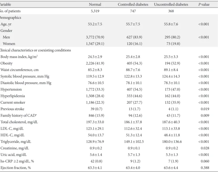
The mean age of study participants was 53.7±7.6 years and 4,694 (73.0%) were male. Of them, 5,319, 747, and 368 partici- pants were categorized as normal, controlled diabetes, and un- controlled diabetes, respectively. The baseline characteristics of the study population according to the diabetes control are listed in Table 1, Supplementary Table 1. In controlled diabetes, the mean FPG and HbA1c were 121.8±17.7 mg/dL and 6.2%±
0.5%. On the other hand, in uncontrolled diabetes, the mean FPG and HbA1c were 158.6±40.8 mg/dL and 8.1%±1.2%.
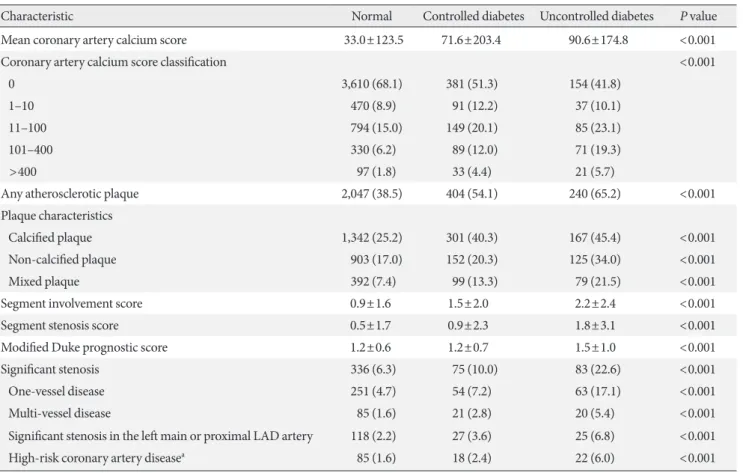
CCTA findings
CCTA findings according to the diabetes control are shown (Table 2, Supplementary Table 2). A total of 236 coronary seg- ments (0.2%) were not interpretable due to artifacts. There was significant difference in CACS according to the diabetes con- trol (P<0.001). The prevalence of any atherosclerotic, calcified, non-calcified, or mixed plaque increased with the diabetes control (P for all<0.001). Plaque burden scores such as seg- ment involvement score, segment stenosis score, and modified Duke prognostic score also increased with the diabetes control (P for all<0.001). Of study participants, 494 (7.7%) had signifi- cant coronary arteries stenosis (≥50% diameter stenosis) in at
Table 1. Baseline characteristics of study participants according to the diabetes control
Variable Normal Controlled diabetes Uncontrolled diabetes P value
No. of patients 5,319 747 368
Demographics
Age, yr 53.2±7.5 55.7±7.5 55.8±7.6 <0.001
Gender
Men 3,772 (70.9) 627 (83.9) 295 (80.2) <0.001
Women 1,547 (29.1) 120 (16.1) 73 (19.8)
Clinical characteristics or coexisting conditions
Body mass index, kg/m2 24.5±2.9 25.4±2.8 25.5±3.3 <0.001
Obesity 2,226 (41.9) 405 (54.3) 194 (52.9) <0.001
Waist circumference, cm 85.2±8.3 88.7±7.6 89.1±8.4 <0.001
Systolic blood pressure, mm Hg 119.5±12.9 122.8±13.3 124.4±14.3 <0.001
Diastolic blood pressure, mm Hg 76.6±10.5 78.1±10.1 78.3±10.1 <0.001
Hypertension 1,772 (33.3) 407 (54.5) 173 (47.0) <0.001
Hyperlipidemia 1,508 (28.4) 333 (44.6) 162 (44.0) <0.001
Current smoker 1,186 (22.3) 207 (27.7) 132 (35.9) <0.001
Previous stroke 39 (0.7) 13 (1.7) 4 (1.1) 0.019
Family history of CADa 846 (15.9) 94 (12.6) 43 (11.7) 0.009
Total cholesterol, mg/dL 197.3±33.0 186.1±37.8 187.6±40.3 <0.001
LDL-C, mg/dL 123.1±29.1 112.6±32.4 113.1±33.8 <0.001
HDL-C, mg/dL 54.0±13.7 51.3±12.4 48.4±11.8 <0.001
Triglyceride, mg/dL 128.9±76.9 149.1±102.5 180.0±136.6 <0.001
Creatinine, mg/dL 0.9±0.2 0.9±0.1 0.9±0.2 0.028
Uric acid, mg/dL 5.6±1.4 5.7±1.3 5.3±1.3 <0.001
hs-CRP ≥2 mg/dL, % 42 (0.8) 9 (1.2) 7 (1.9) 0.060
Ejection fraction, % 63.3±4.1 63.4±4.0 63.6±4.4 0.388
Values are presented as mean±standard deviation or number (%).
CAD, coronary artery disease; LDL-C, low density lipoprotein cholesterol; HDL-C, high density lipoprotein cholesterol; hs-CRP, high-sensitivi- ty C-reactive protein.
aCAD in a first-degree relative of any age.
least one coronary artery on CCTA. The prevalence of signifi- cant stenosis, multi-vessel disease, significant stenosis in the LM or proximal LAD artery, high-risk CAD significantly in- creased according to the diabetes control (P for all<0.001).
Association between the diabetes control and subclinical atherosclerosis
Controlled diabetic individuals had more calcified and mixed plaques than the normal individuals. However, there were no statistically significant differences in the adjusted odds ratios for any atherosclerotic and non-calcified plaque, significant stenosis, multi-vessel disease, significant stenosis in the LM or proximal LAD, and high-risk CAD between the normal and controlled diabetic individuals. On the other hand, uncon- trolled diabetic individuals had significantly associated with any subclinical coronary atherosclerosis compared with nor- mal individuals (P for all<0.05) (Table 3).
Clinical outcomes
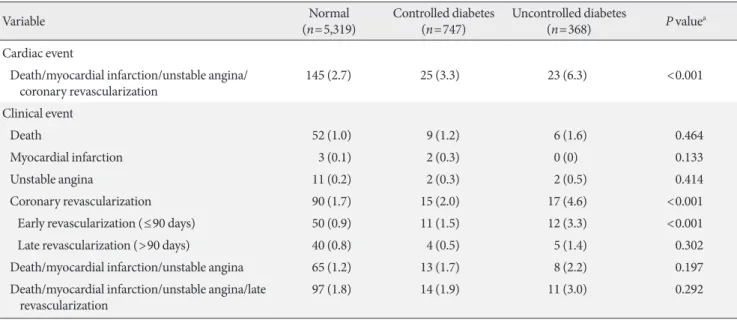
Supplementary Fig. 2 shows the study flow. During the follow- up period (median 5.4 years [interquartile range, 4.4 to 6.4 years]), a total of 209 cardiac events occurred in 193 patients:
67 all-cause deaths, five non-fatal myocardial infarctions, 15 acute coronary syndrome requiring hospitalization, and 122 coronary revascularizations (Table 4, Supplementary Table 3).
Fig. 1 shows Kaplan-Meier survival curves according to the di- abetes control. The 6-year cardiac event-free survival rates were 97.2%±0.2% in normal individuals, 96.4%±0.7% in con- trolled diabetic individuals, and 93.9%±1.3% in uncontrolled individuals (log-rank P<0.001). There was no significant dif- ference in cardiac events between normal and controlled dia- betic individuals (P=0.365). However, uncontrolled diabetes was associated with an increased risk of cardiac events com- pared with normal individuals (P<0.001) and controlled dia- betic individuals (P=0.023).
Table 2. Coronary computed tomographic angiographic findings according to the diabetes control
Characteristic Normal Controlled diabetes Uncontrolled diabetes P value
Mean coronary artery calcium score 33.0±123.5 71.6±203.4 90.6±174.8 <0.001
Coronary artery calcium score classification <0.001
0 3,610 (68.1) 381 (51.3) 154 (41.8)
1–10 470 (8.9) 91 (12.2) 37 (10.1)
11–100 794 (15.0) 149 (20.1) 85 (23.1)
101–400 330 (6.2) 89 (12.0) 71 (19.3)
>400 97 (1.8) 33 (4.4) 21 (5.7)
Any atherosclerotic plaque 2,047 (38.5) 404 (54.1) 240 (65.2) <0.001
Plaque characteristics
Calcified plaque 1,342 (25.2) 301 (40.3) 167 (45.4) <0.001
Non-calcified plaque 903 (17.0) 152 (20.3) 125 (34.0) <0.001
Mixed plaque 392 (7.4) 99 (13.3) 79 (21.5) <0.001
Segment involvement score 0.9±1.6 1.5±2.0 2.2±2.4 <0.001
Segment stenosis score 0.5±1.7 0.9±2.3 1.8±3.1 <0.001
Modified Duke prognostic score 1.2±0.6 1.2±0.7 1.5±1.0 <0.001
Significant stenosis 336 (6.3) 75 (10.0) 83 (22.6) <0.001
One-vessel disease 251 (4.7) 54 (7.2) 63 (17.1) <0.001
Multi-vessel disease 85 (1.6) 21 (2.8) 20 (5.4) <0.001
Significant stenosis in the left main or proximal LAD artery 118 (2.2) 27 (3.6) 25 (6.8) <0.001
High-risk coronary artery diseasea 85 (1.6) 18 (2.4) 22 (6.0) <0.001
Values are presented as mean±standard deviation or number (%).
LAD, left anterior descending.
aDefined as at least 2-vessel coronary disease with proximal left anterior descending artery involvement, 3-vessel disease, or left main disease.
DISCUSSION
The main findings of this study were as follows: (1) in asymp-
tomatic individuals, uncontrolled diabetes was independently associated with significant subclinical coronary atherosclerosis compared with normal and controlled diabetic individuals; (2) Table 3. Univariable and multivariable analyses of each coronary computed tomographic angiography variables, corrected for clinical risk factors
Variable Univariable Multivariable
Odds ratio (95% CI) P value Odds ratio (95% CI) P value Any atherosclerotic plaque
Uncontrolled diabetes 3.00 (2.40–3.74) <0.001 2.16 (1.70–2.75) <0.001
Controlled diabetes 1.88 (1.61–2.20) <0.001 1.16 (0.98–1.38) 0.086
Normal (reference: normal) 1 - 1 -
Calcified plaque
Uncontrolled diabetes 2.46 (1.99–3.05) <0.001 1.77 (1.40–2.23) <0.001
Controlled diabetes 2.00 (1.71–2.34) <0.001 1.29 (1.08–1.53) 0.004
Normal (reference: normal) 1 - 1 -
Non-calcified plaque
Uncontrolled diabetes 2.52 (2.00–3.16) <0.001 2.03 (1.60–2.57) <0.001
Controlled diabetes 1.25 (1.03–1.51) 0.023 0.90 (0.74–1.10) 0.323
Normal (reference: normal) 1 - 1 -
Mixed plaque
Uncontrolled diabetes 3.44 (2.63–4.50) <0.001 2.59 (1.95–3.43) <0.001
Controlled diabetes 1.92 (1.52–2.43) <0.001 1.32 (1.04–1.69) 0.025
Normal (reference: normal) 1 - 1 -
Significant stenosis in at least one coronary artery
Uncontrolled diabetes 4.32 (3.30–5.65) <0.001 3.34 (2.52–4.43) <0.001
Controlled diabetes 1.66 (1.27–2.15) <0.001 1.08 (0.82–1.42) 0.583
Normal (reference: normal) 1 - 1 -
Multi-vessel disease
Uncontrolled diabetes 3.54 (2.15–5.83) <0.001 2.51 (1.50–4.22) 0.001
Controlled diabetes 1.78 (1.10–2.89) 0.019 1.05 (0.63–1.75) 0.844
Normal (reference: normal) 1 - 1 -
Significant stenosis in the left main or proximal left anterior descending artery
Uncontrolled diabetes 3.21 (2.06–5.01) <0.001 2.39 (1.51–3.79) <0.001
Controlled diabetes 1.65 (1.08–2.53) 0.021 1.06 (0.68–1.66) 0.786
Normal (reference: normal) 1 - 1 -
High-risk coronary artery diseasea
Uncontrolled diabetes 3.92 (2.42–6.34) <0.001 2.76 (1.68–4.56) <0.001
Controlled diabetes 1.52 (0.91–2.54) 0.110 0.90 (0.52–1.54) 0.692
Normal (reference: normal) 1 - 1 -
Covariates in the multivariable model include age, sex, obesity, hypertension, hyperlipidemia, current smoking, family history of coronary ar- tery disease, and high-sensitivity C-reactive protein ≥2 mg/L.
CI, confidence interval.
aDefined as at least 2-vessel coronary disease with proximal left anterior descending artery involvement, 3-vessel disease, or left main disease.
Table 4. Clinical outcomes according to the diabetes control
Variable Normal
(n=5,319) Controlled diabetes
(n=747) Uncontrolled diabetes
(n=368) P valuea
Cardiac event
D eath/myocardial infarction/unstable angina/
coronary revascularization 145 (2.7) 25 (3.3) 23 (6.3) <0.001
Clinical event
Death 52 (1.0) 9 (1.2) 6 (1.6) 0.464
Myocardial infarction 3 (0.1) 2 (0.3) 0 (0) 0.133
Unstable angina 11 (0.2) 2 (0.3) 2 (0.5) 0.414
Coronary revascularization 90 (1.7) 15 (2.0) 17 (4.6) <0.001
Early revascularization (≤90 days) 50 (0.9) 11 (1.5) 12 (3.3) <0.001
Late revascularization (>90 days) 40 (0.8) 4 (0.5) 5 (1.4) 0.302
Death/myocardial infarction/unstable angina 65 (1.2) 13 (1.7) 8 (2.2) 0.197
D eath/myocardial infarction/unstable angina/late
revascularization 97 (1.8) 14 (1.9) 11 (3.0) 0.292
Values are presented as number (%).
aP values were calculated using the log-rank test.
Fig. 1. Kaplan-Meier event-free survival curves of (A) 6-year cardiac events and (B) 6-year composite outcomes of all-cause death, myocardial infarction and unstable angina according to the diabetes control. The numbers in each figure represent the 6-year event-free survival rates.
100
95
90
100
95
90
Event-free survival rate (%) Event-free survival rate (%)
0 1 2 3 4 5 6 0 1 2 3 4 5 6
Years after enrollment Years after enrollment
5,319 5,236 4,984 4,614 4,146 3,314 1,719 5,319 5,295 5,046 4,683 4,216 3,384 1,762
747 733 697 643 586 489 255 747 744 707 654 595 497 257
368 353 338 312 285 231 115 368 366 353 327 300 245 120
No. of patients No. of patients
Normal Normal
Controlled diabetes Controlled diabetes
Uncontrolled diabetes Uncontrolled diabetes
Cardiac events Death/Myocaridal infarction/Unstable angina 97.2%±0.2%
P=0.365
98.7%±0.2%
96.4%±0.7%
P=0.023
98.0%±0.6%
93.9%±1.3%
97.9%±0.8%
Log-rank P<0.001 Log-rank P=0.197
Normal
Controlled diabetes Uncontrolled diabetes
Normal
Controlled diabetes Uncontrolled diabetes P<0.001
consequently, uncontrolled diabetic individuals experienced more cardiac events; and (3) there findings suggest that diabe- tes control had a beneficial effect on the risk of subclinical cor- onary atherosclerosis in asymptomatic individuals.
In this study, uncontrolled diabetic individuals had a higher prevalence, extent, and severity of coronary atherosclerosis on CCTA than normal individuals. Even after adjustments for clinical and laboratory variables, uncontrolled diabetes was consistently associated with any subclinical coronary athero-
sclerosis. Moreover, uncontrolled diabetes was an independent risk factor for significant stenosis in the LM or proximal LAD, multi-vessel disease and high-risk CAD, which have been known to be associated with a worse prognosis [23]. As a re- sult, uncontrolled diabetic individuals experienced more car- diac events. By contrast, compared with normal individuals, controlled diabetic individuals were not associated with the in- creased risk of significant subclinical coronary atherosclerosis (e.g., significant stenosis at least one coronary artery, signifi-
A B
cant stenosis in the LM or proximal LAD, multi-vessel disease, or high-risk CAD). Consequently, cardiac event rates may have been comparable. Therefore, our findings support that di- abetes control is important in preventing significant subclini- cal coronary atherosclerosis and cardiac events in asymptom- atic diabetic individuals.
In earlier studies, intensive glucose control has not shown a significant effect on the rates of major cardiovascular events in diabetic patients [4,24]. However, long-term follow-up studies have demonstrated that early intensive glucose control may be effective in decreasing cardiovascular events in these patients [3,11]. A recent 15-year follow-up study also observed a signif- icantly lower risk of major cardiovascular events during the periods of separation of the HbA1c curves [25]. Given that cardiac events are thought to occur after long periods of sub- clinical disease, our study provides some insights into these re- sults. The current study showed that control of diabetes was as- sociated with beneficial effects for significant subclinical coro- nary atherosclerosis which may have led to the differences in cardiac events between uncontrolled and controlled diabetic individuals. Therefore, in asymptomatic diabetic individuals, emphasis should be given on diabetes control to prevent future cardiac events.
To date, randomized trials have failed to demonstrate that routine screening for CAD can decrease cardiac events in rela- tively well-controlled asymptomatic diabetic populations [26,27]. In these trials, the study participants were treated by contemporary medical practice, achieving HbA1c, LDL-C, and systolic blood pressure levels at or near the target ranges (HbA1c 7.0% to 7.5%, LDL-C 86 to 114 mg/dL, and systolic blood pressure 129 to 133 mm Hg). Eventually, intensive inter- vention for current cardiac risk factors resulted in lower cardi- ac event rates in these patients. A previous study also demon- strated resolution of myocardial ischemia resulted from more aggressive treatment of cardiovascular risk factors [28]. In this present study, controlled diabetic individuals also had near tar- geted levels for HbA1c (6.2%), LDL-C (113 mg/dL), and sys- tolic blood pressure (123 mm Hg). As a result, controlled dia- betes was not associated with significant subclinical coronary atherosclerosis on CCTA and an increased risk of cardiac events. Since previous and our studies showed that the adher- ence to current guidelines could improve subclinical coronary atherosclerosis and clinical outcomes in asymptomatic diabetic individuals, further implementation of established guidelines is needed in this population.
Our study has several limitations. First, the current study was based in a single center. Moreover, because all study par- ticipants voluntarily went to the hospital for general health ex- amination, there was a potential for selection bias. Second, since the present study is a retrospective cohort study, there was a limitation that these data did not fully reflect patient out- comes. Additionally, we did not specify the cause of death.
Third, calcified plaques and higher coronary artery calcium score may lead to overestimation of significant coronary arter- ies stenosis. Fourth, our study population was almost Korean men. In addition, ethnic differences and clinical differences in diabetes have been noted between Asian and Western popula- tions. Therefore, the generalization of our findings to female and other ethnic groups may be limited. Fifth, CCTA itself has limitations including radiation hazard, use of contrast, and higher cost. Although our study enrolled only volunteers, the use of CCTA in asymptomatic individuals has not yet been justified. Finally, we did not obtain the specific medical histo- ries about diabetic duration, modalities of diabetic manage- ment, and antiplatelet agents, which could play an important role in potential confounders. Despite these limitations, we be- lieve that the current study may have a clinical implication in unveiling an association between glycemic control and sub- clinical coronary atherosclerosis in asymptomatic individuals.
In this large observational study of asymptomatic individu- als undergoing CCTA, uncontrolled diabetes was associated with significant subclinical coronary atherosclerosis and an in- creased risk for cardiac events. However, controlled diabetes did not associated with significant subclinical coronary athero- sclerosis and an increase in cardiac events. These findings should be validated in additional studies.
SUPPLEMENTARY MATERIALS
Supplementary materials related to this article can be found online at https://doi.org/10.4093/dmj.2019.0073.
CONFLICTS OF INTEREST
No potential conflict of interest relevant to this article was re- ported.
AUTHOR CONTRIBUTIONS
Conception or design: G.M.P., C.H.L., S.W.L.
Acquisition, analysis, or interpretation of data: G.M.P., C.H.L., S.W.L., S.C.Y., Y.H.K., D.H.Y., J.W.K., T.H.L., H.K.K., J.C.
Drafting the work or revising: G.M.P., C.H.L., S.W.L., S.C.Y., Y.G.K., K.B.W., S.H.A., S.J.K., E.H.K., W.J.L., M.S.K., J.Y.P., S.G.L.
Final approval of the manuscript: G.M.P., C.H.L., S.W.L., S.C.Y., Y.H.K., Y.G.K., K.B.W., S.H.A., S.J.K., D.H.Y., J.W.K., T.H.L., E.H.K., W.J.L., M.S.K., J.Y.P., H.K.K., J.C., S.G.L.
ORCID
Gyung-Min Park https://orcid.org/0000-0001-5846-0606 Seung-Whan Lee https://orcid.org/0000-0002-2662-5952
ACKNOWLEDGMENTS
This research was supported by Basic Science Research Program through the National Research Foundation of Korea funded by the Ministry of Education (2018R1D1A3B07043344).
REFERENCES
1. American Diabetes Association. 6. Glycemic targets: standards of medical care in diabetes. 2018. Diabetes Care 2018;41(Suppl 1):S55-64.
2. Diabetes Control and Complications Trial Research Group, Nathan DM, Genuth S, Lachin J, Cleary P, Crofford O, Davis M, Rand L, Siebert C. The effect of intensive treatment of dia- betes on the development and progression of long-term com- plications in insulin-dependent diabetes mellitus. N Engl J Med 1993;329:977-86.
3. Holman RR, Paul SK, Bethel MA, Matthews DR, Neil HA. 10- Year follow-up of intensive glucose control in type 2 diabetes.
N Engl J Med 2008;359:1577-89.
4. Duckworth W, Abraira C, Moritz T, Reda D, Emanuele N, Reaven PD, Zieve FJ, Marks J, Davis SN, Hayward R, Warren SR, Goldman S, McCarren M, Vitek ME, Henderson WG, Huang GD; VADT Investigators. Glucose control and vascular complications in veterans with type 2 diabetes. N Engl J Med 2009;360:129-39.
5. ADVANCE Collaborative Group, Patel A, MacMahon S, Chalmers J, Neal B, Billot L, Woodward M, Marre M, Cooper M, Glasziou P, Grobbee D, Hamet P, Harrap S, Heller S, Liu L, Mancia G, Mogensen CE, Pan C, Poulter N, Rodgers A, Wil- liams B, Bompoint S, de Galan BE, Joshi R, Travert F. Intensive
blood glucose control and vascular outcomes in patients with type 2 diabetes. N Engl J Med 2008;358:2560-72.
6. Ismail-Beigi F, Craven T, Banerji MA, Basile J, Calles J, Cohen RM, Cuddihy R, Cushman WC, Genuth S, Grimm RH Jr, Hamilton BP, Hoogwerf B, Karl D, Katz L, Krikorian A, O’Connor P, Pop-Busui R, Schubart U, Simmons D, Taylor H, Thomas A, Weiss D, Hramiak I; ACCORD trial group. Effect of intensive treatment of hyperglycaemia on microvascular outcomes in type 2 diabetes: an analysis of the ACCORD ran- domised trial. Lancet 2010;376:419-30.
7. Van Werkhoven JM, Cademartiri F, Seitun S, Maffei E, Palum- bo A, Martini C, Tarantini G, Kroft LJ, de Roos A, Weustink AC, Jukema JW, Ardissino D, Mollet NR, Schuijf JD, Bax JJ. Di- abetes: prognostic value of CT coronary angiography. Compar- ison with a nondiabetic population. Radiology 2010;256:83-92.
8. Hadamitzky M, Hein F, Meyer T, Bischoff B, Martinoff S, Scho- mig A, Hausleiter J. Prognostic value of coronary computed to- mographic angiography in diabetic patients without known coronary artery disease. Diabetes Care 2010;33:1358-63.
9. Rana JS, Dunning A, Achenbach S, Al-Mallah M, Budoff MJ, Cademartiri F, Callister TQ, Chang HJ, Cheng VY, Chinnaiyan K, Chow BJ, Cury R, Delago A, Feuchtner G, Hadamitzky M, Hausleiter J, Kaufmann P, Karlsberg RP, Kim YJ, Leipsic J, Labounty TM, Lin FY, Maffei E, Raff G, Villines TC, Shaw LJ, Berman DS, Min JK. Differences in prevalence, extent, severity, and prognosis of coronary artery disease among patients with and without diabetes undergoing coronary computed tomog- raphy angiography: results from 10,110 individuals from the CONFIRM (COronary CT Angiography EvaluatioN For Clin- ical Outcomes): an InteRnational Multicenter Registry. Diabe- tes Care 2012;35:1787-94.
10. Hammoud T, Tanguay JF, Bourassa MG. Management of coro- nary artery disease: therapeutic options in patients with diabe- tes. J Am Coll Cardiol 2000;36:355-65.
11. Writing Group for the DCCT/EDIC Research Group, Orchard TJ, Nathan DM, Zinman B, Cleary P, Brillon D, Backlund JY, Lachin JM. Association between 7 years of intensive treatment of type 1 diabetes and long-term mortality. JAMA 2015;313:45- 53.
12. Park GM, Yun SC, Cho YR, Gil EH, Her SH, Kim SH, Jo MW, Lee MS, Lee SW, Kim YH, Yang DH, Kang JW, Lim TH, Kim BJ, Koh JM, Kim HK, Choe J, Park SW, Park SJ. Prevalence of coronary atherosclerosis in an Asian population: findings from coronary computed tomographic angiography. Int J Cardio- vasc Imaging 2015;31:659-68.
13. Park GM, Han S, Kim SH, Jo MW, Her SH, Lee JB, Lee MS, Kim HC, Ahn JM, Lee SW, Kim YH, Kim BJ, Koh JM, Kim HK, Choe J, Park SW, Park SJ. Model for assessing cardiovas- cular risk in a Korean population. Circ Cardiovasc Qual Out- comes 2014;7:944-51.
14. American Diabetes Association. 2. Classification and diagnosis of diabetes: standards of medical care in diabetes. 2018. Diabe- tes Care 2018;41(Suppl 1):S13-27.
15. Jung CH, Lee MJ, Hwang JY, Jang JE, Leem J, Park JY, Lee J, Kim HK, Lee WJ. Elevated serum ferritin level is associated with the incident type 2 diabetes in healthy Korean men: a 4 year longitudinal study. PLoS One 2013;8:e75250.
16. Raff GL, Abidov A, Achenbach S, Berman DS, Boxt LM, Budoff MJ, Cheng V, DeFrance T, Hellinger JC, Karlsberg RP; Society of Cardiovascular Computed Tomography. SCCT guidelines for the interpretation and reporting of coronary computed to- mographic angiography. J Cardiovasc Comput Tomogr 2009;3:
122-36.
17. Agatston AS, Janowitz WR, Hildner FJ, Zusmer NR, Viamonte M Jr, Detrano R. Quantification of coronary artery calcium us- ing ultrafast computed tomography. J Am Coll Cardiol 1990;
15:827-32.
18. Leber AW, Becker A, Knez A, von Ziegler F, Sirol M, Nikolaou K, Ohnesorge B, Fayad ZA, Becker CR, Reiser M, Steinbeck G, Boekstegers P. Accuracy of 64-slice computed tomography to classify and quantify plaque volumes in the proximal coronary system: a comparative study using intravascular ultrasound. J Am Coll Cardiol 2006;47:672-7.
19. Hausleiter J, Meyer T, Hadamitzky M, Kastrati A, Martinoff S, Schomig A. Prevalence of noncalcified coronary plaques by 64-slice computed tomography in patients with an intermedi- ate risk for significant coronary artery disease. J Am Coll Car- diol 2006;48:312-8.
20. Min JK, Shaw LJ, Devereux RB, Okin PM, Weinsaft JW, Russo DJ, Lippolis NJ, Berman DS, Callister TQ. Prognostic value of multidetector coronary computed tomographic angiography for prediction of all-cause mortality. J Am Coll Cardiol 2007;50:
1161-70.
21. Min JK, Berman DS, Dunning A, Achenbach S, Al-Mallah M, Budoff MJ, Cademartiri F, Callister TQ, Chang HJ, Cheng V, Chinnaiyan K, Chow BJ, Cury R, Delago A, Feuchtner G, Had- amitzky M, Hausleiter J, Kaufmann P, Karlsberg RP, Kim YJ, Leipsic J, Lin FY, Maffei E, Plank F, Raff G, Villines T, Labounty TM, Shaw LJ. All-cause mortality benefit of coronary revascu- larization vs. medical therapy in patients without known coro-
nary artery disease undergoing coronary computed tomo- graphic angiography: results from CONFIRM (COronary CT Angiography EvaluatioN for Clinical Outcomes: an InteRna- tional Multicenter Registry). Eur Heart J 2012;33:3088-97.
22. BARI 2D Study Group, Frye RL, August P, Brooks MM, Hardi- son RM, Kelsey SF, MacGregor JM, Orchard TJ, Chaitman BR, Genuth SM, Goldberg SH, Hlatky MA, Jones TL, Molitch ME, Nesto RW, Sako EY, Sobel BE. A randomized trial of therapies for type 2 diabetes and coronary artery disease. N Engl J Med 2009;360:2503-15.
23. Park GM, Lee JH, Lee SW, Yun SC, Kim YH, Cho YR, Gil EH, Kim TS, Kim CJ, Cho JS, Park MW, Her SH, Yang DH, Kang JW, Lim TH, Koh EH, Lee WJ, Kim MS, Lee KU, Kim HK, Choe J, Park JY. Comparison of coronary computed tomo- graphic angiographic findings in asymptomatic subjects with versus without diabetes mellitus. Am J Cardiol 2015;116:372-8.
24. UK Prospective Diabetes Study (UKPDS) Group. Intensive blood-glucose control with sulphonylureas or insulin com- pared with conventional treatment and risk of complications in patients with type 2 diabetes (UKPDS 33). Lancet 1998;352:
837-53.
25. Reaven PD, Emanuele NV, Wiitala WL, Bahn GD, Reda DJ, McCarren M, Duckworth WC, Hayward RA; VADT Investiga- tors. Intensive glucose control in patients with type 2 diabetes:
15-year follow-up. N Engl J Med 2019;380:2215-24.
26. Young LH, Wackers FJ, Chyun DA, Davey JA, Barrett EJ, Taille- fer R, Heller GV, Iskandrian AE, Wittlin SD, Filipchuk N, Rat- ner RE, Inzucchi SE; DIAD Investigators. Cardiac outcomes after screening for asymptomatic coronary artery disease in patients with type 2 diabetes: the DIAD study. A randomized controlled trial. JAMA 2009;301:1547-55.
27. Muhlestein JB, Lappe DL, Lima JA, Rosen BD, May HT, Knight S, Bluemke DA, Towner SR, Le V, Bair TL, Vavere AL, Ander- son JL. Effect of screening for coronary artery disease using CT angiography on mortality and cardiac events in high-risk pa- tients with diabetes: the FACTOR-64 randomized clinical trial.
JAMA 2014;312:2234-43.
28. Wackers FJ, Chyun DA, Young LH, Heller GV, Iskandrian AE, Davey JA, Barrett EJ, Taillefer R, Wittlin SD, Filipchuk N, Rat- ner RE, Inzucchi SE; Detection of Ischemia in Asymptomatic Diabetics (DIAD) Investigators. Resolution of asymptomatic myocardial ischemia in patients with type 2 diabetes in the De- tection of Ischemia in Asymptomatic Diabetics (DIAD) study.
Diabetes Care 2007;30:2892-8.