HIGHLIGHTS
• The purpose of this study is to investigate the effects of white matter hyperintensity (WMH) on functional improvement using the Korean version of Modified Barthel Index during inpatient stroke rehabilitation.
• There was a significant association between the severity of WMH and the poor functional improvement of ischemic stroke patients.
• The WMH might be considered as one of many factors that can affect functional recovery during rehabilitation of stroke.
Brain Neurorehabil. 2019 Sep;12(2):e14 https://doi.org/10.12786/bn.2019.12.e14 pISSN 1976-8753·eISSN 2383-9910
Original Article
Received: Mar 28, 2019 Revised: Jun 21, 2019 Accepted: Aug 14, 2019 Correspondence to Jinmann Chon
Department of Rehabilitation Medicine, Kyung Hee University Medical Center, 23 Kyungheedae-ro, Dongdaemun-gu, Seoul 02447, Korea.
E-mail: [email protected]
Miryeong Yang, Seung Ah Lee, Yunsoo Soh, Yong Kim, Eun Jeong Lee, Yeocheon Yun, Jae Hoon Kim, Jinmann Chon
Effect of White Matter Hyperintensity on the Functional Outcome of
Ischemic Stroke Patients after Inpatient Stroke Rehabilitation
Brain & NeuroRehabilitation
Copyright © 2019. Korea Society for Neurorehabilitation i
ABSTRACT
The aim of the study is to investigate the association between cerebral white matter
hyperintensity (WMH) and the functional improvement using the Korean version of Modified Barthel Index (K-MBI) score during inpatient stroke rehabilitation. One hundred sixty participants were divided into 2 groups based on the severity of WMH according to Fazekas scale: Mild WMH group was defined as patients with Fazekas scale 0 and 1, and severe WMH group was defined as Fazekas scale 2 and 3. Functional status was assessed using the K-MBI score, and functional gains were calculated from the K-MBI score. The absolute functional gain in mild WMH group was significantly higher compared to severe WMH group (p < 0.05). In addition, patients in mild WMH had higher absolute functional efficiency, rehabilitation effectiveness, and relative functional efficiency. In the generalized linear model analyses, there was an association between functional outcomes and severity of WMH. In this study, the severity and extent of WMH are significantly correlated with poor functional improvement in patients with ischemic stroke. The WMH could be considered as one of many factors that can influence functional recovery during rehabilitation of stroke.
Keywords: White Matter; Stroke Rehabilitation; Recovery of Function; Inpatients
INTRODUCTION
Cerebral white matter hyperintensity (WMH) is a usual finding on magnetic resonance imaging (MRI) in elderly persons and patients with cerebrovascular diseases. WMH is found in 21% of individuals 64 years of age, and 94% of individuals 82 years of age [1]. The etiology of WMH is secondary to small-vessel ischemic changes. Arteriolosclerosis, myelin pallor, and Braak score were significantly associated with increased WMH accumulation [2]. Previous studies have suggested that WMH is a risk factor dementia after stroke [3]. In addition, one study determined that WMH is related with executive dysfunction [4].
Stroke rehabilitation is a progressive, goal-orientated practice to achieve optimal
physical, cognitive, emotional and functional activity level [5]. Many factors can affect the
Original Article
Received: Mar 28, 2019 Revised: Jun 21, 2019 Accepted: Aug 14, 2019 Correspondence to Jinmann Chon
Department of Rehabilitation Medicine, Kyung Hee University Medical Center, 23 Kyungheedae-ro, Dongdaemun-gu, Seoul 02447, Korea.
E-mail: [email protected] Copyright © 2019. Korea Society for Neurorehabilitation
This is an Open Access article distributed under the terms of the Creative Commons Attribution Non-Commercial License (https://
creativecommons.org/licenses/by-nc/4.0) which permits unrestricted non-commercial use, distribution, and reproduction in any medium, provided the original work is properly cited.
ORCID iDs Miryeong Yang
https://orcid.org/0000-0001-6167-5042 Seung Ah Lee
https://orcid.org/0000-0002-3426-6259 Yunsoo Soh
https://orcid.org/0000-0001-8368-4900 Yong Kim
https://orcid.org/0000-0003-0950-3774 Eun Jeong Lee
https://orcid.org/0000-0002-0451-6026 Yeocheon Yun
https://orcid.org/0000-0002-8688-8041 Jae Hoon Kim
https://orcid.org/0000-0001-6770-3606
Miryeong Yang ,1 Seung Ah Lee ,2,3 Yunsoo Soh ,1,3 Yong Kim ,1,3 Eun Jeong Lee ,1 Yeocheon Yun ,1 Jae Hoon Kim ,2 Jinmann Chon 1,3
1Department of Rehabilitation Medicine, Kyung Hee University Medical Center, Seoul, Korea
2Department of Rehabilitation Medicine, Kyung Hee University Hospital at Gangdong, Seoul, Korea
3Department of Medicine, Kyung Hee University, Seoul, Korea
Effect of White Matter Hyperintensity on the Functional Outcome of
Ischemic Stroke Patients after
Inpatient Stroke Rehabilitation
Jinmann Chon
https://orcid.org/0000-0002-4186-6623 Conflict of Interest
The authors have no potential conflicts of interest to disclose.
rehabilitation outcome after stroke such as medical history, site of lesion, age, sex, ethnicity, visual impairments, aphasia, depression, and cognition [6-9].
There have been few studies investigating the association between WMH and functional gains during inpatient rehabilitation. We assumed that WMH causes cognitive and executive dysfunction that can affect functional outcomes in ischemic stroke patients. Therefore, the aim of the study is to examine the effect of WMHs on the functional improvement using the Korean version of Modified Barthel Index (K-MBI) during inpatient stroke rehabilitation.
MATERIALS AND METHODS
Subjects
This was a retrospective study of patients with middle cerebral artery (MCA) territory ischemic stroke. The data were collected from medical records of patients who were admitted to Kyung Hee Hospital for ischemic infarction, between January 2015 and May 2017. The patients were delivered from an acute care department (e.g., internal medicine, neurology, neurosurgery, and intensive care unit) at Kyung Hee Hospital or from other neighboring hospitals. This study was approved by the Institutional Review Board (IRB) of Kyung Hee University Hospital (IRB No. 2019-05-074).
The inclusion criteria for the study were 1) MCA territory ischemic stroke confirmed by MRI;
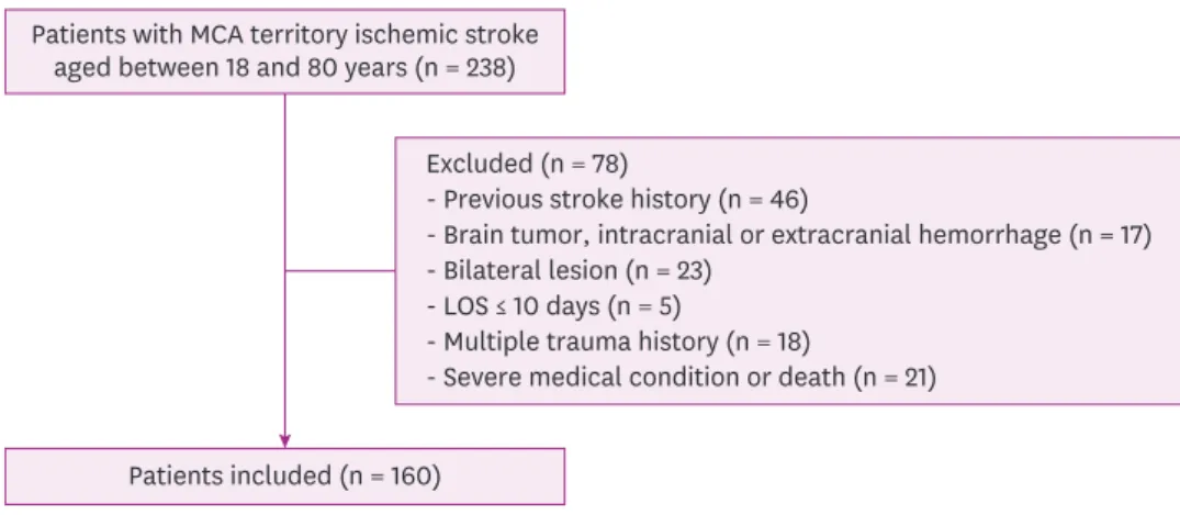
2) Patients' age between 18 and 80 years; and 3) Stable medical status being able to receive active rehabilitation treatment (Fig. 1). Exclusion criteria were 1) Evidence of a previous stroke by patient's history or computed tomography scan; 2) Evidence of brain tumor, trauma, intracranial, or extracranial hemorrhage; 3) Bilateral lesions; 4) Length of stay (LOS) in the rehabilitation department less than 10 days assuming that the extent of rehabilitation in such a short period is limited; 5) Presence of other acute disabilities such as multiple trauma history; and 6) Presence of additional severe medical conditions restricting active rehabilitation (e.g., cardiac failure with New York Heart Association functional capacity stage III–IV, severe chronic lung disease necessitating a constant use of oxygen), or transfer to an acute care departments due to complications and/or death while hospitalized at the rehabilitation department.
2/9 https://doi.org/10.12786/bn.2019.12.e14
Effect of White Matter Hyperintensity Brain & NeuroRehabilitation
https://e-bnr.org
Patients included (n = 160) Patients with MCA territory ischemic stroke
aged between 18 and 80 years (n = 238)
Excluded (n = 78)
- Previous stroke history (n = 46)
- Brain tumor, intracranial or extracranial hemorrhage (n = 17) - Bilateral lesion (n = 23)
- LOS ≤ 10 days (n = 5)
- Multiple trauma history (n = 18)
- Severe medical condition or death (n = 21)
Fig. 1. Inclusion flow chart.
MCA, middle cerebral artery; LOS, length of stay.
The patients undergo, on average, 6 hours of physical therapy and 6 hours of occupational therapy a week (6 days/week). Demographic data and history of premorbid risk factors were collected from medical records.
Outcome measure
Functional and cognitive status assessment
Functional status was assessed by the K-MBI score. The K-MBI is a measure of the activities of daily living (ADL) scoring from 0 to 100 and is valid and reliable in a patient with stroke [10].
Cognitive status was evaluated by Korean version of Mini-Mental State Examination (K-MMSE) which score ranged from 0 to 30. This tool is a brief instrument used to assess cognitive abilities [11].
The K-MBI and K-MMSE were administered to all patients within 72 hours after admission by the rehabilitation team (composed of a physiotherapist, an occupational therapist). The same team evaluated K-MBI and K-MMSE 3 days before discharge. The team administering the K-MBI and K-MMSE did not include the treating physicians. Other variables that were examined included sex, age, the time interval from stroke onset to admission, laterality of stroke, LOS, National Institutes of Health Stroke Scale (NIHSS), and comorbidities such as hypertension, diabetes, dyslipidemia.
K-MMSE gain
The cognitive improvement was assessed by the difference between the K-MMSE score at admission and K-MMSE score at discharge.
Absolute functional gain (AFG) and absolute functional efficiency (AFE)
Functional gains were demonstrated by using absolute and relative methods determined from the K-MBI score. AFG is the K-MBI score gain (ΔK-MBI). AFE reflects AFG per day [12].
AFG = DC(K − MBI) − adm(K − MBI)
(DC, discharge; adm, admission)
Rehabilitation effectiveness (REs) and relative functional efficiency (RFE)
The REs was used to calculate relative gains. REs was suggested firstly by Heinemann et al.
[13] expressed as a percentage indicating the proportion of potential improvement achieved during rehabilitation.
(max, maximum possible score)
AFE = 𝐴𝐴𝐴𝐴𝐴𝐴𝐴𝐴𝐴𝐴𝐴𝐴 𝐿𝐿𝐿𝐿𝐿𝐿𝐿𝐿𝐿𝐿𝐿𝐿
𝑅𝑅𝑅𝑅𝑅𝑅𝑅𝑅𝑅𝑅𝑅𝑅 = 𝐷𝐷𝐷𝐷𝐷𝐷𝐷𝐷(𝐾𝐾𝐾𝐾 − 𝑀𝑀𝑀𝑀𝑀𝑀𝑀𝑀𝑀𝑀𝑀𝑀) − 𝑎𝑎𝑎𝑎𝑎𝑎𝑎𝑎𝑎𝑎𝑎𝑎(𝐾𝐾𝐾𝐾 − 𝑀𝑀𝑀𝑀𝑀𝑀𝑀𝑀𝑀𝑀𝑀𝑀)
𝑎𝑎𝑎𝑎𝑎𝑎𝑎𝑎𝑚𝑚𝑚𝑚(𝐾𝐾𝐾𝐾 − 𝑀𝑀𝑀𝑀𝑀𝑀𝑀𝑀𝑀𝑀𝑀𝑀) − 𝑎𝑎𝑎𝑎𝑎𝑎𝑎𝑎𝑎𝑎𝑎𝑎(𝐾𝐾𝐾𝐾 − 𝑀𝑀𝑀𝑀𝑀𝑀𝑀𝑀𝑀𝑀𝑀𝑀) × 100 (%) RFE = 𝑅𝑅𝑅𝑅𝑅𝑅𝑅𝑅𝑅𝑅𝑅𝑅
𝐿𝐿𝐿𝐿𝐿𝐿𝐿𝐿𝐿𝐿𝐿𝐿
WMH classification
Each MRI study included fluid-attenuated inversion recovery images at the acute stroke phase. We applied the simple modified Fazekas rating scale [14]. The modified Fazekas ranges from grade 0 to grade 3. Grade 1 was characterized by punctate lesions in the deep white matter. The maximum diameter is 9 mm for a single lesion and 20 mm for grouped lesions. Grade 2 was an early confluent lesion of 10–20 mm single lesion and grouped lesions with a diameter of > 20 mm with no bridges connecting individual lesions. Grade 3 was defined by single lesions or confluent lesions of ≥ 20 mm in diameter. All participants were divided into mild and severe groups based on the severity of WMH according to Fazekas scale. Patients with Fazekas scale 0, 1 was sorted as mild WMH group. Severe WMH group was classified by Fazekas scale 2, 3. It was performed by one skilled physician in the department of rehabilitation medicine.
Statistical analysis
Statistical analyses were carried out using the Statistical Package for the Social Sciences (SPSS) version 18.0 for Windows (SPSS Inc., Chicago, IL, USA). The Kolmogorov Smirnov test was applied to assess whether the parametric values were normally distributed. All parametric data were shown to be normally distributed.
We compared the values between the 2 groups (mild WMH group and severe WMH group) according to the severity of WMH. Independent t-tests were performed for continuous variables to compare values between the mild WMH group and severe WMH group. Chi-square tests were conducted for categorical variables. Association between the functional and cognitive outcomes and severity of WMH was analyzed using generalized linear model adjusted for age, sex, stroke lesion side, initial NIHSS, and LOS. A p value < 0.05 was considered significant.
RESULTS
General characteristics
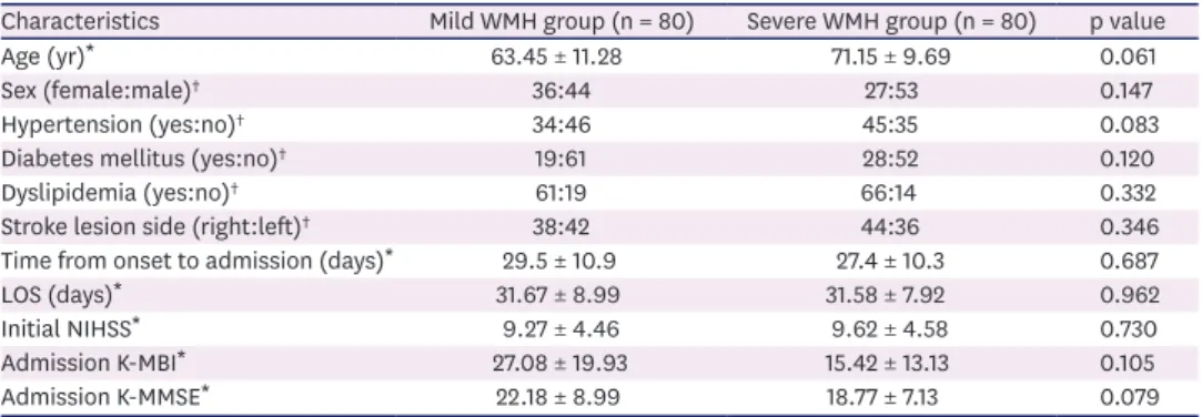
A total of 559 patients were consecutively admitted to Kyung Hee Hospital between January 2015 and May 2017. Of 559 patients, 160 patients fulfilled the inclusion and exclusion criteria and were classified to mild WMH group (n = 80) and severe WMH group (n = 80). In a mild group, 16 patients were Fazekas scale grade 0 and 64 patients were grade 1. In a severe group, 59 patients were grade 2 and 21 patients were grade 3. There was no significant difference in age, sex, stroke lesion side, hypertension, diabetes, and dyslipidemia between mild and severe groups (Table 1).
The mean duration of time from the onset of stroke to admission was 29.5 ± 10.9 days in mild WMH group and 27.4 ± 10.3 days in severe WMH group. LOS was 31.67 ± 8.99 in mild WMH group and 31.58 ± 7.92 in severe WMH group. There were no significant differences between the 2 groups. Initial NIHSS score also showed no significant differences between the 2 groups (p = 0.730). In mild WMH group, admission K-MBI score was 27.08 ± 19.93 and admission K-MMSE was 22.18 ± 8.99. In severe WMH group, admission K-MBI score was 15.42 ± 13.13 and admission K-MMSE was 18.77 ± 7.13, which was lower than mild WMH group. However, there were no statistical differences. At discharge, K-MBI score is 47.75 ± 21.25 in mild WMH group and 22.83 ± 15.23 in the severe group. Discharge K-MMSE is 25.76 ± 5.07 in mild WMH group and 20.20 ± 6.84 in the severe group. There was a significant difference in discharge K-MBI and discharge K-MMSE between mild and severe WMH group (Table 2).
4/9 https://doi.org/10.12786/bn.2019.12.e14
Effect of White Matter Hyperintensity Brain & NeuroRehabilitation
https://e-bnr.org
Cognitive improvement between the mild WMH group and the severe WMH group
K-MMSE gain during inpatient rehabilitation was 3.57 ± 5.70 in mild WMH group and 1.43
± 1.50 in severe WMH group, which showed significantly higher cognitive improvement in mild WMH group than severe WMH group (p < 0.05) (Table 2). The generalized linear model analyses showed association between discharge K-MMSE and severity of WMH. However, there was no significant association between K-MMSE gain and severity of WMH (Table 3).
Table 1. General characteristics between mild WMH and severe WMH groups
Characteristics Mild WMH group (n = 80) Severe WMH group (n = 80) p value
Age (yr)* 63.45 ± 11.28 71.15 ± 9.69 0.061
Sex (female:male)† 36:44 27:53 0.147
Hypertension (yes:no)† 34:46 45:35 0.083
Diabetes mellitus (yes:no)† 19:61 28:52 0.120
Dyslipidemia (yes:no)† 61:19 66:14 0.332
Stroke lesion side (right:left)† 38:42 44:36 0.346
Time from onset to admission (days)* 29.5 ± 10.9 27.4 ± 10.3 0.687
LOS (days)* 31.67 ± 8.99 31.58 ± 7.92 0.962
Initial NIHSS* 9.27 ± 4.46 9.62 ± 4.58 0.730
Admission K-MBI* 27.08 ± 19.93 15.42 ± 13.13 0.105
Admission K-MMSE* 22.18 ± 8.99 18.77 ± 7.13 0.079
Values are presented as number of patients or mean ± standard deviation.
WMH, white matter hyperintensities; LOS, length of stay; NIHSS, National Institutes of Health Stroke Scale; K-MBI, Korean version of Modified Barthel Index; K-MMSE, Korean version of Mini-Mental Status Examination.
*Independent t-tests was conducted for continuous variables; †The χ2 tests was conducted for categorical variables.
Table 2. Functional and cognitive outcomes between mild WMH and severe WMH groups
Characteristics Mild WMH group (n = 80) Severe WMH group (n = 80) p value*
Discharge K-MBI 47.75 ± 21.25 22.83 ± 15.23 0.003
Discharge K-MMSE 25.76 ± 5.07 20.20 ± 6.84 < 0.001
K-MMSE gain 3.57 ± 5.70 1.43 ± 1.50 0.033
AFG 20.67 ± 10.48 7.42 ± 6.46 0.001
AFE 0.67 ± 0.36 0.25 ± 0.23 0.003
RES 0.31 ± 0.17 0.09 ± 0.07 0.001
RFE 0.010 ± 0.004 0.003 ± 0.002 < 0.001
Values are presented as mean ± standard deviation.
WMH, white matter hyperintensities; K-MBI, Modified Barthel Index; K-MMSE, Korean Mini-Mental Status Examination; AFG, absolute functional gain; AFE, absolute functional efficiency; RES, rehabilitation effectiveness;
RFE, relative functional efficiency.
*p < 0.05, between mild WMH and severe WMH groups.
Table 3. Generalized linear model on association of functional and cognitive outcomes with severity of WMH
Dependent variable Independent variable B estimate 95% CI p value*
Discharge K-MBI Mild WMH group (ref. severe WMH group) 24.92 10.76, 39.08 0.001 Discharge K-MMSE Mild WMH group (ref. severe WMH group) 4.72 0.12, 9.33 0.040 K-MMSE gain Mild WMH group (ref. severe WMH group) 2.01 −0.50, 4.52 0.120 AFG Mild WMH group (ref. severe WMH group) 13.25 6.58, 19.92 < 0.001 AFE Mild WMH group (ref. severe WMH group) 0.42 0.18, 0.65 < 0.001 RES Mild WMH group (ref. severe WMH group) 0.21 0.11, 0.32 < 0.001 RFE Mild WMH group (ref. severe WMH group) 6.45 3.61, 9.28 < 0.001 Adjusted by age, sex, stroke lesion side, initial NIHSS, and LOS.
WMH, white matter hyperintensities; CI, confidence interval; K-MBI, Modified Barthel Index; K-MMSE, Korean Mini-Mental Status Examination; AFG, absolute functional gain; AFE, absolute functional efficiency; RES, rehabilitation effectiveness; RFE, relative functional efficiency; NIHSS, National Institutes of Health Stroke Scale;
LOS, length of stay.
*p < 0.05.
Functional outcome between the mild WMH group and the severe WMH group AFG were 20.67 ± 10.48 in mild WMH group and 7.42 ± 6.46 in severe WMH group. AFG in mild WMH group was significantly higher compared to severe WMH group (p = 0.001) (Table 2). In addition, patients in mild WMH showed significantly higher AFE compared to severe WMH group (p = 0.003). RES and RFE showed a significantly higher score in mild WMH group compared to severe WMH group. In the generalized linear model analyses, there was an association between functional outcomes such as discharge K-MBI, AFG, AFE, RES, RFE and severity of WMH after adjusting age, sex, stroke lesion side, initial NIHSS, and LOS (Table 3).
DISCUSSION
This study demonstrated that functional improvement in ischemic stroke patients with severe WMH patients was significantly lower compared to those with mild WMH. The association between cognitive impairment and WMH has been reported [3]. However, there are few studies investigating the impact of WMH on the functional gain during inpatient rehabilitation. To our knowledge, this is the first study that examines the effect of WMH on rehabilitation outcomes of ischemic stroke patients during inpatient rehabilitation by using K-MBI for assessing outcome.
Clear evidence exists that WMH leads to cognitive decline and is associated with an increased risk of Alzheimer's disease in elderly persons [15]. WMH, together with lacunar infarcts and cerebral microhemorrhages, is considered to be the primary pathology in subcortical ischemic vascular dementia [16]. In non-demented elderly subjects, WMH has been associated with cognitive impairment. The Rotterdam Scan study examined the relationship between periventricular and subcortical WMH and cognitive function in 1,077 elderly population [17]. They suggested that both periventricular and subcortical WMH were related to all neuropsychological measures such as psychomotor speed, memory performance, and global cognitive function [17]. Our findings are also consistent with previous studies. In our study, discharge K-MMSE during inpatient stroke was higher in mild WMH group than severe WMH group. This result indicates that the severity of WMH may affect the cognitive improvement of ischemic stroke patient during inpatient rehabilitation.
One study investigated the impact of WMH on short-term functional outcomes at 3 months and long-term mortality in ischemic stroke patients with large artery atherosclerosis [18]. There was no significant difference in functional outcome between patients with and without severe WMH at 3 months [18]. To assess the functional outcome, they used the modified Rankin scale, which does not take into account specific basic ADL, as the K-MBI does. Moreover, they did not consider the presence of rehabilitation. In our study, we used K-MBI to consider ADL.
The results showed that the functional gain of the mild WMH group was better than that of the severe WMH group after adjusting age, sex, stroke lesion side, initial NIHSS, and LOS.
In addition to cognitive impairment, global functional decline consistently has been reported to be associated with age-related changes in white matter [19]. The Leukoaraiosis And DISability study, a multi-center European collaboration established in 2000, investigated whether age-related groups in white matter are an independent determinant of functional decline in older people [19]. They used instrumental ADL scale that included eight items: the ability to use the telephone, shopping, preparation of food, housework, laundry, transportation, responsibilities for own medications, and ability to handle finances. This study suggested that
6/9 https://doi.org/10.12786/bn.2019.12.e14
Effect of White Matter Hyperintensity Brain & NeuroRehabilitation
https://e-bnr.org
age related changes in white matter independently predicted rapid global functional decline in non-disabling older adults. Other studies reported significant associations between gait and balance dysfunction and WMH [20,21]. A 4-year-follow-up study suggested that in the eighth and ninth decades of life, gait and balance dysfunction and falls were associated with increasing periventricular WMH [22]. In this study, there was a difference in functional gain measured by K-MBI according to WMH severity. This suggests that the severity of WMH may affect daily living activity during rehabilitation in stroke patients.
In patients with the same diagnosis, the rehabilitation outcome varies [6]. Some patients show little improvement after comprehensive rehabilitation. On the other hand, some other patients show great functional recovery after the same treatment. This is because stroke rehabilitation can be affected by many factors such as medical comorbidities, site of lesion, cognitive status, and many demographic variables [6-9]. In addition to this, our study emphasized that the severity of WMH must be considered when determining the rehabilitation aims and predicting the outcome.
In our study, risk factors that can affect the prevalence of WMH such as age, arterial hypertension, diabetes, and dyslipidemia showed no statistically significant differences between groups, so we assumed that their impact on functional outcome was little. In previous studies, cardiovascular risk factors such as age, hypertension, and diabetes have been reported to be associated with the presence of WMH [23-25]. Our study did not divide the group into the presence of WMH. Because we divided the group according to the severity of WMH, we think that it differs from the previous study results.
A Korean study in patients with stroke found an association between the frequency of severe WMH and stroke subtype [26]. Patients with large artery disease had a higher prevalence (55.4%) of WMH than those with lacunar stroke (30.3%) or cardioembolic stroke (14.3%).
Because stroke subtype may influence the presence of WMH in patients with ischemic stroke, we selectively analyzed patients who had ischemic strokes in the MCA territory, which is one of the strengths of our study.
This study has limitations. First, this is a single center study. The sample size in this study was small but the effectiveness was still significant according to the effect size. Second, all other potential correlates or confounding factors that could affect outcomes were not considered. Third, stroke lesion location and size affect stroke rehabilitation. To minimize this, stroke lesion was restricted to MCA territory. However, it was difficult to assess size of stroke lesion accurately. The lack of precise control of this point is a limitation of the study.
Additional studies will be needed in the future. Finally, we focused on inpatient rehabilitation outcome, so long-term functional outcome needs to be assessed in future studies.
In conclusion, our study emphasized the impact of the severity of WMHs on functional improvement in ischemic stroke patients during inpatient stroke rehabilitation. The WMHs might be considered as one of many factors that can affect functional recovery in the rehabilitation of stroke patients.
ACKNOWLEDGMENT
The authors convey heartfelt thanks to all the participants of the study.
REFERENCES
1. Breteler MM, van Swieten JC, Bots ML, Grobbee DE, Claus JJ, van den Hout JH, van Harskamp F, Tanghe HL, de Jong PT, van Gijn J, Hofman A. Cerebral white matter lesions, vascular risk factors, and cognitive function in a population-based study: the Rotterdam Study. Neurology 1994;44:1246-1252.
PUBMED | CROSSREF
2. Erten-Lyons D, Woltjer R, Kaye J, Mattek N, Dodge HH, Green S, Tran H, Howieson DB, Wild K, Silbert LC. Neuropathologic basis of white matter hyperintensity accumulation with advanced age. Neurology 2013;81:977-983.
PUBMED | CROSSREF
3. Liu CK, Miller BL, Cummings JL, Mehringer CM, Goldberg MA, Howng SL, Benson DF. A quantitative MRI study of vascular dementia. Neurology 1992;42:138-143.
PUBMED | CROSSREF
4. Jokinen H, Kalska H, Mäntylä R, Ylikoski R, Hietanen M, Pohjasvaara T, Kaste M, Erkinjuntti T. White matter hyperintensities as a predictor of neuropsychological deficits post-stroke. J Neurol Neurosurg Psychiatry 2005;76:1229-1233.
PUBMED | CROSSREF
5. Hebert D, Lindsay MP, McIntyre A, Kirton A, Rumney PG, Bagg S, Bayley M, Dowlatshahi D, Dukelow S, Garnhum M, Glasser E, Halabi ML, Kang E, MacKay-Lyons M, Martino R, Rochette A, Rowe S, Salbach N, Semenko B, Stack B, Swinton L, Weber V, Mayer M, Verrilli S, DeVeber G, Andersen J, Barlow K, Cassidy C, Dilenge ME, Fehlings D, Hung R, Iruthayarajah J, Lenz L, Majnemer A, Purtzki J, Rafay M, Sonnenberg LK, Townley A, Janzen S, Foley N, Teasell R. Canadian stroke best practice recommendations: stroke rehabilitation practice guidelines, update 2015. Int J Stroke 2016;11:459-484.
PUBMED | CROSSREF
6. Heruti RJ, Lusky A, Dankner R, Ring H, Dolgopiat M, Barell V, Levenkrohn S, Adunsky A. Rehabilitation outcome of elderly patients after a first stroke: effect of cognitive status at admission on the functional outcome. Arch Phys Med Rehabil 2002;83:742-749.
PUBMED | CROSSREF
7. Roth EJ, Mueller K, Green D. Stroke rehabilitation outcome: impact of coronary artery disease. Stroke 1988;19:42-47.
PUBMED | CROSSREF
8. Westling B, Norrving B, Thorngren M. Survival following stroke. A prospective population-based study of 438 hospitalized cases with prediction according to subtype, severity and age. Acta Neurol Scand 1990;81:457-463.
PUBMED | CROSSREF
9. Andrews K, Brocklehurst JC, Richards B, Laycock PJ. Stroke: does side matter? Rheumatol Rehabil 1982;21:175-178.
PUBMED | CROSSREF
10. Shah S, Vanclay F, Cooper B. Improving the sensitivity of the Barthel Index for stroke rehabilitation. J Clin Epidemiol 1989;42:703-709.
PUBMED | CROSSREF
11. Han C, Jo SA, Jo I, Kim E, Park MH, Kang Y. An adaptation of the Korean mini-mental state examination (K-MMSE) in elderly Koreans: demographic influence and population-based norms (the AGE study). Arch Gerontol Geriatr 2008;47:302-310.
PUBMED | CROSSREF
12. Shah S, Vanclay F, Cooper B. Efficiency, effectiveness, and duration of stroke rehabilitation. Stroke 1990;21:241-246.
PUBMED | CROSSREF
13. Heinemann AW, Roth EJ, Cichowski K, Betts HB. Multivariate analysis of improvement and outcome following stroke rehabilitation. Arch Neurol 1987;44:1167-1172.
PUBMED | CROSSREF
14. Fazekas F, Chawluk JB, Alavi A, Hurtig HI, Zimmerman RA. MR signal abnormalities at 1.5 T in Alzheimer's dementia and normal aging. AJR Am J Roentgenol 1987;149:351-356.
PUBMED | CROSSREF
15. Brickman AM, Zahodne LB, Guzman VA, Narkhede A, Meier IB, Griffith EY, Provenzano FA, Schupf N, Manly JJ, Stern Y, Luchsinger JA, Mayeux R. Reconsidering harbingers of dementia: progression of parietal lobe white matter hyperintensities predicts Alzheimer's disease incidence. Neurobiol Aging 2015;36:27-32.
PUBMED | CROSSREF
8/9 https://doi.org/10.12786/bn.2019.12.e14
Effect of White Matter Hyperintensity Brain & NeuroRehabilitation
https://e-bnr.org
16. Gorelick PB, Scuteri A, Black SE, Decarli C, Greenberg SM, Iadecola C, Launer LJ, Laurent S, Lopez OL, Nyenhuis D, Petersen RC, Schneider JA, Tzourio C, Arnett DK, Bennett DA, Chui HC, Higashida RT, Lindquist R, Nilsson PM, Roman GC, Sellke FW, Seshadri S; American Heart Association Stroke Council, Council on Epidemiology and Prevention, Council on Cardiovascular Nursing, Council on Cardiovascular Radiology and Intervention, and Council on Cardiovascular Surgery and Anesthesia. Vascular
contributions to cognitive impairment and dementia: a statement for healthcare professionals from the American Heart Association/American Stroke Association. Stroke 2011;42:2672-2713.
PUBMED | CROSSREF
17. de Groot JC, de Leeuw FE, Oudkerk M, van Gijn J, Hofman A, Jolles J, Breteler MM. Cerebral white matter lesions and cognitive function: the Rotterdam Scan Study. Ann Neurol 2000;47:145-151.
PUBMED | CROSSREF
18. Baik M, Kim K, Yoo J, Kim HC, Jeong SH, Kim KH, Park HJ, Kim YD, Heo JH, Nam HS. Differential impact of white matter hyperintensities on long-term outcomes in ischemic stroke patients with large artery atherosclerosis. PLoS One 2017;12:e0189611.
PUBMED | CROSSREF
19. Inzitari D, Pracucci G, Poggesi A, Carlucci G, Barkhof F, Chabriat H, Erkinjuntti T, Fazekas F, Ferro JM, Hennerici M, Langhorne P, O'Brien J, Scheltens P, Visser MC, Wahlund LO, Waldemar G, Wallin A, Pantoni L; LADvIS Study Group. Changes in white matter as determinant of global functional decline in older independent outpatients: three year follow-up of LADIS (leukoaraiosis and disability) study cohort.
BMJ 2009;339:b2477.
PUBMED | CROSSREF
20. Guttmann CR, Benson R, Warfield SK, Wei X, Anderson MC, Hall CB, Abu-Hasaballah K, Mugler JP 3rd, Wolfson L. White matter abnormalities in mobility-impaired older persons. Neurology 2000;54:1277-1283.
PUBMED | CROSSREF
21. Kerber KA, Enrietto JA, Jacobson KM, Baloh RW. Disequilibrium in older people: a prospective study.
Neurology 1998;51:574-580.
PUBMED | CROSSREF
22. Whitman GT, Tang Y, Lin A, Baloh RW. A prospective study of cerebral white matter abnormalities in older people with gait dysfunction. Neurology 2001;57:990-994.
PUBMED | CROSSREF
23. Brundel M, Kappelle LJ, Biessels GJ. Brain imaging in type 2 diabetes. Eur Neuropsychopharmacol 2014;24:1967-1981.
PUBMED | CROSSREF
24. Wardlaw JM, Valdés Hernández MC, Muñoz-Maniega S. What are white matter hyperintensities made of ? Relevance to vascular cognitive impairment. J Am Heart Assoc 2015;4:001140.
PUBMED | CROSSREF
25. van Dijk EJ, Breteler MM, Schmidt R, Berger K, Nilsson LG, Oudkerk M, Pajak A, Sans S, de Ridder M, Dufouil C, Fuhrer R, Giampaoli S, Launer LJ, Hofman A; CASCADE Consortium. The association between blood pressure, hypertension, and cerebral white matter lesions: cardiovascular determinants of dementia study. Hypertension 2004;44:625-630.
PUBMED | CROSSREF
26. Lee SJ, Kim JS, Lee KS, An JY, Kim W, Kim YI, Kim BS, Jung SL. The leukoaraiosis is more prevalent in the large artery atherosclerosis stroke subtype among Korean patients with ischemic stroke. BMC Neurol 2008;8:31.
PUBMED | CROSSREF