30
www.jkfs.or.kr
비구 횡 골절에서 골편들 사이 포착으로 인한 상둔 동맥의 잠복 손상 - 증례 보고 -
최혁진ㆍ김병철ㆍ권 훈*ㆍ장재훈
†부산대학교병원 신경외과, 영상의학과*, 정형외과†
Latent Superior Gluteal Artery Injury by Entrapment between the Fragments in Transverse Acetabular Fracture
- A Case Report -
Hyuk Jin Choi, M.D., Byung Chul Kim, M.D., Hoon Kwon, M.D.*, Jae Hoon Jang, M.D.†
Departments of Neurosurgery, Radiology*, and Orthopaedic Surgery†, Bio-Medical Research Institute, Pusan National University Hospital, Busan, Korea
Received October 30, 2020 Revised November 18, 2020 Accepted November 18, 2020 Correspondence to:
Jae Hoon Jang, M.D.
Department of Orthopaedic Surgery, Pusan National University Hospital, 179 Gudeok-ro, Seo-gu, Busan 49241, Korea
Tel: +82-51-240-7248 Fax: +82-51-247-8395 E-mail: [email protected] Financial support: This work was supported by clinical research grant in 2019 from Pusan National University Hospital.
Conflict of interests: None.
The superior gluteal artery is branched from the internal iliac artery and is located outside the pelvis through a greater sciatic notch. This anatomical characteristic makes the artery vulnerable to injury when pelvic fracture involves the sciatic notch. In the case of a superior gluteal artery injury, hemody- namic instability can occur, and appropriate evaluation and management are mandatory in the acute phase. On the other hand, if the initial detection of the injury is neglected due to a masked pattern, it can cause massive bleeding during surgery, resulting in difficult hemostasis. This paper reports an ex- perience of a latent superior gluteal artery injury by entrapment between the fragments of a transverse acetabular fracture.
Key Words: Pelvic fracture, Sciatic notch, Superior gluteal artery injury
Copyright © 2021 The Korean Fracture Society. All rights reserved.
This is an Open Access article distributed under the terms of the Creative Commons Attribution Non-Commercial License (http://creativecommons.org/licenses/by-nc/4.0) which permits unrestricted non-commercial use, distribution, and reproduction in any medium, provided the original work is properly cited.
CASE REPORT
J Korean Fract Soc 2021;34(1):30-33ISSN 1225-1682 (Print)ㆍISSN 2287-9293 (Online) https://doi.org/10.12671/jkfs.2021.34.1.30
상둔 동맥(superior gluteal artery)은 내 장골 동맥(internal iliac artery)에서 분지하여 좌골 절흔(greater sciatic notch)을 통해 골반 외측으로 나와 둔근(gluteus muscle)의 혈액 공급 을 담당한다. 이러한 해부학적 특성으로 좌골 절흔 주위를 침 범한 골반 골절에서 상둔 동맥 손상의 위험이 있으며, 손상이 있는 경우 그 양상에 따라 혈역학적 불안정성을 유발할 수 있
어 초기 처치부터 적절한 평가와 치료가 이루어져야 한다.1,2) 그러나 잠복 양상으로 인해 술 전에 상둔 동맥 손상을 발견하 지 못한다면 수술 중에 출혈이 발생할 수 있고 지혈에 큰 어 려움을 겪을 것이다. 저자들은 좌골 절흔 주위를 침범한 비구 횡 골절의 골편 사이에 상둔 동맥이 포착되어 손상이 간과되 었던 이례적인 증례를 경험하였고, 술 전 재평가를 통해 이를
Latent Superior Gluteal Artery Injury in Transverse Acetabular Fracture Hyuk Jin Choi, et al.
31
발견하고 적절한 조치 후 수술을 시행했던 증례를 기술해 보 고자 한다.
증례 보고
56세 남자 환자가 교통사고 이후 발생한 의식 저하를 주소 로 본원 외상센터 응급실에 내원하였다. 내원 당시 수축기/이 완기 혈압 110/70 mmHg, 심박수 84회, 호흡수 16회 및 97%
의 산소포화도를 보였으며, Glasgow Coma Scale (GCS) 및 Injury Severity Score (ISS)는 각각 14점 및 38점이었다. 초기 영상 검사상 외상성 경막외혈종(epidural hematoma), 좌측 비구 횡 골절과 후벽 골절, 좌측 경-비골 간부 골절이 확인 되었다. 조영 증강 복부 및 골반 컴퓨터 단층촬영(computed tomography, CT)상 복강 및 골반 주위로 급성 출혈 소견은 보이지 않아 추가적인 조치 없이 신경외과 중환자실로 입원 하였다. 입원 중 저명한 신경학적 증상이 없고, 추시 뇌 CT상 뇌출혈량이 늘지 않았으며, 의식 수준 회복 등의 양호한 경과 를 보임으로써 신경외과적으로 보존적 치료가 결정되어 경- 비골 골절과 비구 골절에 대해 예정 수술 계획하였다. 술 전
평가상 3차원 CT에서 비구 횡 골절의 원위 골편이 좌골 절흔 후방으로 끼어있는 양상이 관찰되었고(Fig. 1), 상둔 동맥 손 상이 의심되는 상황이었으나 조영 증강 골반 CT상 조영제의 저명한 혈관 외 유출이나 출혈로 인한 혈종은 그 부위에서 관 찰되지 않았다. 그러나 CT의 동맥 조영기(arterial phase)에서 상둔 동맥이 골절 부위에서 조영이 사라지는 소견이 관찰되 었고(Fig. 2), 상둔 동맥이 골편 사이에 포착되어 급성 출혈은 없지만 손상은 있었을 것으로 의심되었다. 이는 수술장에서 골편의 정복 중 지연성 출혈을 야기할 수 있을 것이라 판단되 어 인터벤션 영상의학과에 의뢰하여 혈관조영술을 시행하였 다. 혈관조영술상 상둔 동맥에서의 박리(dissection) 혹은 절 단(transection) 등의 소견이 관찰되어 coil을 이용하여 색전 술을 시행하였다(Fig. 3). 이후 환자의 생체 징후가 안정적으 로 유지되어 수상 5일에 Kocher-Langenbeck 접근법을 통해 관혈적 정복술 및 금속판을 이용한 내고정술을 시행하였고 골절 정복 중 상둔 동맥 손상으로 인한 출혈은 발생하지 않았 다(Fig. 4). 이후 경과 관찰 중 지연성 출혈 등의 유의한 합병 증이 없이 골절이 치유된 것을 확인할 수 있었다(Fig. 5).
A B C
Fig. 1. (A) Antero-posterior radiograph of the pelvis obtained in the emergency room showed a transverse acetabular fracture. (B) Iliac oblique radiograph shows the same findings, which involved the sciatic notch. (C) The distal fragment of the fracture appeared to be stuck in the proximal fragment at the sciatic notch in three-dimensional computed tomography (arrow).
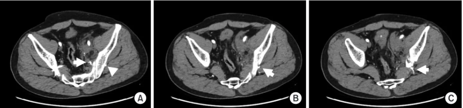
A B C
Fig. 2. Serial cut of contrast-enhanced pelvic computed tomography (CE-CT). (A) Arterial phase of CE-CT shows the proximal portion (arrow) and distal portion (arrowhead) of the superior gluteal artery above the sciatic notch. (B) The proximal portion disappeared, and only the distal portion (arrow) was observed near the sciatic notch. (C) Only the distal portion (arrow) was observed at the fractured sciatic notch. On the other hand, contrast leakage or hematoma was not observed.
Journal of the Korean Fracture Society Vol. 34, No. 1, January 2021
32
고 찰
상둔 동맥 손상은 주로 외상으로 인한 골반환 손상이나 비구 골절에서 동반되는 경우가 흔하며, 골반 외상에서 동 맥 색전술이 흔히 이루어지는 혈관 중 하나이다.3) 최근 인터 벤션 영상의학의 발달로 응급실에서의 초기 평가에서 소생 술에 반응하지 않는 혈역학적 불안정성, 조영 증강 골반 CT 에서 동맥 출혈이 의심되는 등의 징후가 관찰될 때 혈관 조영 술 후 동맥 색전술을 통해 이를 처치한다.4-7) 그러나 본 증례 와 같이 손상이 있었음에도 불구하고 골편 사이에 혈관이 포 착되는 등으로 인해 출혈이 숨겨진 잠복 양상의 경우, 초기 평 가에서 세밀한 주의를 기울이지 않는다면 동맥 손상이 간과 될 수 있으며, 이로 인해 수술장에서 골절 정복 중 출혈이 발 생할 수 있어 지혈에 어려움을 겪을 수 있다. 특히 Kocher- Langenbeck 접근법과 같이 골반의 외측으로 접근했을 때 좌
골 절흔 내측에서 혈관 손상이 발생한 경우라면 내측 절단부 로의 접근이 힘들어 지혈에 큰 어려움을 겪을 수 있다. 본 증 례에서는 초기 평가 시에 상둔 동맥이 골편 사이에 끼어 출혈 을 유발하지 않아 생체 징후에는 큰 이상이 없었으며, 수상 후 5일 동안 유의한 생체 징후 변화가 없었던 터라 자연 지혈 이 이뤄졌을 수도 있다. 그러나 수술 중 골편을 정복할 때 손 상이 가중되어 출혈이 발생했다면 수술 시간이 연장되고 지 혈에 어려움을 겪을 수 있어, 수상 후 시간이 충분히 경과했 다 해도 출혈로 인한 문제가 발생할 수 있으니 수술 전 적절 한 평가가 이루어져야 한다. 따라서 좌골 절흔 주위의 골절이 있다면 술 전에 상둔 동맥 손상을 염두에 두고 골절 양상을 면밀히 분석해야 하며, 조영 증강 CT의 동맥 조영기를 정확 하게 확인해야 하겠다. 의심이 될 경우에는 동맥 조영술을 통 해 보다 정확한 평가가 이루어져야 하며, 손상이 확인되면 동 맥 색전술을 통해 수술장에서의 출혈을 예방하는 조치가 필
A B C
Fig. 3. (A) Digital subtraction angiography (DSA) demonstrates cutoff with a filling defect in the left superior gluteal artery. (B) Selective emboliza- tion of the left superior gluteal artery was performed with microcoils using a sandwich technique. The configuration of microcoils in the superior gluteal artery shows compression of the left superior gluteal artery by the fracture fragment. (C) Post-embolization DSA shows complete exclusion of the cutoff segment of the left superior gluteal artery.
Fig. 4. Postoperative anteroposterior and iliac oblique radiograph shows a reduced fracture, and coils for embolization across the sciatic notch. No bleeding occurred during the surgery.
Fig. 5. Antero-posterior and iliac oblique radiograph at 15 months after surgery. Union was achieved without complications, such as delayed bleeding.
Latent Superior Gluteal Artery Injury in Transverse Acetabular Fracture Hyuk Jin Choi, et al.
33
요하겠다.
상둔 동맥 손상에 대한 연구는 여러 상황에서 발생한 증 례 보고들이 대부분이며 발병률, 손상 양상, 골절 양상과의 연관성 및 위험도에 대한 보고는 제한적인 실정이다.8-10) 좌골 절흔 주위에 골절이 있는 경우 항상 상둔 동맥 손상에 대한 가능성을 염두에 두고 이에 대한 평가 및 처치가 면밀히 시행 되어야 하며, 필요 시 시설이 갖춰진 기관으로 이송도 고려해 야 하겠다. 또한, 골절 양상을 적절히 분석하여 본 증례와 같 이 숨겨진 손상에 대해서도 가능성을 두고 초기 및 술 전 평 가가 이루어져야 할 것이다. 추후 좌골 절흔 주위를 침범한 골반 골절에 대해 골절 양상 및 형태와 상둔 동맥 손상과의 연관성 및 위험도에 대한 추가적인 연구도 필요하겠다.
요 약
상둔 동맥은 내 장골 동맥으로부터 분지되어 좌골 절흔을 통해 골반 외측으로 위치하여 좌골 절흔을 침범한 골반 골절 에서 손상의 위험이 있다. 손상이 있는 경우 혈역학적 불안정 성을 보일 수 있으며 이에 대한 적절한 평가와 처치가 이루어 져야 한다. 그러나 잠복 양상의 손상으로 초기에 진단이 간과 된다면 수술 중에 대량의 출혈을 야기할 수 있어 지혈에 어려 움을 겪을 수 있다. 이에 저자들은 좌골 절흔 주위를 침범한 비구 횡 골절에서 골편 사이에 상둔 동맥이 포착되어 초기에 손상이 간과되었던 증례를 경험하였고, 이에 대해 보고하고 자 한다.
색인 단어:
골반 골절, 좌골 절흔, 상둔 동맥 손상ORCID
최혁진, https://orcid.org/0000-0001-9806-8676 김병철, https://orcid.org/0000-0002-0439-1382
권 훈, https://orcid.org/0000-0003-4055-5863 장재훈, https://orcid.org/0000-0002-2636-7957
References
1. Stephen DJ, Kreder HJ, Day AC, et al: Early detection of arterial bleeding in acute pelvic trauma. J Trauma, 47: 638-642, 1999.
2. Miller PR, Moore PS, Mansell E, Meredith JW, Chang MC:
External fixation or arteriogram in bleeding pelvic fracture: ini- tial therapy guided by markers of arterial hemorrhage. J Trauma, 54: 437-443, 2003.
3. Vaidya R, Waldron J, Scott A, Nasr K: Angiography and em- bolization in the management of bleeding pelvic fractures. J Am Acad Orthop Surg, 26: e68-e76, 2018.
4. Yoon W, Kim JK, Jeong YY, Seo JJ, Park JG, Kang HK: Pelvic arterial hemorrhage in patients with pelvic fractures: detection with contrast-enhanced CT. Radiographics, 4: 1591-1605; dis- cussion 1605-1606, 2004.
5. Tanizaki S, Maeda S, Hayashi H, et al: Early embolization without external fixation in pelvic trauma. Am J Emerg Med, 30: 342-346, 2012.
6. Brun J, Guillot S, Bouzat P, et al: Detecting active pelvic arterial haemorrhage on admission following serious pelvic fracture in multiple trauma patients. Injury, 45: 101-106, 2014.
7. Lai YC, Wu CH, Chen HW, Wang LJ, Wong YC: Predictors of active arterial hemorrhage on angiography in pelvic fracture pa- tients. Jpn J Radiol, 36: 223-230, 2018.
8. Maled I, Velez R, Lopez R, Batalla L, Caja VL: Pseudoaneurysm of the superior gluteal artery during iliosacral screw fixation.
Acta Orthop Belg, 73: 544-547, 2007.
9. Songur M, Şahin E, Zehir S, Oz II, Kalem M: Gluteal com- partment syndrome secondary to superior gluteal artery injury following pelvis fracture: a case report and review of literature.
Turk J Emerg Med, 16: 29-31, 2016.
10. Kim WY, Lee SW, Kim KS, Lee JY: Superior gluteal artery pseudoaneurysm caused by pelvic C-clamp blind application: a case report. Hip Pelvis, 29: 145-149, 2017.