I
n the mid-1980s, the all-ceramic restoration was reborn, partially because of the general public inter- est in “esthetic dentistry”, as well as increased research and development of materials and meth- ods. Castable ceramics and “high-strength”porcelains were introduced as esthetic alternatives to porcelain fused to metal crowns with varying degrees of clinical success. But breakage and recurrent decay continued to be persistent prob- lems with many ceramic inlays and crowns.
Feldspathic porcelain crowns particularly those that remain in the mouth for any length of time, can suffer from static fatigue and can fracture because of water sorption into the surface faults in the porcelain
In 1990, a new all ceramic system was introduced
to the dental profession as an alternative technique for the fabrication of all ceramic crowns, inlay- onlays and veneers. IPS Empress 1,2is a pressed ceramic system with its base components being leucite crystals 5 ㎛ long that are homogeneous- ly dispersed in a glass phases. This technology offered a high strength ceramic system with wear characteristics similar to natural dentition, and the beauty and translucency of natural den- tition and enamel. With all these advantages, there were still limitations to the system. Because of the low flexural strength of the ceramic and its inability to withstand occlusal forces where mul- ti-unit bridges are indicated, this system, as well as others, was limited to single unit restorations.
To overcome the limitations associated with
Esthetic restoration of anterior dentition using Empress 2 system: A clinical report
Min-Ho Kim, DDSa, and Jae-Ho Yang, DDS, MSD, PhDb
College of Dentistry, Seoul National University, Seoul, Korea
Metal - ceramic restorations have been the standard of fixed oral rehabilitation for over 30 years, and while many advances have been made in materials and research, the esthetic chal- lenges of the light reflective metal substructure will always put clinician in an esthetic dilem- ma, for only a very talented ceramist / dentist team can create esthetics that rival nature on a repeatable basis. All ceramic restorations have also been a choice in our restorative armamentarium, but in the past, the materials have also had their own limitation, number on being question- able strength to withstand occlusal forces generated during mastication. IPS Empress 2 offers dentistry a metal free alternative to traditional restorative techniques. This clinical report describes the treatment of patients with esthetic problem of anterior dentition using Empress 2 system.
J Korean Acad Prosthodont : Volume 38, Number 6, 2000
aResident, Department of Prosthodontics.
bProfessor, Department of Prosthodontics.
current all ceramic systems, IPS Empress 23,4 (Ivoclar North America, Amherst, NY) was intro- duced. This new ceramic technology offers the strength necessary for utilization in multi-unit ante- rior bridges restorations. This lithium disilicate glass ceramic substructure, combined with its sintered glass-ceramic layering ceramic, offers excep- tional aesthetics, high translucency, even greater wear compatibility, and an etchable internal sur- face that incorporates adhesive bonding to the underlying tooth structure. As a result of this ceramic’s high strength, conventional cementa- tion techniques may also be used when clinical con- ditions prohibit bonding adhesive techniques.
IPS Empress 2
IPS Empress 2 is not just a modification of IPS Empress, but an entirely new glass technology. The substructure ceramic is made up of lithium dis- ilicate crystals suspended within a glass matrix.
The strength is achieved by the amount of crys- tals within the glass matrix, which is about 60 to 80 percent, compared to 30 to 40 percent with IPS Empress. Lithium disilicate crystals also have a sim- ilar index of light refraction as the glass matrix, which offers translucency of the framework as opposed to the opacity of other ceramic framework systems. The layering ceramic is also a new type of glass ceramic. This powdered overlay is applied directly to the framework, and is a sintered glass-
ceramic that also exhibits a crystalline content. The crystals within the layering ceramic are called flu- orapatite. These needle like crystals are identical in shape and composition of those found in nat- ural tooth enamel. This material functions and resembles natural dentition in its physical and opti- cal properties, and provides natural wear com- patibility, translucency, opalescence and bright- ness found in natural teeth.
CASE 1 : Veneer case
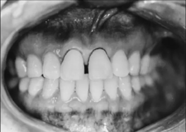
A 27-year-old woman presented with a chief complaint of diastema between her maxillary right central incisor and left central incisor (Fig. 1).
An ideal chamfer preparation of approximate- ly 0.5 to 0.6 mm is usually advised for ceramic veneers; But to confine the preparation to the enamel wherever possible, especially at the fin- ishing line area, differentiate the preparation depth according to the enamel thickness available in each area (cervical: 0.3 mm, incisal: 1 mm).
The preparation was extended slightly to the interproximal areas to hide the restoration mar- gins and increase the amount of enamel available for adhesion. Final impression was made using a polysiloxane impression material (Examix, GC, Japan): To obtain optimal gingival displacement during this stage, a retraction cord #00 (Ultrapack, ultradent) soaked with hemostatic solution (Hemodent, Premier Dental) was used. An irre-
Fig. 1. Facial view of the patient’s condition. Fig. 2. Final preparation and impression taking
versible hydrocolloid impression of the opposing dentition was made (Fig. 2).
Although veneer preparation was almost always confined to enamel thickness and no more than 4 or 5 days elapsed from preparation to final application, acrylic resin provisional restorations were positioned in this case. Because provision- al restorations are important prototype for the final veneers, and essential for maintaining the origi- nal tooth position whenever interproximal areas are involved in the preparation.
CLINICAL PROCEDURE
After checking proximal contacts, shade match, contour, and marginal adaptation of the veneers, the color and viscosity of the cement to be used were established through the try-in pastes. With nondiscolored teeth in this case, a translucent cement ensures the best results.
Veneers were etched using 4 % hydrofluoric acid for 2 minutes, rinsed with water, and dried. A silane agent (monobond S, Ivoclar) was then applied and blown dry. Silane was necessary to alter the surface chemistry of the ceramic and make it more compatible to the resin composite cement.
Concurrently the enamel surface of the prepared tooth was etched with 35 % phosphoric acid; in the presence of dentinal exposure a dentinal adhesive (Syntac, Ivoclar) was applied. A dual-polymerizing
resin composite cement (VariolinkⅡ, Ivoclar, Liechtenstein) was used for luting veneers.
The excess cement was removed interproxi- mally with a brush and dental floss. The veneer margins were covered with glycerin gel, and the resin composite cement was light polymerized from each side for 40 seconds. A gingival cord was placed into sulcus prior to luting to allow resid- ual cement to be removed completely from the crevice. Occlusion was evaluated after luting (Fig. 3).
CASE 2 : All ceramic crown
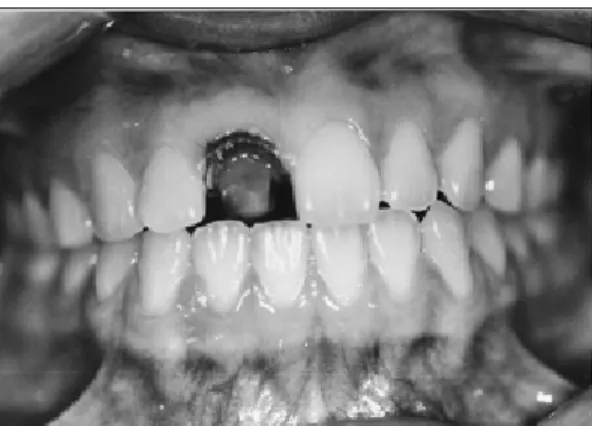
A 21-year-old woman presented with a chief complaint of poor esthetics of her maxillary right central incisor, particularly the discoloration of the tooth (Fig. 4). The tooth was endodontically treated two years ago. A complete examination was performed, including an occlusal analysis and a periodontal examination. It was determined that the patient’s occlusion and periodontal condition would allow the option of an all-ceramic crown.
Using Vita shade guide the anticipated shade of the restoration was chosen. Preparation of the tooth was done according to manufacturer’
(a) (b)
Fig. 3. (a) Post-operative view -facial. (b) Empress veneers were luted with a dual cured resin luting cement (Variolink Ⅱ).
s recommendation. And provisional restoration was fabricated. Impression taking was done according to the existing guidelines for conven- tional fixed restoration (Fig. 5).
CLINICAL PROCEDURE
A dual-curing resin cement, Variolink Ⅱ was chosen to lute the crown to the tooth. In this case, the tooth was discolored, so that more opaque cement is preferred to hide the underly- ing structure.
After cleaning the crown in an ultrasonic clean- er. The inner surface of the crown and tooth were treated in the same manner as veneer case.
Variolink Ⅱ was then mixed in equal portions and placed inside the crown, and the crown was gently placed onto the tooth. Excess resin was
removed with a brush and dental floss. The crown margins were covered with glycerin gel, and the resin composite cement was light polymerized from each side for 40 seconds.
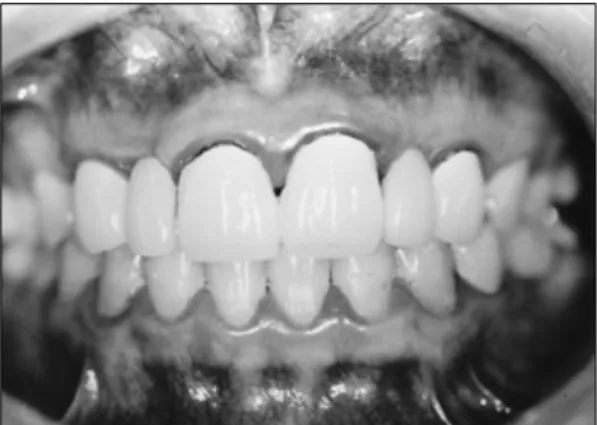
The occlusion was checked in centric relation and centric occlusion, as well as in all excursions, including crossover. The esthetic results were acceptable and the patient was given the usual postoperative instructions regarding bonded ceramic restorations (Fig. 6).
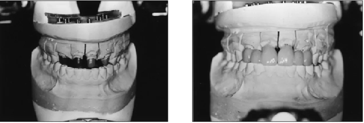
CASE 3 : All ceramic bridge
A 36-year-old female patient was presented with a chief complaint of poor esthetics of her max- illary anterior 6-unit bridge, particularly the met- al visible at the gingival margin (Fig. 7). The existing ceramometal bridge was originally placed Fig. 4.Facial view of the patient’s condition. Fig. 5.Gingival retraction for impression
(a) (b)
Fig. 6.(a) Facial view of Empress 2 crown after insertion and finishing (b) at rest position.
in 1991. A complete examination was performed, including an occlusal analysis and a periodontal examination. It was determined that the patient’
s occlusion and periodontal condition would allow the option of an all ceramic bridge, which would eliminate the exposure of metal at the gingival margin of the existing bridge. After dis- cussing therapy options with the patient, the decision was made to remove old porcelain fused to metal restoration on tooth and replaces it with two IPS Empress 2 bridges. The old bridge was removed without complication, and the tooth preparations were refined to receive all-ceramic bridge (Fig. 8). Teeth number 11,21,13 and 23 were examined and deemed favorable to use as abutments for three-unit bridge. The teeth were prepared and provisional restorations were
placed (Fig. 9).
An attempt was made to keep the margins supragingival, the internal line angles rounded, the depth of preparation at 1.5 mm minimum, and to keep the margin a butt joint for thickness of the ceramic.
After complete healing of the pontic site, the pro- visional bridge was removed, and final preparation was completed for the abutment teeth. Preparation requirements for Empress 2 are 1.5 mm buccal-lin- gual, and 2.0 mm occlusal. Supra-gingival cham- fer margins were place to provide definitive fin- ish lines for the restorations, soft tissue health was re-verified and final impressions were made
Detailed information, which included a written prescription, master impressions, pre-operative impression, impression of provisional restora- tions, full-face and natural smile photographs of 35 mm slides was sent to the laboratory.
LABORATORY FABRICATION
Impressions were verified for accuracy and poured with a die stone (DieKeen Ivory, Modern Materials). Interocclusal registrations were record- ed, and a facebow was used to relate the master casts to a semi-adjustable articulator (Hanau).
Casts were mounted on the Hanau Modula artic- ulator system. The maxillary working, approved provisional and opposing casts were mounted and an incisal edges wax matrix was fabricated to Fig. 7.Facial view of patient’s existing prosthesis
Fig. 9.After soft tissue healing.
Fig. 8.Removal of old prosthesis
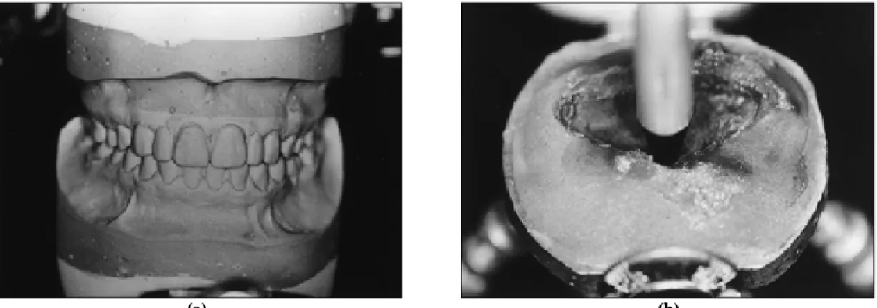
insure correct placement of incisal edge posi- tion of the final restoration. Finally, a custom incisal guide table was fabricated and the master dies were trimmed and sealed (Fig. 10).
Framework Design
The framework was then designed into the matrix, and the final framework was created fol- lowing manufacturer’s recommended design parameters. The wax pattern dimensions were 0.8 mm coping thickness and an interproximal con- nector size of 5.0 mm vertical height by 4.0 mm horizontal thickness. The wax frameworks were then sprued, invested and pressed. The IPS Empress 2 lithium disilicate framework ingots were pressed at a temperature of 920℃
The framework was then lightly blasted with alu- minum oxide and steam-cleaned to insure a clean surface. The framework was then seated on a solid model to insure a correct fit.
Ceramic Application
To insure a good bond between the frame- work and layering ceramic, a thin wash of the appropriate dentin shade was applied, along with the modification shades for the interproxi- mal, occlusal and gingival areas. This ceramic application was then fired at 800℃. The ceramic framework was placed back on a solid cast for
application of dentin ceramics. These ceramics were sculpted to correct anatomic shape, and modifi- cation ceramics were placed in the body and incisal areas, and fired in a ceramic furnace at 800
℃. After cooling, the restoration was fitted to the model and contour adjustments were made to facilitate accurate placement of enamel ceramics.
Once dentin recontouring was completed, stains were placed incisally, occlusally and anywhere else internal color was deemed necessary. These stain were fired at 780℃. Restorations were then sculpted to final coronal contour with appro- priate enamel ceramics.
Following the enamel application, the occlusal surfaces of the posterior unit were completed using a higher value and opacity enamel to con- trast with the high chroma dentins that were previously placed. The restoration was fired in a ceramic furnace at 800℃ contoured to final con- tours and naturally glazed to create the satin sheen of natural dentition. After glazing and verification of final fit and form to the solid cast, the internal surfaces were etched for 20 seconds with IPS Ceramic Etch Gel (Fig. 11).
CLINICAL PROCEDURE
The inner surface of bridge and teeth were treated in the same manner as Case 1 and 2.
Final finishing was completed in the traditional
(a) (b)
Fig. 10.Laboratory procedure. After mounting customized anterior guide table was fabricated (a),(b).
manner (Fig. 12).
DISCUSSION
IPS Empress 2 offers dentistry a metal free alternative to traditional restorative techniques.
The combination of an all-ceramic lithium disil- icate framework and the fluorapatite crystalline layering ceramic produce biocompatible restora- tions that offer nearly exact optical properties of natural dentition with regard to translucency, value, opalescence and fluorescence. As an addi- tional benefit, this lithium disilicate-fluorapatite ceramic system is the only metal free multiunit sys- tem that allows adhesive bonding, as well as conventional cementation
The use of ceramic veneers for esthetic restora-
tion of anterior teeth has increased considerably in recent years.5Problems such as unaesthetic old composites, fractured teeth, diastemas, incisal wear, and discoloration can be successfully cor- rected through the use of a conservative restora- tive technique: Porcelain veneers.6
This restoration technique offers numerous advantages over direct composite resin restora- tions, including chromatic stability and wear resistance. Porcelain veneers can also serve as a valid alternative to all-ceramic crowns when the correct indications exist, and thus can avoid sig- nificant tooth reduction that may cause irre- versible pulp damage. The use of ceramic veneers is without doubt strictly linked to the development of adhesive luting systems,7,8 which presently
(a) (b)
Fig. 12.(a) Post-operative view - facial (b) at rest position.
(a) (b)
Fig. 11.Frontal view of Empress 2 bridge.
feature very low polymerization contraction and are available in a variety of shades and opacity, Glass ceramics can ensure significantly improved strength and excellent translucency that is simi- lar to that of natural teeth.
IPS Empress 2 staining ingot (Ivoclar), a leucite- reinforced glass-ceramic, allows good esthetic restoration results to be obtained. Empress is a heat pressed ceramic; the glass ceramic ingot is pre- cerammed by the manufacturer and then processed in the laboratory using either the painting or layering technique. With the layering technique, after the wax is burned out and the glass ceram- ic is pressed into a preheated muffle, the sub- structure is covered with dentin and enamel porcelain and a final glaze firing is carried out. With the painting technique, the veneer is obtained directly from the wax-up and then finished with surface coloration.9Using a lost wax process, heated cylinders of leucite containing ceramic material are pressed into a special flask system.
Leucite crystals only a few microns in diameter are formed in the ceramic by a controlled crystallization process. The leucite crystals strengthen the ceram- ic and help reduce crack propagation. In a recent clinical study, Kreji, et al.10reported excellent initial marginal adaptation and no fractures, porosities, or occlusal wear after 18 months. If the clinician’s goal is to have a restoration of more than one shade, the wax pattern can be cut back and additional feldspathic porcelain can be added after the pressing process. Because of its ceram- ic nature, IPS Empress 2 can be etched and bond- ed with an appropriate resin cement technique.
SUMMARY
A case reports using IPS Empress 2 were pre- sented. IPS Empress 2 is a new lithium disili- cate reinforced ceramic material that can be used for veneers, inlays, crowns and anterior bridges.
Although only limited long-term data are available, preliminary reports show excellent marginal adap- tation, superior esthetics, and improved strength over other strengthened porcelains.
The search for the ultimate restorative materi- al continues. At present, the system described seems to offer the flexibility, ease of use, and strength needed to be an esthetic option in certain intraoral clinical situations. Preliminary reports and personal communications of clinical suc- cess indicate that further long term, in vivo, clin- ical trials are warranted and necessary before this material is more widely used.
REFERENCES
1. Wohlwend A, Schaerer P. The Empress technique for the fabrication of full ceramic crowns, inlays and veneers. Quintessenz Zahntech 1990; 16:966-978.
2. Lenher CR, Studer S, Schaerer P. Empress full ceramic crowns: First clinical results. J Dent Res 1992;
71:685. Abstract No. 1143.
3. Severance G. Introducing a lithium disilicate restorative: All Ceramic Alternative. Signature 1998; 5(3):1-3.
4. Sorensen J, Cruz M, Mitto W. Research Evaluations of a Lithium Disilicate Restorative system: IPS Empress 2. Signature 1998; 5(3): 4-10.
5. Friedman MJ. Augmenting restorative dentistry with porcelain veneers. J Am Dent Assoc 1991; 122:29- 34.
6. Garber DA, Goldstein RE, Feinman RA. Porcelain laminate veneers. Chicago: Quintessence, 1988:14- 23.
7. Simonsen RJ, Calamia JR. Tensile bond strength of etched porcelain (abstract). J Dent Res 1983; 62:297 8. Calamia JR, Simonsen RJ. Effect of coupling agents on bond strength of etched porcelain (abstract). J Dent Res 1984;63:179.
9. Fradeani M, Barducci G. Versatility of IPS Empress restorations. Part Ⅰ: Crowns. J Esthet Dent 1996;8:127-135.
10. Kreji I, Kreji D, Lutz G. Clinical evaluation of a new pressed ceramic inlay material after 1.5 years.
Quintessence Int 23: 181-186, 1992.
Reprint request to:
DR.JAE-HOYANG
DEPARTMENT OFPROSTHODONTICS, COLLEGE OFDENTISTRY, SEOULNATIONALUNIVERSITY
28 YONKON-DONG, CHONGRO-KU, SEOUL, 110-749 KOREA
E-mail: [email protected]