submit.radiology.or.kr J Korean Soc Radiol 2013;68(3):221-224
221 INTRODUCTION
Inflammatory myofibroblastic tumor (IMT) is a rare mesen- chymal neoplasm that is typified by a myofibroblastic and mixed inflammatory cell infiltrate (1). The tumor presents itself most commonly in children and young adults, and it occurs most fre- quently in the lungs (1). Although IMT has been reported in various extrapulmonary sites, its occurrence in the ileum is un- common. Furthermore, its presentation as intussusceptions is rare and thus, only a few cases have been reported (2). To the best of our knowledge, there is no report in the radiology litera- ture regarding IMT-induced intussusception.
We report multidetector computed tomography (CT) imag- ing findings of an IMT of the ileum in an adult causing an il- eoileal intussusception, and further discuss its radiological and pathological features.
CASE REPORT
A 55-year-old man was admitted to our hospital with a one- week history of abdominal pain. He had no history of any surgi- cal operation and systemic disease. A physical examination re- vealed a distended abdomen with tenderness in the entire abdomen as well as hyperactive bowel sounds. Plain abdominal radiographs revealed dilated small bowel loops with different heights of air-fluid level and a thickening of valvulae conniven- tes with mechanical ileus. Contrast-enhanced abdominal CT demonstrated dilated small bowel loops and invagination of a segment of the distal ileum with mesentery into the adjacent thick-walled intussuscipiens (Fig. 1A, B). A 2.5 cm sized lead mass with enhancing thin wall in the distal portion of the intus- susceptum was identified. The CT attenuation value of the mass was measured at about 34 Hounsfield units (HU) on the unen-
Case Report
pISSN 1738-2637
J Korean Soc Radiol 2013;68(3):221-224
Received October 29, 2012; Accepted January 2, 2013 Corresponding author: Il Young Kim, MD Department of Radiology, Soonchunhyang University College of Medicine, Cheonan Hospital,
31 Suncheonhyang 6-gil, Dongnam-gu, Cheonan 330-721, Korea.
Tel. 82-41-570-3515 Fax. 82-41-579-9026 E-mail: ilykim@schmc.ac.kr
Copyrights © 2013 The Korean Society of Radiology Inflammatory myofibroblastic tumor (IMT) of the ileum rarely develops and more-
over, intussusception of the tumor is even rarer. We report a case of IMT of the ileum with ileoileal intussusception in which we obtained multi-detector computed to- mography images that revealed dilatation of the proximal ileum as well as luminal mass lesion in the distal ileum with intussusceptum.
Index terms Abdomen Neoplasm
Inflammatory Myofibroblastic Tumor Small Bowel Intussusception CT
Multi-Detector Computed Tomography Findings of Inflammatory Myofibroblastic Tumor of the Ileum with Ileoileal Intussusception:
A Case Report
염증성근섬유모세포종양에 의해 유발된 회장장중첩증의 다중검출 전산화단층촬영 소견: 증례 보고
Jeong Ah Hwang, MD, Il Young Kim, MD, Young Tong Kim, MD, Hyeong Cheol Shin, MD
Department of Radiology, Soonchunhyang University College of Medicine, Cheonan Hospital, Cheonan, Korea
Multi-Detector Computed Tomography Findings of Inflammatory Myofibroblastic Tumor of the Ileum with Ileoileal Intussusception
submit.radiology.or.kr
J Korean Soc Radiol 2013;68(3):221-224
222
cells was positive for the smooth muscle actin (Fig. 1H). The re- sected tumor of the ileum was diagnosed as polypoid IMT.
DISCUSSION
IMT is a rare mesenchymal neoplasm that is typified by a myo- fibroblastic and mixed inflammatory cell infiltrate (1). Since its first description in 1937, the understanding of IMT has evolved from a reactive inflammatory process to a neoplasm of an uncer- tain malignant potential (2). Disagreement and uncertainty about the histogenesis of IMTs has resulted in a number of synonyms, which include plasma cell granuloma, plasma cell pseudotumor, inflammatory myofibrohistiocytic proliferation, omental-mesen- teric myxoid hamartoma, and, most commonly, inflammatory pseudotumor (3).
The etiology of IMT is poorly understood. Since IMT may be seen following abdominal surgery and trauma, it may represent an infectious or inflammatory process (4). However, based on the role of oncogenic viruses and cytogenetic abnormalities, in- cluding the anaplastic lymphoma kinase gene rearrangements hanced image and approximately 41 HU on the enhanced im-
age (Fig. 1C, D). There was a small amount of pelvic ascites and no abnormal lymphadenopathy. The differential diagnosis in- cluded Meckel diverticulum, duplication cyst, gastrointestinal stromal tumor, or leiomyoma with intussusception.
An exploratory laparotomy revealed ileoileal intussusceptions at the distal ileum along with edematous small bowel proximal to the lesion. The patient underwent a segmental resection of the intussuscepted ileum as well as an ileoileostomy.
Grossly, the resected specimen of the ileum measured 8 cm in length and 3.5 cm in circumference. There was a solid, well- defined, and intraluminal pedunculated mass measuring 2.5 × 2.0 cm in the ileum (Fig. 1E). The cut surface of the mass re- vealed a whitish tissue and there was no hemorrhage or necrosis in the mass (Fig. 1F). Microscopically, the well-demarcated tu- mor involved a submucosa and a muscle layer. The tumor was composed of an admixture of proliferating the spindle cells and inflammatory cells embedded in a highly vascularized stroma.
There were mild cellular atypia and mitotic figures (4/50 high power field) (Fig. 1G). Immunohistochemistry of the tumor
F B
G C
H D
E A
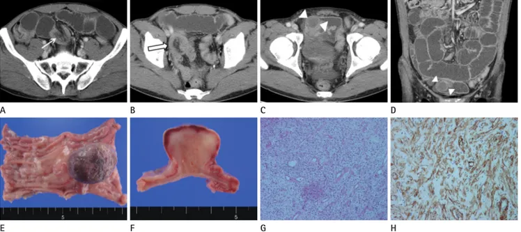
Fig. 1. A 55-year-old man with abdominal pain for one week.
A, B. Contrast enhanced axial CT scan demonstrates invaginated mesenteric fat and vessels (arrow) as well as bowel wall thickening of the intus- susceptum (open arrow).
C, D. Axial (C) and coronal (D) reformatted images show a 2.5 cm sized lead mass (arrowheads) with enhancing thin wall in the distal portion of the intussusceptum. The CT attenuation value of the mass is measured about 34 Hounsfield unit (HU) on unenhanced image (not shown) and about 41 HU on enhanced image.
E, F. Gross specimen showing intra-luminal pedunculated solid mass measuring 2.5 × 2.0 cm. Cut surface shows myxoid component and there is no evidence of hemorrhage or necrosis.
G, H. Microscopic view of the lesion shows that the tumor is composed proliferation of spindle-shaped myofibroblastic cells and infiltration of inflammatory cells embedded in a highly vascularized stroma (H&E staining, × 40). Immunohistochemistry was positive for smooth muscle actin.
Jeong Ah Hwang, et al
submit.radiology.or.kr J Korean Soc Radiol 2013;68(3):221-224
223
pearance of IMT is variable. The mass may be hypoattenuated or isoattenuated on unenhanced scans; moreover, calcification has been observed in the cases of the pancreas, stomach, and liver. Enhancement usually occurs but is not pronounced, and a variety of patterns have been noted including early peripheral, with delayed central filling, and heterogeneous, homogeneous, and no enhancement (8). In our case, the CT attenuation value of the mass measured at about 34 HU on the unenhanced image and approximately 41 HU on the enhanced image.
The combination of conventional hematoxylin and eosin stain- ing and appropriate immunohistochemical staining pathological studies can reliably distinguish IMTs with other mesenchymal tu- mors (9). The confirmation of the anaplastic lymphoma kinase protein/gene rearrangement is also useful in distinguishing IMT from other spindle cell neoplastic mimickers (3).
The mainstay of treatment for this tumor is surgical resection with wide margins. Incompletely resected tumors may have lo- cal recurrence within 1 year; however, recurrence has been seen up to 9 years after resection of the primary tumor (10).
In conclusion, ileoileal intussusceptions rarely occur in adults, particularly that due to IMT. It is hard to detect the exact under- lying disease of intussusceptions on radiologic examination. Al- though intra-luminal IMT of small bowel loop may be a rare cause of intussusception, IMT may be considered as one of the causes of intestinal obstruction.
REFERENCES
1. Coffin CM, Watterson J, Priest JR, Dehner LP. Extrapulmo- nary inflammatory myofibroblastic tumor (inflammatory pseudotumor). A clinicopathologic and immunohistochemi- cal study of 84 cases. Am J Surg Pathol 1995;19:859-872 2. Milne AN, Sweeney KJ, O’Riordain DS, Pauwels P, Debiec-
Rychter M, Offerhaus GJ, et al. Inflammatory myofibro- blastic tumor with ALK/TPM3 fusion presenting as ileoco- lic intussusception: an unusual presentation of an unusual neoplasm. Hum Pathol 2006;37:112-116
3. Ntloko S, Gounden A, Naidoo M, Madiba TE, Singh Y, Ramdial PK, et al. Intestinal inflammatory myofibroblastic tumour. S Afr J Surg 2011;49:190-193
4. Demirkan NC, Akalin T, Yilmaz F, Ozgenc F, Ozcan C, Al- kanat MB, et al. Inflammatory myofibroblastic tumor of on chromosome 2p23, clonal chromosome abnormalities, and
DNA aneuploidy, a neoplastic origin for IMT has been support- ed more recently (2, 3).
IMT occurs most commonly in the lungs. Extrapulmonary sites have also been reported, which include the mesentery and omentum, genitourinary tract, gastrointestinal tract, retroperito- neum, pelvis, head and neck, trunk, and extremities (1). Among extrapulmonary IMTs, 43% of the sites arise from mesentery and omentum whereas the small bowel accounts for only 1.2%
of the affected sites (1). Moreover, IMTs arising from the small bowel causing intussusception are rare (2, 3). Intussusception is primarily a disease among infants and children, and about 5%
of the cases occur in adults. Whereas 90% of childhood-type in- tussusceptions are idiopathic, an underlying pathologic process is usually identifiable in over 90% of adult cases (5). Small bowel intussusception without a lead point is more common than in- tussusception with a lead point. A lead point intussusception in- volving the small bowel is generally due to a benign condition and less often due to a neoplasm, which is usually a metastatic lesion (6).
IMTs arising from small bowel causing intussusception are extremely rare. Although it is hard to discern the exact underly- ing disease on radiologic examination, as long as IMT can cause an intussusception, the tumor should be considered as one of the causes of intestinal obstruction.
The clinical presentation of the IMT depends to some extent on its site of origin. Patients with intra-abdominal tumors may also be present with abdominal mass, abdominal pain, vomit- ing, constipation, and bowel obstruction. IMT may be a cause of chronic obstruction and of acute obstruction owing to intussus- ception (3).
Although the radiological examinations confirm obstructive features and the presence of intra-abdominal mass lesions, IMT is not a radiologic distinct entity (7). There are many factors that affect the appearance of an intussusception. These include the presence of a lead point, the configuration of the lead mass, the degree of bowel wall edema, and the amount of invaginated mesenteric fat (6). Identification of a lead mass that is separate and distinct from bowel loops is not easy. However, a mass that is seen on CT can serve as a reliable radiologic indicator of in- tussusceptions with a lead point, even though it is hard to dis- cern the exact underlying disease in most cases (6). The CT ap-
Multi-Detector Computed Tomography Findings of Inflammatory Myofibroblastic Tumor of the Ileum with Ileoileal Intussusception
submit.radiology.or.kr
J Korean Soc Radiol 2013;68(3):221-224
224
diatr Surg 1998;33:1843-1845
8. Narla LD, Newman B, Spottswood SS, Narla S, Kolli R. In- flammatory pseudotumor. Radiographics 2003;23:719- 729
9. Greenson JK. Gastrointestinal stromal tumors and other mesenchymal lesions of the gut. Mod Pathol 2003;16:
366-375
10. Kim KA, Park CM, Lee JH, Cha SH, Park SW, Hong SJ, et al.
Inflammatory myofibroblastic tumor of the stomach with peritoneal dissemination in a young adult: imaging find- ings. Abdom Imaging 2004;29:9-11
small bowel wall in childhood: Report of a case and a re- view of the literature. Pathol Int 2001;51:47-49
5. Agha FP. Intussusception in adults. AJR Am J Roentgenol 1986;146:527-531
6. Kim YH, Blake MA, Harisinghani MG, Archer-Arroyo K, Hahn PF, Pitman MB, et al. Adult intestinal intussuscep- tion: CT appearances and identification of a causative lead point. Radiographics 2006;26:733-744
7. Ciftci AO, Akçören Z, Tanyel FC, Senocak ME, Cagˇlar M, Hiçsönmez A. Inflammatory pseudotumor causing intesti- nal obstruction: diagnostic and therapeutic aspects. J Pe-
염증성근섬유모세포종양에 의해 유발된 회장장중첩증의 다중검출 전산화단층촬영 소견: 증례 보고
황정아 · 김일영 · 김영통 · 신형철
염증성근섬유모세포종양이 회장에 대단히 드물게 발생할 수 있다. 더욱이 이 종양에 의한 장중첩증은 더욱 드물다. 저자들 은 염증성근섬유모세포종양에 의해 유발된 장중첩증을 경험하였기에 이에 대한 multi-detector computed tomography
(MDCT) 영상소견을 보고하고자 한다. MDCT에서 회장말단부의 폐쇄에 의한 확장과 종양에 의한 장중첩증의 영상소견
을 보였다.
순천향대학교 의과대학 천안병원 영상의학과