리스프랑 관절 골절 및 탈구의 수상 기전과 치료 방법에 따른 임상적 결과와 예후 분석
박현우, 이형석
단국대학교병원 정형외과
Analysis of Clinical Outcome and Prognosis for Lisfranc Joint Fracture and Dislocation according to the Injury Mechanism and
Treatment Method
Hyun-Woo Park, Hyung Suk Yi
Department of Orthopaedic Surgery, Dankook University Hospital, Cheonan, Korea
Purpose: The purpose of this study was to assess the treatment outcomes and prognosis of Lisfranc joint fracture and dislocation ac- cording to the mechanism of injury and treatment method.
Materials and Methods: Twenty six patients with Lisfranc fracture-dislocation who had been treated surgically were included in this ret- rospective study. The patients were divided into two groups according to mechanism of injury: direct crushing injury (16 patients) and indirect rotational or compressive injury (10 patients). The patients were also divided into three groups according to the surgical meth- ods. The parameters used were radiographic evaluation, patients’ subjective satisfaction levels, length of hospital stay, and the American Orthopaedic Foot and Ankle Society (AOFAS) midfoot score. Statistical analysis was performed.
Results: The mean postoperative AOFAS midfoot score was 78.7. The mean length of stay was 39.6 days. Statistically significant differenc- es in subjective satisfaction, AOFAS midfoot score, and length of hospital stay were observed between the two groups (p<0.05). However, no significance differences were observed between the three groups who were divided according to the different surgical methods (p>0.05).
Conclusion: Mechanism of trauma and the severity of soft-tissue injury were significant prognostic factors affecting the surgical out- comes of Lisfranc joint fracture and dislocation.
Key Words: Lisfranc joint, Fracture and dislocation, Injury mechanism
체 노동자에서 빈도가 높다.1) 이러한 리스프랑 관절의 골절 및 탈 구는 장기적 예후가 좋지 않은 경우가 많아 정확한 진단과 치료가 중요한 골절이다. 1909년 Quenu와 Kuss에 의한 분류부터 시작하 여 1982년 Hardcastle 등에 의한 분류, 1986년 Myerson 등의 여러 저자들이 리스프랑 관절 손상에 대한 분류를 제시하였고, 실제로 임상의들 사이의 소통에 많은 도움을 주고 있는 것은 사실이다.1,2) 하지만 이러한 방사선학적인 분류만으로는 치료계획 설정 및 예후 예측에 제한점이 있었다. Kuo 등3)에 의하면 리스프랑 관절 손상은 특히 족부의 축성 압박 또는 족배부의 강한 압박으로 발생하거나, 족부의 압궤 손상과 함께 발생한다. 본 저자들도 치료 경험상 같은 리스프랑 관절 손상의 진단이더라도 손상 기전에 따라 손상 양상
서 론
족부의 리스프랑 관절(Lisfranc joint) 골절 및 탈구는 대부분 추 락 손상, 교통사고 또는 작업 관련 손상 등의 고에너지 손상에 기 인한다. 20∼40대 연령의 젊은 남자가 약 90%를 차지하고 특히 육
Received March 31, 2014 Revised April 26, 2014 Accepted May 29, 2014 Corresponding Author: Hyun-Woo Park
Department of Orthopaedic Surgery, Dankook University Hospital, 201 Manghyang-ro, Dongnam-gu, Cheonan 330-715, Korea
Tel: 82-41-550-3296, Fax: 82-41-556-0524, E-mail: [email protected] Financial support: None.
Conflict of interest: None.
This is an Open Access article distributed under the terms of the Creative Commons Attribution Non-Commercial License (http://creativecommons.org/licenses/CC
by-nc/3.0) which permits unrestricted non-commercial use, distribution, and reproduction in any medium, provided the original work is properly cited.
Copyright 2014 Korean Foot and Ankle Society. All rights reserved.ⓒ
www.jkfas.org 점, 불량 2점, 매우 불량 1점), 총 재원기간, American Orthopaedic Foot and Ankle Society (AOFAS) 중족부 평가법을 이용하였다. 통 계적 분석은 환자의 모집단이 26명으로 정규분포를 따르지 않으므 로, 수상 기전에 따른 분석에서는 Mann-Whitney U test를 사용하 였고 수술 방법에 따른 분석에서는 Kruskal-Wallis test를 시행하였 다(IBM SPSS Statistics 19.0; IBM Co., Armonk, NY, USA). 본 연구 는 단국대학교병원 의학연구윤리심의위원회(Institutional Review Board)의 승인을 받았다.
결 과
총 26명의 환자들을 Myerson 분류법을 기준으로 분류한 결과 B2형이 8명으로 가장 많은 수를 차지하였으며, 그 뒤를 이어서 B1 형이 총 6명을 나타내었다(Table 2). 전체 환자 중 만족스러운 정 복 23예, 불만족스러운 정복 3예를 나타냈으며, 불만족스러운 정 복을 보인 예에서는 골 소실이나 심한 복합골절을 동반하였다.
AOFAS 중족부 점수는 평균 78.7점(범위 48∼93점)으로 측정되었 으며, 평균 재원 기간은 39.6일(범위 6∼127일)이었다. 특히 연부 조직 손상이 심한 경우 재원 기간이 연장되었다. 직접적 손상을 받 은 군과 압박 및 비틀림 손상을 받은 군에서 주관적 만족도를 분 석해본 결과 직접적 손상을 받은 군에서 3.3점(범위 2∼5점), 압박 및 동반된 연부조직 손상의 정도, 그리고 그 예후가 서로 달라 이
를 비교 분석하고자 하였으며, 추가적으로 수술적 치료 방법에 따 른 예후 차이에 대한 연구를 하고자 하였다.
대상 및 방법
2006년부터 2012년까지 본원에서 리스프랑 관절 골절 및 탈구 로 진단하 수술적 치료를 받았던 환자 중 외래 추시 기간이 최소 1 년 이상인 총 26예의 환자를 대상으로 하였다(Table 1). 평균 나이 는 41.1세였고 남자 21명, 여자 5명으로 집계되었다. 심한 압궤 손 상 등으로 인하여 추후 부분적 족부 및 족지 절단술을 시행하였 던 환자는 모두 제외하였다. 환자들의 수상 기전에 따라서 축성 압 박 및 비틀어짐 손상 등을 받은 환자군(10명) 및 족배부의 직접적 인 압박을 받은 환자군(16명)의 두 군으로 분류하여 결과를 분석 하였다. 정복의 정확도를 나타내는 지표로는 Myerson 등1)의 지침 에 따라, 족부 전후방 방사선 사진상 제 1, 2중족골 기저부의 해리 가 2 mm 이하이고 족부 측면 사진상 거골과 제 1중족골 간의 각 도가 15도 이하인 경우를 만족스러운 정복이라 판정하였다.3,4) 환 자의 치료 결과에 대한 평가는 후향적인 의무기록지 및 방사선 자 료의 검토 및 분석을 통하여 시행하였다. 평가 기준으로는 방사선 학적 평가, 환자의 주관적 만족도(매우 만족 5점, 만족 4점, 보통 3
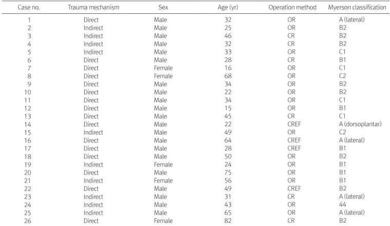
Table 1. Summary of Cases
Case no. Trauma mechanism Sex Age (yr) Operation method Myerson classification
1 2 3 4 5 6 7 8 9 10 11 12 13 14 15 16 17 18 19 20 21 22 23 24 25 26
Direct Indirect Indirect Indirect Indirect Direct Direct Direct Direct Direct Direct Direct Direct Direct Indirect Direct Direct Direct Indirect Direct Indirect Direct Indirect Indirect Indirect Direct
Male Male Male Male Male Male Female Female Male Male Male Male Male Male Male Male Male Male Female Male Female Male Male Male Male Female
32 25 46 32 33 28 16 68 34 22 34 15 45 22 49 64 28 50 24 75 56 49 31 43 65 82
OR OR CR CR OR CR OR OR OR OR OR OR CR CREF OR CREF CREF OR OR OR OR CREF CR OR OR CR
A (lateral) B2 B2 B2 C1 B1 C1 C2 B2 B2 C1 B1 C1
A (dorsoplantar) C2
A (lateral) B1 B2 B1 B1 B1 B2 A (lateral) 44 A (lateral) B2 OR: open reduction and internal fixation, CR: closed reduction and percutaneous pinning, CREF: closed reduction and external fixation.
Table 2. Reduction Accuracy and Clinical Outcomes of All Patients Case
no.
Reduction accuracy
AOFAS
midfoot score LOM Subjective satisfaction LOS 1
2 3 4 5 6 7 8 9 10 11 12 13 14 15 16 17 18 19 20 21 22 23 24 25 26
Anatomical Anatomical Anatomical Anatomical Anatomical Anatomical Satisfactory Unsatisfactory Satisfactory Anatomical Anatomical Anatomical Anatomical Anatomical Unsatisfactory Anatomical Satisfactory Satisfactory Anatomical Satisfactory Anatomical Anatomical Satisfactory Anatomical Satisfactory Unsatisfactory
81 82 87 92 90 72 68 65 72 81 81 64 82 93 51 82 92 75 92 79 92 72 89 82 82 48
+
―
―
―
― + + + + +
―
―
―
― + + +
―
―
―
― +
―
―
―
―
4 4 5 5 5 3 2 2 3 4 4 2 4 5 2 4 5 3 5 3 5 3 5 5 5 2
16 18 11 23 19 33 63 127 41 114 8 84 24 79 28 18 24 87 7 79 15 23 6 21 17 45 AOFAS: American Orthopaedic Foot and Ankle Society, LOM: limitation of motion, LOS: length of hospital stay.
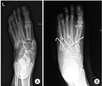
Figure 1. (A) Preoperative anteroposterior radiograph of left foot showed fracture and dislocation of Lisfranc joint. (B) Postoperative an- teroposterior radiograph of left foot showed well reduced fracture and dislocation with cannulated screw fixation.
Figure 2. (A) Preoperative anteroposterior radiograph of left foot showed fracture and dislocation of Lisfranc joint. (B) Postoperative an- teroposterior radiograph of left foot showed well reduced fracture and dislocation of Lisfranc joint using percutaneous K-wire fixation.
Figure 3. (A) Preoperative anteroposterior radiograph of right foot showed fracture and dislocation of Lisfranc joint. (B) Preoperative lat- eral radiograph of right foot showed fracture and dislocation of Lisfranc joint. (C) Postoperative anteroposterior radiograph of right foot showed well reduced fracture and dislocation of Lisfranc joint using Ilizarov ex- ternal fixator. (D) Postoperative lateral radiograph of right foot showed well reduced fracture and dislocation of Lisfranc joint using Ilizarov
www.jkfas.org 한적 관혈적 정복술, 금속 핀 고정술을 이용한 수술적 치료 방법이 리스프랑 관절 골절 및 탈구의 치료로 부상하게 되었고,6,8,11,12) Ahn 등13)은 리스프랑 골절 및 탈구 환자에서의 제한적 관혈적 정복술 및 일리자로프를 이용한 외고정이 추가적인 치료적인 선택이 될 수 있음을 제시한 바 있다. 이와 같이, 심한 연부조직 손상이 발생 한 리스프랑 관절 및 골절 환자에서 관혈적 정복술 및 내고정술보 다는 제한적 관혈적 정복술 및 경피적 핀 삽입술 등 추가적인 연부 조직 손상을 최소화하는 수술적 치료 방법이 좋은 대안이 될 수 있 을 것으로 생각되었으며, 본 연구에서의 수술적 치료 방법에 따른 통계적 유의한 차이가 없음은 이를 뒷받침한다. Demirkale 등14)은 리스프랑 골절 및 탈구에서 동반된 연부조직 손상 정도가 치료 결 과 및 예후에 음성적 예후 인자로 작용한다고 보고하였으며, 본 연 구에서도 이와 같은 맥락으로 손상 기전에 따른 리스프랑 관절의 골절 및 탈구 환자의 치료 결과 및 임상적 예후가 다른 것을 알 수 있었고, 연부조직 손상을 동반한 직접적 손상에서 예후가 좋지 않 았음을 비교해서 알 수 있었다. 따라서 손상 기전에 따라 치료 방 법도 서로 달리 접근해야 하므로 수상 시 방사선학적인 소견만으 로 무조건적인 수술적 치료 방침 설정은 지양해야 할 것으로 보이 며, 수상 기전에 따른 연부조직 손상을 충분히 고려하여야 좋은 임 상적 결과를 얻을 수 있을 것으로 생각된다. 이번 후향적인 연구는 제한된 환자수, 짧은 추시 관찰기한이라는 제한점을 가지고 있다.
수술적 치료 방법의 결정에서 이미 환자의 방사선학적 상태 및 연 부조직 손상을 고려하여 결정하였으므로 표본선정편향이 발생했 을 가능성을 배제할 수 없으나 각 환자군에서 임상적 결과 및 예후 가 유의한 차이를 보이지 않았다는 것에서는 임상적 의의를 둘 수 있을 것이다. 향후 압궤 손상과 동반한 리스프랑 관절의 골절 및 탈구에 대해서는 충분한 표본수 확보를 위한 다기관 연구 및 연부 조직 손상을 반영하는 추가적인 분류법에 대한 연구가 필요할 것 으로 생각된다.
결 론
리스프랑 관절의 골절 및 탈구 환자의 치료에 있어 수술 방법에 따른 임상적 결과는 유의한 차이가 없었으나 심한 연부조직 손상 이 동반한 경우는 그 예후가 유의하게 나빴다. 따라서 연부조직 손 상 정도를 고려하여 수술적 치료 방법을 선택하는 것이 좋은 결과 를 얻을 것으로 생각된다.
REFERENCES
11 Myerson MS, Fisher RT, Burgess AR, Kenzora JE. Fracture dislo- cations of the tarsometatarsal joints: end results correlated with pathology and treatment1 Foot Ankle1 1986;6:225-421
21 Hardcastle PH, Reschauer R, Kutscha-Lissberg E, Schoffmann W.
Injuries to the tarsometatarsal joint1 Incidence, classification and 및 비틀림 손상을 받은 군에서 4.6점(범위 2∼5점)으로 나타났으
며, 통계적으로 유의한 차이를 나타내었다(p=0.003). 두 군 간 평 균 재원 기간을 비교해보면 직접적 손상을 받은 군에서 평균 54.1 일(범위 8∼127일), 압박 및 비틀림 손상을 받은 군에서 16.5일(범 위 6∼28일)로 나타났으며, 두 군 간 재원기간은 통계적으로 유의 하였다(p=0.001). 술 후 1년째 검사한 AOFAS 중족부 점수의 평균 은 직접적 손상을 받은 군에서 75.4점(범위 48∼93점), 압박 및 비 틀림 손상을 받은 군에서는 83.9점(범위 51∼92점)으로 직접적 손 상을 받은 군에서 통계적으로 유의하게 낮게 측정됨을 알 수 있었 다(p=0.014). 수술적 치료에 따른 예후 분석에서는 관혈적 정복술 및 내고정술(Fig. 1)을 시행한 16예, 도수 정복술 및 K-강선을 이용 한 경피적 내고정술(Fig. 2)을 시행한 6예, 그리고 외고정술(Fig. 3) 을 시행한 4예에서 세 가지 수술 방법에 따른 통계적 분석 시 주관 적 만족도(p=0.574), 평균 재원 기간(p=0.731) 및 평균 AOFAS 중 족부 점수(p=0.334)에서 모두 유의한 차이를 보이지 않았다.
고 찰
리스프랑 관절의 골절 및 탈구에 대한 치료 방침으로 도수 정복 술 및 경피적 핀 고정술, 관혈적 정복술 및 견고한 금속 내고정술 등이 있다. 1986년 Myerson 등1)은 관혈적 정복 및 견고한 금속 내 고정술로 치료한 결과가 기존의 도수 정복 및 석고 붕대 고정 방법 보다 탁월한 것으로 발표하였으며, Schepers 등5)에 의하면 나사 또 는 금속판을 이용한 내고정술이 보다 나은 정복 및 정복의 유지에 도움이 준다고 하였고, Kuo 등3)의 연구에서 또한 해부학적인 정복 이 AOFAS 점수 및 장기적인 예후 및 관절염 발생에 있어서 통계적 으로 유의하게 우수한 성적을 나타내고 있다. 이러한 연구 결과에 근거하여 최근의 리스프랑 관절의 골절 및 탈구의 치료 흐름은 관 혈적인 정복을 통하여 가능한 한 해부학적 정복을 이루고 견고한 내고정술로 치료하는 것이며, 본 저자 또한 이러한 추세에 동의하 는 바이다. 하지만 족배부의 직접적인 압궤 손상에 의해 리스프랑 관절 및 골절이 발생한 경우에는 대부분 심한 연부조직의 손상을 동반하는 경우가 많아서, 관혈적 정복 및 금속 내고정술이 족부의 연부조직 치유에 부정적인 영향을 줄 수 있으며 이차적인 감염, 내 고정물의 이완 등 예기치 못한 합병증을 일으킬 수 있다.6-8) Peru- gia 등9)은 도수 정복 후 전후방 방사선 사진상 족근 중족 관절에 2 mm 미만 간격의 정복을 이룬 경우를 근접한 해부학적 정복(nearly anatomical reduction)이라 분류하고 경피적 핀 삽입술을 시행하였 으며 그 장기적인 결과가 완전한 해부학적 정복 및 금속 내고정술 로 치료한 경우의 장기적인 결과와 큰 차이가 없음을 보고하였다.
또한 해부학적 정복, 근접한 해부학적 정복, 비해부학적 정복을 이 룬 환자군에 대한 비교를 한 임상 연구에서는 단지 비해부학적 정 복을 이룬 환자군에서만 임상적으로 유의하게 외상 후 관절염의 발생 빈도가 높았다고 한다.3,10) 이러한 연구 결과에 기초해서 제
20001 p11265-801
91 Perugia D, Basile A, Battaglia A, Stopponi M, De Simeonibus AU.
Fracture dislocations of Lisfranc's joint treated with closed re- duction and percutaneous fixation1 Int Orthop1 2003;27:30-51 101 Myerson MS. The diagnosis and treatment of injury to the tarso-
metatarsal joint complex1 J Bone Joint Surg Br1 1999;81:756-631 111 Richter M, Wippermann B, Krettek C, Schratt HE, Hufner T,
Therman H. Fractures and fracture dislocations of the midfoot:
occurrence, causes and long-term results1 Foot Ankle Int1 2001;
22:392-81
121 Thompson MC, Mormino MA. Injury to the tarsometatarsal joint complex1 J Am Acad Orthop Surg1 2003;11:260-71
131 Ahn GY, Yoo YS, Yun HH, Yun KP, Nam IH. Treatment of frac- ture and dislocation of Lisfranc joint with limited open reduc- tion, pin fixation and Ilizarov external fixation1 J Korean Foot Ankle Soc1 2004;8:182-901
141 Demirkale I, Tecimel O, Celik I, Kilicarslan K, Ocguder A, Dogan M. The effect of the Tscherne injury pattern on the outcome of operatively treated Lisfranc fracture dislocations1 Foot Ankle Surg1 2013;19:188-931
treatment1 J Bone Joint Surg Br1 1982;64:349-561
31 Kuo RS, Tejwani NC, Digiovanni CW, Holt SK, Benirschke SK, Hansen ST Jr, et al. Outcome after open reduction and internal fixation of Lisfranc joint injuries1 J Bone Joint Surg Am1 2000;82:
1609-181
41 Mulier T, Reynders P, Dereymaeker G, Broos P. Severe Lisfrancs injuries: primary arthrodesis or ORIF? Foot Ankle Int1 2002;23:
902-51
51 Schepers T, Oprel PP, Van Lieshout EM. Influence of approach and implant on reduction accuracy and stability in lisfranc fracture-dislocation at the tarsometatarsal joint1 Foot Ankle Int1 2013;34:705-101
61 Adelaar RS. The treatment of tarsometatarsal fracture-disloca- tion1 Inst Cours Lect1 1990;39:141-51
71 Heckman JD. Fracture and dislocation of the foot1 In: Rockwood CA, Green DP, Bucholz RD, editors1 Rockwood and Green's fractures in adults1 4th ed1 Philadelphia: Lippincott-Raven;
19961 p12363-731
81 Thordarson DB. Fractures of the midfoot and forefoot1 In: Myer- son MS, editor1 Foot and ankle disorders1 Philadelphia: Saunders;