HR Lee, et al
536 Ann Dermatol
Received September 29, 2010, Revised December 20, 2010, Accepted for publication December 20, 2010
Corresponding author: June Hyunkyung Lee, M.D., Department of Der- matology, Eulji Hospital, College of Medicine, Eulji University, 280-1 Hagye 1-dong, Nowon-gu, Seoul 139-711, Korea. Tel: 82-2-970-8278, Fax: 82-2-974-1577, E-mail: [email protected]
This is an Open Access article distributed under the terms of the Creative Commons Attribution Non-Commercial License (http://
creativecommons.org/licenses/by-nc/3.0) which permits unrestricted non-commercial use, distribution, and reproduction in any medium, provided the original work is properly cited.
Ann Dermatol Vol. 23, No. 4, 2011 http://dx.doi.org/10.5021/ad.2011.23.4.536
CASE REPORT
Squamous Cell Carcinoma Developing within Lesions of Disseminated Superficial Actinic Porokeratosis
Hyung Rae Lee, M.D., Tae Young Han, M.D., Sook-Ja Son, M.D., June Hyunkyung Lee, M.D.
Department of Dermatology, Eulji Hospital, College of Medicine, Eulji University, Seoul, Korea
Disseminated superficial actinic porokeratosis (DSAP) consists of multiple annular, hyperkeratotic lesions that have a bilateral distribution on sun-exposed areas, particularly the extremities. DSAPs have a wider distribution than poro- keratosis of Mibelli and usually develop during the 3rd or 4th decade of life. Squamous cell carcinoma that arises in the classical type of porokeratosis of Mibelli is well-docu- mented, but there are only a few reports of squamous cell carcinoma in DSAP. Here, we describe a 62-year-old man with DSAP who developed squamous cell carcinoma on his right forearm. (Ann Dermatol 23(4) 536∼538, 2011) -Keywords-
Disseminated superficial actinic porokeratosis, Squamous cell carcinoma
INTRODUCTION
Disseminated superficial actinic porokeratosis (DSAP) is the most common form of porokeratosis, and is inherited as an autosomal dominant condition, with reduced pene- trance at a younger age1. The characteristic lesions of DSAP represent multiple annular, hyperkeratotic, brown- ish macules measuring 2∼5 mm in diameter. The center of a macule is minimally atrophic or depressed and the
border spreads centrifugally in raised ridges1,2. The dis- tribution is symmetric and usually affects sun-exposed areas.
Development of squamous cell carcinoma within the clas- sic type of porokeratosis of Mibelli is well- documented, but there are only a few reports of squamous cell carcino- ma in DSAP3-7. We describe a case of squamous cell carci- noma developing within lesions of DSAP.
CASE REPORT
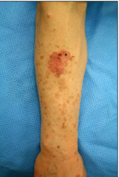
A 62-year-old male presented with pruritic eruptions on sun-exposed portions of both forearms that had gradually increased in number over a period of 5 years (Fig. 1). The lesions were exacerbated during the summer months.
Along with these lesions, an erythematous, irregular, marginated, scaly, crusted plaque had developed on the right forearm 3 years earlier. There was no significant medical or family history.
On physical examination, a 2×3 cm erythematous, irregular, marginated, scaly, crusted plaque was noted on the right forearm. In addition, the patient had numerous annular, brown, atrophic, and symmetric macules sur- rounded by well-demarcated, raised ridges on extensor aspects of both forearms, which are characteristics of DSAP. Complete blood count, as well as liver and kidney function tests were all within normal limits.
A skin biopsy specimen of the multiple, brown, annular lesions showed histologic changes of typical DSAP. There was a cornoid lamella composed of a column of para- keratosis with underlying hypogranulosis and perivascular lymphocytic infiltrations in the dermis localized beneath the cornoid lamella. A skin biopsy obtained from the erythematous plaque on the right forearm showed dysregulated keratinocytes with hyperchromatic, atypical nuclei, consistent with squamous cell carcinoma. A cornoid lamella was observed in the lesion of the
Squamous Cell Carcinoma Developing within Lesions of Disseminated Superficial Actinic Porokeratosis
Vol. 23, No. 4, 2011 537 Fig. 1. An irregular, marginated, erythematous plaque and
multiple, brown, atrophic macules surrounded by well-demar- cated, raised ridges on the right forearm.
Fig. 2. Biopsy specimen obtained from the erythematous plaque on the right arm. In epidermis, acanthosis and dysregulated keratinocytes with hyperchromatic, atypical nuclei are observed.
A cornoid lamella composed of a column of parakeratosis is seen in the lesion of the squamous cell carcinoma (H&E, ×100).
Fig. 3. Overexpression of p53 in the epidermis of a disseminated superficial actinic porokeratosis lesion. A column of parakeratosis with underlying hypogranulosis is observed. Perivascular lym- phocytic infiltrations are localized beneath the cornoid lamella (p53 immunohistochemical stain, ×200).
squamous cell carcinoma (Fig. 2).
Positron emission tomography-computed tomography revealed no evidence of distant metastasis. The squamous cell carcinoma was treated by total excision and split-thickness skin graft and radiotherapy. The patient is currently being treated with topical sunscreens.
DISCUSSION
Porokeratosis has several clinical varieties including poro- keratosis of Mibelli, giant porokeratosis, DSAP, poroker- atosis palmaris et plantaris, punctuate porokeratosis, and
linear porokeratosis8. The most common form of poroker- atosis is DSAP, which was first described as a clinical en- tity by Chernosky in 1966. The distribution of typical le- sions is symmetric and usually affects sun-exposed areas.
The lesions generally spare the face, palms, soles, and mu- cosal surfaces1,7. DSAP can affect people of all ages, but manifests during the 3rd or 4th decade of life.
The pathogenesis of DSAP is not clearly understood, but frequent p53 overexpression in the epidermis of poroker- atotic lesions have been detected9. Overexpression of p53 can be induced by p53 gene mutations and other DNA damaging agents, such as ultraviolet (UV) light and ioniz- ing radiation. In a previous study, mutations of the p53 gene were not detected in porokeratotic lesions9. This finding suggests that other causative mechanisms exist for overexpression of p53 in the epidermis of porokeratotic lesions. The patient in this case showed overexpression of p53 in the epidermis of DSAP lesions (Fig. 3).
The occurrence of malignancies in porokeratotic lesions is clinical evidence of the pre-cancerous nature of this disease. Malignancies have been reported for porokera- tosis of Mibelli, linear porokeratosis, porokeratosis palma- ris et plantaris and DSAP3-7,10,11. Associated malignancies are squamous cell carcinoma, Bowen’s disease and basal cell carcinoma. Squamous cell carcinoma arising in the classic type of porokeratosis of Mibelli is well-docu- mented, but there are only a few reports of squamous cell carcinoma in DSAP3-7. All of the reported squamous cell carcinoma cases arising from DSAP lesions have origi- nated in the distal extremities. These findings indicate that there is a significant role of UV light on the evolution of squamous cell carcinoma from DSAP. In addition, results from previous reports show that p53 gene mutations are
HR Lee, et al
538 Ann Dermatol
responsible for the progression of porokeratosis to SCC in at least some cases9.
In our case, a cornoild lamella was observed in the squ- amous cell carcinoma, strongly suggesting the possibility that the carcinoma developed from clones of abnormal epithelial cells. However, not all cases of squamous cell carcinoma in DSAP have cornoid lamellae in lesions of the carcinoma6. These cases cannot exclude the possibility that squamous cell carcinoma developed simply from chronic sun exposure unrelated to DSAP lesions.
However, even when a cornoid lamella directly over the tumor is not identified, the presence of a cornoid lamella in close proximity to the tumor suggests the possibility that the carcinoma has arisen from the DSAP lesions6,7. Porokeratosis does not usually need treatment, but in some cases, treatment is necessary due to potential for progression to a malignancy and for cosmetic purposes. In contrast, treatment of DSAP lesions is unsatisfactory. As in this case, the potential for progression to cancer induced by UV light exists, thus sun avoidance must be empha- sized when treating DSAP.
The prognosis of squamous cell carcinoma in DSAP le- sions has not been reported yet. However, the reported cases have had no recurrences after excision of the can- cerous lesions.
REFERENCES
1. Shumack SP, Commens CA. Disseminated superficial actinic porokeratosis: a clinical study. J Am Acad Dermatol 1989;
20:1015-1022.
2. Schwarz T, Seiser A, Gschnait F. Disseminated superficial
"actinic" porokeratosis. J Am Acad Dermatol 1984;11:724- 730.
3. Yang HY, Nam TS, Kim YT, Kim JH. A case of squamous cell carcinoma and Bowen’s disease associated with superficial disseminated porokeratosis. Ann Dermatol 1990;2:31-34.
4. James WD, Rodman OG. Squamous cell carcinoma arising in porokeratosis of mibelli. Int J Dermatol 1986;25:389-391.
5. Shrum JR, Cooper PH, Greer KE, Landes HB. Squamous cell carcinoma in disseminated superficial actinic porokeratosis. J Am Acad Dermatol 1982;6:58-62.
6. Chernosky ME, Rapini RP. Squamous cell carcinoma in le- sions of disseminated superficial actinic porokeratosis: a re- port of two cases. Arch Dermatol 1986;122:853-855.
7. Leache A, Soto de Delás J, Vázquez Doval J, Lozano MD, Quintanilla E. Squamous cell carcinoma arising from a le- sion of disseminated superficial actinic porokeratosis. Clin Exp Dermatol 1991;16:460-462.
8. Won JH, Lee MJ, Park JS, Chung H. A case of the giant and hyperkeratotic variant of porokeratosis. Korean J Dermatol 2009;47:101-103.
9. Ninomiya Y, Urano Y, Yoshimoto K, Iwahana H, Sasaki S, Arase S, et al. p53 gene mutation analysis in porokeratosis and porokeratosis-associated squamous cell carcinoma. J Dermatol Sci 1997;14:173-178.
10. Lozinski AZ, Fisher BK, Walter JB, Fitzpatrick PJ. Metastatic squamous cell carcinoma in linear porokeratosis of Mibelli. J Am Acad Dermatol 1987;16:448-451.
11. Guss SB, Osbourn RA, Lutzner MA. Porokeratosis plantaris, palmaris, et disseminata. A third type of porokeratosis. Arch Dermatol 1971;104:366-373.