ISSN 2234-3806 • eISSN 2234-3814
https://doi.org/10.3343/alm.2018.38.4.306
Galectin-3 Reflects the Echocardiographic Grades of Left Ventricular Diastolic Dysfunction
Uzair Ansari, M.D.1*, Michael Behnes, M.D.1*, Julia Hoffmann, M.S.1, Michele Natale, M.S.1, Christian Fastner, M.D.1, Ibrahim El-Battrawy, M.D.1, Jonas Rusnak, M.D.1, Seung-Hyun Kim, M.D.1, Siegfried Lang, Ph.D.1, Ursula Hoffmann, M.D.1, Thomas Bertsch, M.D.2, Martin Borggrefe, M.D.1, and Ibrahim Akin, M.D. 1
First Department of Medicine1, University Medical Center Mannheim, University of Heidelberg, Mannheim, Germany: DZHK (German Center for Cardiovascular Research) partner site Mannheim; Institute of Clinical Chemistry, Laboratory Medicine and Transfusion Medicine2, General Hospital Nuremberg, Paracelsus Medical University, Nuremberg, Germany
Background: The level of Galectin-3 (Gal-3) protein purportedly reflects an ongoing car- diac fibrotic process and has been associated with ventricular remodeling, which is instru- mental in the development of heart failure with preserved ejection fraction (HFpEF) syn- drome. The aim of this study was to investigate the potential use of Gal-3 in improved char- acterization of the grades of diastolic dysfunction as defined by echocardiography.
Methods: Seventy HFpEF patients undergoing routine echocardiography were prospec- tively enrolled in the present monocentric study. Blood samples for measurements of Gal-3 and amino-terminal pro-brain natriuretic peptide (NT-proBNP) were collected within 24 hours pre- or post-echocardiographic examination. The classification of patients into sub- groups based on diastolic dysfunction grade permitted detailed statistical analyses of the derived data.
Results: The Gal-3 serum levels of all patients corresponded to echocardiographic indi- ces, suggesting HFpEF (E/A, P =0.03 and E/E’, P =0.02). Gal-3 was also associated with progressive diastolic dysfunction, and increased levels corresponded to the course of dis- ease (P =0.012). Detailed analyses of ROC curves suggested that Gal-3 levels could dis- criminate patients with grade III diastolic dysfunction (area under the curve [AUC]=0.770, P =0.005).
Conclusions: Gal-3 demonstrates remarkable effectiveness in the diagnosis of patients suffering from severe grade diastolic dysfunction. Increasing levels of Gal-3 possibly reflect the progressive course of HFpEF, as classified by the echocardiographic grades of diastolic dysfunction.
Key Words: Galectin-3, Preserved ejection fraction, NT-proBNP, Diastolic dysfunction
Received: July 26, 2017
Revision received: October 19, 2017 Accepted: January 15, 2018 Corresponding author: Uzair Ansari University Medical Center Mannheim, First Department of Medicine, Theodor- Kutzer-Ufer 1-3, 68167 Mannheim, Germany
Tel: +49-621-383-5229 Fax: +49-621-383-2012 E-mail: [email protected]
* These authors contributed equally to this study.
© Korean Society for Laboratory Medicine This is an Open Access article distributed under the terms of the Creative Commons Attribution Non-Commercial License (http://creativecom- mons.org/licenses/by-nc/4.0) which permits unrestricted non-commercial use, distribution, and reproduction in any medium, provided the original work is properly cited.
INTRODUCTION
The prevalence of heart failure with preserved ejection fraction (HFpEF) has increased significantly over the past decade [1, 2].
Hypothesis suggesting that HFpEF is an independent syndrome typically characterized by the presence of diastolic dysfunction has not ameliorated the ominous prognosis of this presentation.
The reported mortality rates are as high as ~65% at five years post initial hospitalization [3]. It is estimated that >50% of all patients presenting with signs and symptoms of heart failure could have a preserved ejection fraction (EF) [4]. Myocardial stiffening from hypertrophy and fibrosis are central to the tradi- tional paradigm of HFpEF; however, the roles of abnormal cal- cium handling and venous turgor have also been exposed in re-
2017-03-16 https://crossmark-cdn.crossref.org/widget/v2.0/logos/CROSSMARK_Color_square.svg
cent studies [5-7]. The pathophysiological factors implicated in the development of this syndrome include impaired relaxation (attributed to low-grade inflammation, extracellular matrix accu- mulation, and fibrosis), increased left ventricular stiffness and reduced compliance, atrial dysfunction, chronotropic incompe- tence, pre- and post-capillary pulmonary hypertension, and vas- cular stiffening [4, 8].
The use of serum biomarkers has helped provide vital infor- mation regarding the pathogenesis of HFpEF and is a proven clinical tool for the identification of at-risk patients, syndrome di- agnosis, risk stratification, as well as therapy monitoring [3]. Ga- lectin-3 (Gal-3), a similar serum biomarker, is a soluble β-galac- toside-binding protein secreted by activated macrophages and is a key component in chronic inflammation facilitating fibrogen- esis and organ scarring [9]. The hypothesis that Gal-3 influences the onset of heart failure has been corroborated by infusing Gal-3 into the pericardial sac of wild-type rats, which triggered exten- sive myocardial fibrosis. It has also been suggested that Gal-3 induces cardiac fibrosis via activation of cyclin D1, thus enabling a macrophage derived mediator to affect the myocardium [10].
Additionally, Gal-3 complements other similar heart failure bio- markers, such as aminoterminal pro-brain natriuretic peptide (NT-proBNP) and troponin T and troponin I (TnT/TnI), by pro- viding an upstream signal of the myocardial fibrotic state, ven- tricular adverse remodeling, and cardiomyopathy progression [11].
The predominant use of echocardiography to assess impaired myocardial diastolic function is naturally influenced by patient compliance, adiposity, and pulmonary diseases such as emphy- sema. The use of biomarkers to determine grades of diastolic dysfunction is not subject to these limitations. Recent studies have attempted to delineate the potential relationship between Gal-3 levels and transthoracic echocardiographic indices such as left ventricular EF (LVEF) and right ventricular (RV) systolic pressure [12]. Expanding on this idea, our study attempted to explore the hitherto poorly investigated hypothesis that quantita- tive levels of Gal-3 could also reflect other echocardiographic in- dices defined in the evolving stages of HFpEF.
METHODS
1. Study design and population characteristics
The Cardiovascular Imaging and Biomarker Analyses (CIBER) study (clinicaltrails.gov identifier: NCT 03074253) is a clinically prospective, controlled, and mono-centric study conducted at the University Medical Center Mannheim, Germany. The research
adhered to the principles outlined in the Declaration of Helsinki and was approved by a regional ethics committee (the medical ethics commission II of the Faculty of Medicine in Mannheim, University of Heidelberg, Germany). Written informed consent was obtained from all patients.
The present study incorporated a population subset derived from a patient cohort who underwent routine echocardiography at the University Medical Centre Mannheim, Germany between 2014 and 2016. A total of 70 patients diagnosed with HFpEF were included consecutively in this mono-centric, prospective study with an all-comers design. As this was a non-interven- tional, observational study, diagnostic procedures and treatment plans were not modified.
The relevant clinical data of each patient were ascertained and compiled in a database at index presentation, with significant aspects of their medical history, laboratory work-up, and medi- cal/surgical therapy efficiently earmarked for further reference.
Patients under the age of 18 years or those with LVEF <50%, tricuspid annular plane systolic excursion (TAPSE) <17 mm, and/or valvular heart disease were excluded. Blood samples, collected from all patients, were preserved and processed throu- ghout the study. All patients were contacted at the scheduled 6- and 12-month follow-up period for standardized telephonic in- terviews to ascertain the incidence of re-hospitalization due to heart failure as well as all-cause and cardiovascular mortality.
2. Echocardiography
A detailed transthoracic echocardiographic examination was performed for all patients included, and standard techniques were implemented to acquire every reproducible image [13, 14]. The results were analyzed and interpreted by observers with no knowledge of the patients’ clinical and biomarker data.
The assessed structural indices included LVEF (using Simpsons biplane method), left ventricular (LV) end-systolic and end-dia- stolic volume, LV wall and septal thickness, atrial and ventricular size and volume, TAPSE, markers of early and late trans-mitral diastolic velocities (E and A), deceleration time, and early and late diastolic tissue velocities at the lateral mitral annulus (E’) [12]. Patients were classified according to the grade of diastolic dysfunction, and grading guidelines were based on the Ameri- can Society of Echocardiography and European Association of Cardiovascular Imaging (ASE/EACVI) Guidelines recommended by Nagueh [13]. A flow diagram depicting our diagnostic ap- proach for the inclusion of patients is shown in Supplemental Data Fig. S1.
The echocardiographic inclusion criteria specified that pa-
tients have a preserved LV and right ventricular (RV) function.
Consequently, patients with an LVEF <50% and TAPSE <17 mm were excluded. Additionally, patients with moderate and severe heart valve disorders, classified as either stenosis or re- gurgitation, were excluded.
3. Laboratory analysis
Blood samples were collected from all patients (at rest) at a sin- gle assessment time point upon study inclusion by venepuncture with serum monovettes and centrifuged at 2,500 g at 20°C for 10 minutes. The aliquoted samples were cooled down in liquid nitrogen before being stored at –80°C for further analysis. After thawing, the samples were mixed gently by inverting and centri- fuged at 2,500 g for 10 minutes at 20°C.
Gal-3 levels were assessed using the Gal-3 assay on an Archi- tect i1000 analyzer (Abbott, Wiesbaden, Germany). The limit of blank for this assay was 0.8 ng/mL, as specified in the user in- structions (Galectin-3, Architect System, © 2012, 2013 Abbott Laboratories). Serum creatinine concentrations were measured using the Creatinine Jaffe Gen.2 assay on a Cobas c 702 ana- lyzer (Roche Diagnostics, Mannheim, Germany), and the glo- merular filtration rate (eGFR) was estimated using the Modifica- tion of Diet in Renal disease (MDRD) formula (Instructions for use, Cobas c 702 analyzer). The serum level of NT-proBNP, used as a reference biomarker, was measured using a proBNP II STAT assay on a Cobas e 602 analyzer (Roche Diagnostics). The limit of detection (LoD) for this assay was 5 pg/mL (proBNP II STAT, Cobas®, © 2014, Roche Diagnostics).
4. Statistical analysis
The Student t-test was applied for data with a normal distribu- tion, and the Kruskal-Wallis method was used as a non-para- metric test. Abnormally distributed scaled variables with signifi- cant deviations from the Gaussian distribution were compared using the Kolmogorov-Smirnov test. Spearman’s rank correlation for non-parametric data was used to test the association of Gal-3 serum levels with cardiac indices and other parameters, as de- fined by transthoracic echocardiography. The data are presented as the mean with a confidence interval (CI) or median with in- terquartile ranges (IQRs; 25th to 75th percentiles), depending on the distribution. P <0.05 was considered statistically signifi- cant.
The effectiveness of Gal-3 in grading the various stages of HFpEF was evaluated using the Hanley and McNeil method [17], with the reference biomarker, NT-proBNP, plotted simulta- neously for comparison. The data were further log-transformed
for analysis. Potential confounding factors were defined using multivariable linear or logistic regression analyses with backward elimination, and clinical parameters or biomarkers were adjusted depending on the outcome variable (binary or numeric).
Statistical analyses were performed using SPSS Statistics (IBM, Armonk, NY, USA) and GraphPad Prism (GraphPad Software, Table 1. Baseline characteristics of study patients
Characteristics HF-PEF (N=70)
Age, mean (range; 95% CI) 65 (22–97; 84)
Gender, N (%)
Male 36 (51)
Female 34 (49)
Cardiovascular risk factors, N (%)
Arterial hypertension 56 (80)
Hypercholesterinemia 26 (37)
Cardiac family history 11 (16)
Smoking status 23 (33)
Diabetes mellitus 17 (24)
Adipositas 16 (23)
Laboratory parameters, median (IQR)
Creatinine (mg/dL) 0.93 (0.77–1.15)
eGFR (mL/min/1.73 m2) 70.47
Medical history, N (%)
Chronic heart failure 50 (71)
NYHA I 22 (31)
NYHA II 10 (14)
NYHA III 17 (24)
NYHA IV 1 (1)
Atrial fibrillation 27 (38)
Paroxysmal 15 (21)
Persistent 9 (13)
Permanent 3 (4)
Coronary artery disease 35 (50)
1 vessel disease 4 (6)
2 vessel disease 12 (17)
3 vessel disease 19 (27)
Myocardial infarction 5 (7)
Chronic kidney disease 12 (17)
COPD 8 (11)
Asthma 2 (3)
Cancer 18 (26)
Abbreviations: HFpEF, heart failure with preserved ejection fraction; COPD, chronic obstructive pulmonary disease; NYHA, New York Heart Association;
GFR, glomerular filtration rate.
Inc., La Jolla, CA, USA). All patient data resulting from these analyses were subsequently interpreted and classified into the three sub-groups defined by HFpEF grade (I, II, III). The details of this classification and the corroborating echocardiographic measurements are provided in Tables 1 and 2.
RESULTS
1. Study populationThe baseline clinical characteristics of the 70 patients are de- scribed in Table 1. A detailed analysis of the data revealed that the mean age of the patients was 65 years (range 22–97 years) with an equal gender distribution (male 51%, N =36; female 49%, N=34). Arterial hypertension was identified as a predomi- nant risk factor in this group, with 80% (N=56) of the patients
diagnosed as having this disease. Patients suffering from heart failure-related symptoms represented 71% (N=50) of the study population, and their sub-classification into New York Heart As- sociation (NYHA) sub-groups yielded an almost even distribu- tion across classes I, II, and III. Coronary artery disease was iden- tified as a pre-existing condition in at least 50% (N=35) of the patients, while atrial fibrillation was documented in 38% (N=27).
Patients suffering from diabetes mellitus (24%, N=17) or chronic kidney disease (17%, N=12) were also represented. The me- dian creatinine value was estimated at 0.93 mg/dL (range 0.77–
1.15 mg/dL) and the median eGFR according to MDRD formula was 70.47 mL/min/1.73 m2.
2. Echocardiographic characteristics
The distribution of echocardiographic indices according to HF- Table 2. Distribution of echocardiographic indices according to HFpEF sub-groups
All Patients
(N=70) Good Diastolic Function
(N=14) Diastolic Dysfunction I
(N=15) Diastolic Dysfunction II
(N=30) Diastolic Dysfunction III
(N=11) P
LVEF (%) 59.00 (56.00–65.00) 61.00 (56.00–65.00) 58.00 (57.00–62.00) 62.00 (56.00–67.00) 56.00 (54.00–58.00) 0.109 LVEDD (mm) 45.00 (43.00–50.00) 45.00 (42.00–46.00) 44.00 (43.00–48.00) 46.00 (40.00–50.00) 50.00 (45.00–58.00) 0.112 LVESD (mm) 30.00 (27.00 -34.00) 30.00 (26.00–32.00) 29.00 (27.00–35.00) 28.00 (27.00–32.00) 31.00 (28.00–37.00) 0.39 LVPW (mm) 11.00 (9.00–12.00) 10.00 (8.00–11.00) 10.00 (9.00–12.00) 11.00 (10.00–12.00) 12.00 (11.00–14.00) 0.045 LVIS (mm) 12.00 (10.00–13.00) 11.00 (9.00–12.00) 11.00 (10.00–12.00) 12.00 (10.00–13.00) 13.00 (12.00–13.00) 0.065 RA (mm) 36.00 (33.00–40.00) 35.00 (31.00–40.00) 37.00 (35.00–38.00) 35.00 (32.00–40.00) 36.00 (34.00–41.00) 0.911 RA (cm³) 14.00 (12.00–17.00) 13.00 (11.00–16.00) 15.00 (13.00–16.00) 14.00 (12.00–15.00) 17.00 (14.00–18.00) 0.172 LA (mm) 39.00 (35.00–43.00) 35.00 (34.00–38.00) 40.00 (36.00–45.00) 39.00 (35.00–41.00) 43.00 (40.00–47.00) 0.044 LA (cm2) 17.00 (15.00–19.00) 15.00 (11.00–17.00) 17.00 (16.00–18.00) 17.00 (15.00–20.00) 24.00 (20.00–27.00) 0.002 RV-area (cm2) 18.00 (16.00–21.00) 18.00 (14.00–21.00) 18.00 (17.00–20.00) 18.00 (16.00–21.00) 19.00 (15.00–22.00) 0.926 RV-volume (mL) 37.00 (29.00–49.00) 34.00 (25.00–43.00) 40.00 (31.00–44.00) 37.00 (29.00–52.00) 38.00 (26.00–53.00) 0.634 LV-area (cm2) 35.00 (31.00–40.00) 33.00 (28.00–35.00) 39.00 (31.00–44.00) 34.00 (31.00–39.00) 37.00 (33.00–42.00) 0.099 LV-volume (mL) 116.00 (92.00–151.00) 114.00 (89.00–130.00) 149.00 (99.00–170.00) 111.00 (90.00–137.00) 128.00 (92.00–176.00) 0.218 Aorta (mm) 29.00 (27.00–33.00) 28.00 (26.00–31.00) 28.00 (26.00– 38.00) 30.00 (27.00–33.00) 31.00 (29.00–32.00) 0.466 AortaPmean (mmHg) 8.50 (7.00–11.00) 8.00 (8.00–8.00) 9.00 (9.00–9.00) 9.80 (5.50–14.00) 9.00 (7.00–11.00) 0.986 AortaPmax (mmHg) 7.00 (6.00–10.00) 6.00 (5.00–7.00) 9.00 (6.00–10.00) 8.00 (6.00–10.00) 7.00 (6.00–13.00) 0.236 TAPSE (mm) 22.00 (20.00–25.00) 22.00 (21.00–25.00) 21.00 (19.00–23.00) 23.00 (21.00–26.00) 20.00 (19.00–18.00) 0.41 E/A (cm/s) 0.90 (0.70–1,20) 1.20 (1.10–1.50) 0.80 (0.60–0.90) 0.80 (0.70–1.10) 0.90 (0.80–1.30) 0.0001 E’med (m/s) 0.06 (0.05–0.08) 0.11 (0.09–0.13) 0.07 (0.05–0.08) 0.06 (0.05–0.08) 0.04 (0.04–0.06) 0.002 E’lat (m/s) 0.07 (0.06–0.09) 0.10 (0.08–0.13) 0.09 (0.07–0.11) 0.08 (0.06–0.09) 0.05 (0.05–0.06) 0.009 E/E’ 10.00 (8.00–13.00) 6.00 (4.00–7.00) 7.00 (6.00–8.00) 11.00 (10.00–12.00) 19.00 (16.00–26.00) 0.0001 DT (ms) 237.00 (181.00–296.00) 214.00 (164.00–237.00) 243.00 (187.00–299.00) 240.00 (205.00–296.00) 236.00 (192.00–373.00) 0.119 Data are presented as medians with interquartile ranges (IQRs); bold type indicates statistical significance (P <0.05).
Abbreviations: LVEF, left ventricular ejection fraction; LVEDD, left ventricular end diastolic diameter; LVESD, left ventricular end systolic diameter; LVEDV, LV end-diastolic volume; LVESV, LV end-systolic volume; LVPW, LV posterior wall; IVSD, interventricular septal diameter; RA, right atrium; LA, left atrium; RV, right ventricle; LV, left ventricle; TAPSE, tricuspid annular plane systolic excursion; E/A, ratio of the early (E) to late (A) ventricular filling velocities; E/E’, ratio of mitral inflow (E) velocity to tissue Doppler (E’); DT, deceleration time; IVRT, isovolumetric relaxation time.
pEF sub-group is outlined in Table 2. Patients expressed either good diastolic function (N=14) or grade I (N=15), grade II (N=30), or grade III (N=11) diastolic dysfunction. LVEF values were >50%
and TAPSE was >17 mmHg. Detailed analysis of these data re- vealed that LVEF values ranged between 54% and 67%. Indices considered statistically significant included LV posterior wall thickness (P =0.045), left atrial dimensions (P =0.044), left atrial volume (P =0.002), E/A ratio (P =0.0001), E’ lateral (E’ lat) (P = 0.009), and E/E’ ratio (P =0.0001).
3. Characteristics of Gal-3
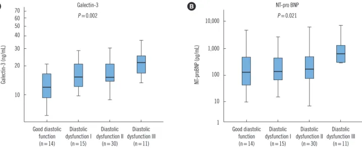
Gal-3 mirrored progressive diastolic dysfunction and increased levels corresponded with the course of disease (P =0.012). The distribution of Gal-3 and NT-proBNP levels is graphically pre- sented in Fig. 1.
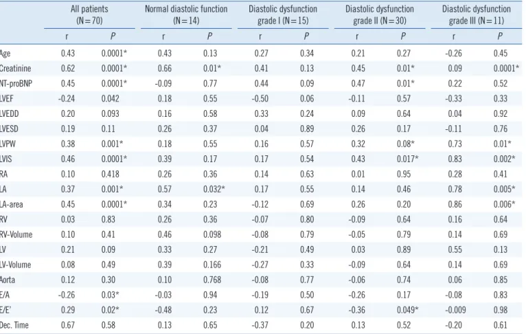
4. Correlation of Gal-3 with baseline characteristics and echocardiographic indices
Univariate linear regression analysis demonstrating the correla- tion of Gal-3 with the baseline characteristics, as well as echo- cardiographic indices is detailed in Table 3. The Gal-3 levels of all patients corresponded with age (P =0.0001), as well as with echocardiographic indices, suggesting HFpEF (E/A, P =0.03 and E/E’, P =0.02). Additionally, there was a significant correla- tion between Gal-3 levels and indices measuring left atrial and LV dimensions in the case of grade III diastolic dysfunction (P values ranging from 0.006 to 0.01). However, this relationship
was blurred in patients with normal or lower grades of diastolic dysfunction. Gal-3 levels also demonstrated a strong association with serum creatinine levels across all HFpEF sub-groups (P val- ues ranging from 0.01 to 0.0001).
Interestingly, although a definitive relationship with NT-proBNP was observed in all patients (P =0.0001), this was statistically nonsignificant across the various HFpEF sub-groups defined by varying degrees of diastolic dysfunction.
5. Gal-3 level discriminates patients with Grade III diastolic dysfunction
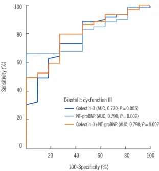
Detailed analyses ROC curves suggested that Gal-3 level dis- criminates patients with grade III diastolic dysfunction (area un- der the curve [AUC]=0.770, P =0.005; Fig. 2). NT-proBNP also revealed such characteristics, but with a numerically greater AUC (AUC=0.798, P =0.002). This revealed no significant dif- ference between the two AUCs. Additionally, the combination of Gal-3 and NT-proBNP exhibited a similar discrimination for this sub-group of HFpEF patients (AUC=0.798, P =0.002).
6. Multivariate logistic regression models
Data were log-transformed for this analysis. Log-transformed Gal-3 and NT-proBNP levels were initially adjusted to multivari- ate logistic regression models with each other and then subse- quently for age, gender, and serum creatinine (Table 4). Patients with Gal-3 levels ≥17.0 ng/mL were six times more likely to suf- fer from grade III diastolic dysfunction (adjusted odds ratio [OR]=
Fig. 1. Box plots showing increased levels of Galectin-3 (A) and increased levels of NT-proBNP (B) in patients with different grades of dia- stolic dysfunction. Significantly highest levels of Galectin-3 and NT-proBNP were noted in Grade III diastolic dysfunction, indicating a pro- gressive increase in their levels along the course of the disease.
Abbreviation: NT-proBNP, amino-terminal pro-brain natriuretic peptide.
Galectin-3 P =0.002 70
60 50 40 30 20
10
Good diastolic function (n=14)
Diastolic dysfunction I
(n=15)
Diastolic dysfunction II
(n=30)
Diastolic dysfunction III
(n=11)
Galectin-3 (ng/mL)
A NT-pro BNP
P =0.021 10,000
1,000
100
10
1 Good diastolic function (n=14)
Diastolic dysfunction I
(n=15)
Diastolic dysfunction II
(n=30)
Diastolic dysfunction III
(n=11)
NT-proBNP (pg/mL)
B
95% CI 0.601–11.830, P =0.197; Table 4). An analysis of log- transformed NT-proBNP levels alone showed statistical insignifi- cance in this scenario.
DISCUSSION
This study aimed to determine the relationship between Gal-3 levels and the echocardiographic indices corresponding to vari- ous stages of HFpEF (LVEF >50%). At the outset, Gal-3 proved effective in the diagnosis of patients suffering from a severe grade of diastolic dysfunction. The ability to diagnose patients with HF- pEF exhibited some similarity with the reference biomarker, NT- proBNP. Interestingly, increasing levels of Gal-3 also possibly re- flected the progressive course of HFpEF, as evident in the echo- cardiographic examination; indices measuring atrial and ven- tricular dimensions were noticeably altered from their normal Table 3. Univariate correlations between Galectin-3 and patient characteristics, biomarkers, and echocardiographic parameters according to HF-pEF sub-groups
All patients
(N=70) Normal diastolic function
(N=14) Diastolic dysfunction
grade I (N=15) Diastolic dysfunction
grade II (N=30) Diastolic dysfunction grade III (N=11)
r P r P r P r P r P
Age 0.43 0.0001* 0.43 0.13 0.27 0.34 0.21 0.27 -0.26 0.45
Creatinine 0.62 0.0001* 0.66 0.01* 0.41 0.13 0.45 0.01* 0.09 0.0001*
NT-proBNP 0.45 0.0001* -0.09 0.77 0.44 0.09 0.47 0.01* 0.22 0.52
LVEF -0.24 0.042 0.18 0.55 -0.50 0.06 -0.11 0.57 -0.33 0.33
LVEDD 0.20 0.093 0.16 0.58 0.33 0.24 0.09 0.64 0.04 0.92
LVESD 0.19 0.11 0.26 0.37 0.04 0.89 0.26 0.17 -0.11 0.76
LVPW 0.38 0.001* 0.18 0.55 0.16 0.57 0.32 0.08* 0.73 0.01*
LVIS 0.46 0.0001* 0.39 0.17 0.17 0.54 0.43 0.017* 0.83 0.002*
RA 0.10 0.418 0.26 0.36 0.14 0.63 0.01 0.95 0.28 0.41
LA 0.37 0.001* 0.57 0.032* 0.17 0.55 0.14 0.46 0.78 0.005*
LA-area 0.45 0.0001* 0.34 0.23 -0.12 0.69 0.26 0.20 0.86 0.006*
RV 0.03 0.83 0.26 0.36 -0.07 0.80 -0.09 0.64 0.16 0.64
RV-Volume 0.10 0.41 0.46 0.098 -0.08 0.79 -0.05 0.79 0.14 0.69
LV 0.21 0.09 0.33 0.27 -0.21 0.49 0.03 0.89 0.55 0.13
LV-Volume 0.08 0.49 0.39 0.166 -0.27 0.33 -0.09 0.64 0.14 0.69
Aorta 0.12 0.30 0.10 0.768 -0.08 0.77 -0.06 0.74 0.06 0.85
E/A -0.26 0.03* -0.03 0.94 -0.19 0.50 -0.26 0.17 -0.08 0.83
E/E’ 0.29 0.02* -0.48 0.23 0.12 0.67 -0.36 0.049* -0.009 0.98
Dec. Time 0.67 0.58 0.13 0.65 -0.37 0.20 0.13 0.52 -0.20 0.61
*Statistical significance (P <0.05).
Abbreviations: LVEF, left ventricular ejection fraction; LVEDD, left ventricular end diastolic diameter; LVESD, left ventricular end systolic diameter; LVPW, left ventricular posterior wall; LVIS, left ventricular interventricular septum; RA, right atrium; LA, left atrium; E/A, markers of early and late trans-mitral diastolic velocities (E and A), early and late diastolic tissue velocities at the lateral mitral annulus (E’); E/E’, ratio of mitral inflow (E) velocity to tissue Doppler (e’); Hf- pEF, Heart failure with preserved ejection fraction.
Table 4. Multivariable logistic regression for evaluating the ability of galectin-3 to identify patients with diastolic dysfunction grade III
Adjusted odds
ratio 95% CI Adjusted P Galectin-3 (≥17.0 ng/mL) 6.19 1.489–25.744 0.012*
NT-proBNP (≥290.6 pg/mL) 2.667 0.601–11.830 0.197
Gender 1.244 0.295–5.246 0.767
Creatinine 1.33 0.367–4.825 0.664
LA (>45 mm) 1.855 0.411–8.379 0.422
*Values were adjusted to creatinine, so no cut-off values were required.
Abbreviation: CI, confidence interval; LA, left atrium; NT-proBNP, amino-ter- minal pro-brain natriuretic peptide.
6.19, 95% CI 1.489–25.744, P =0.012). The likelihood of de- veloping this severe form of HFpEF was two-to-three times greater in patients with NT-proBNP levels ≥290.6 pg/mL (OR=–2.667,
Fig. 2. Receiver-operating characteristic (ROC) curve revealing an effective discrimination of patients with diastolic dysfunction III. ROC=
diastolic dysfunction grade 3 curve. AUC–Area under the Curve.
Abbreviation: NT-proBNP, amino-terminal pro-brain natriuretic peptide.
100
80
60
40
20
0
20 40 60 80 100 100-Specificity (%)
Sensitivity (%)
Diastolic dysfunction III
Galectin-3 (AUC, 0.770; P =0.005) NT-proBNP (AUC, 0.798; P =0.002) Galectin-3+NT-proBNP (AUC, 0.798; P =0.002)
range. Furthermore, the combination of Gal-3 and NT-proBNP values was equally effective in identifying patients with grade III diastolic dysfunction.
Several clinical studies have attempted to elucidate the role of Gal-3 in HFpEF patients [17]. These include the Coordinating study evaluating Outcomes of Advising and Counselling in Heart Failure (COACH) trial [34], the Controlled Rosuvastatin Multina- tional Trial in Heart Failure (CORONA) cohort [34], and studies by Carrasco-Sanchez et al [16], which described the strong prog- nostic value of Gal-3 in HFpEF patients [18]. However, the po- tential of Gal-3 to reflect the evolving stages of diastolic dysfunc- tion remains poorly understood; our study has attempted to dem- onstrate this distinct correlation.
Gal-3 is essentially a product of active macrophages with bind- ing sites on cardiac-resident fibroblasts, mechanistically influ- encing increased myocardial collagen expression, interstitial fi- brosis, TGF-β activation, and subsequent LV dysfunction [19- 21]. Its role in response to injury and inflammation in heart fail- ure is further supplemented by a significant contribution to ven- tricular remodeling [12]. This study provides useful information corroborating the association between LV structure and function and Gal-3 level, essentially mirroring the hypothesized link be- tween cardiac fibrosis, hypertrophy, and evolving heart failure.
Higher LV filling pressures (E/Ea ratio) and the varying degrees of diastolic dysfunction (Ea velocity, early diastolic myocardial re-
laxation velocity below the baseline as the annulus ascends away from the apex) are probable effects of cardiac stiffness mediated by Gal-3.
The identification of Gal-3 as a potential predictive biomarker of HFpEF was suggested by a sub-study of the Pro-BNP Investi- gation of Dyspnea in the Emergency Department (PRIDE) trial, in which echocardiographic indices reflecting diastolic dysfunc- tion showed a strong correlation with measured serum levels [22]. Subsequently, Zile et al [18] demonstrated that Gal-3 lev- els were significantly elevated in an HFpEF patient cohort. Fur- ther studies by de Boer et al [17], involving cohorts enlisted in the COACH study, also garnered substantial interest by suggest- ing that patients with HFpEF had a much stronger correlation with Gal-3 than those with reduced EF. The contribution of arte- rial hypertension in the development of HFpEF is undisputed, and it has been suggested that this interaction could also be in- fluenced by Gal-3. Large community-based cohorts in which Gal-3 levels were measured (Prevention of Renal and Vascular End-Stage Disease and Framingham Offspring-PREVEND) dem- onstrated a convincing relationship with blood pressure values, as well as long-term mortality [23]. We believe that the present study has also successfully validated this relationship.
Although tangible data from the Deventer-Alkmaar HF Project (DEAL-HF) and Aldosterone Receptor Blockade in Diastolic Heart Failure (ALDO-DHF) trials complement the view that Gal-3 level demonstrates a prognostic value regardless of heart failure se- verity [24, 25], early interpretation of these results has raised some criticism. For example, the association of Gal-3 level and risk of new-onset heart failure among patients included in the Framingham Heart study was statistically nonsignificant in pa- tients suffering from chronic kidney disease [26]. Furthermore, a short summary of published trials (RELAX–Phosphodiester- ase-5 Inhibition to Improve Clinical Status and Exercise Capacity in Heart Failure with Preserved Ejection Fraction Trial and ALDO- HF) by deFilippi and Christenson [26, 27], has suggested that Gal-3 levels are not associated with significant cardiac structural or functional abnormalities, as determined by cardiovascular imaging, and are principally linked to renal function. Although results from new studies (REGAL-HF) are still awaited, there ex- ists a growing body of evidence questioning this assertion. To better understand this dilemma, a retrospective study was initi- ated to examine the correlation between Gal-3 level and GFR.
This study revealed a continual relationship after adjustment for age, LVEF, and NT-proBNP [28] and further implied that it is re- nal impairment (with signs of inflammation and fibrosis) that contributes to the prognostic properties of Gal-3 in HFpEF. Sup-
plementary work by Gurel et al [29] suggested the potential use of Gal-3 in detecting HFpEF in patients undergoing hemodialy- sis. The present results demonstrate a significant relationship between Gal-3 levels and patient serum creatinine values across the entire HFpEF spectrum of patients. Additionally, the correla- tion between Gal-3 level and diastolic dysfunction has also been shown to evolve along the course of the HFpEF syndrome. It is well known that renal dysfunction is one of the most powerful predictors of prognosis in heart failure, suggesting the possible existence of an overlap between these syndromes, mediated at a pathophysiological level by Gal-3, which could explain this con- tradictory evidence [30].
Another point for debate is the common use of NT-proBNP, the standard biomarker for diagnosing different forms of heart failure. Our study showed no significant advantage for Gal-3 over NT-proBNP in diagnosing HFpEF, thus raising questions regard- ing the need for Gal-3 as an additional biomarker. As HFpEF and the various grades of diastolic dysfunction are dynamic and di- agnosed based on echocardiographic measurements, it is perti- nent to accurately ascertain the level of dysfunction in the pa- tient so as to tailor pharmacological therapy. Additionally, early diagnosis plays a key role in syndrome management. Based on our results, it could be argued that the combined use of both these biomarkers would enable more effective diagnosis and categorization of heart failure stage. Furthermore, the effective- ness of Gal-3 as a serological biomarker is cemented by the hy- pothesis that the ongoing cardiac fibrotic process is irreversible;
Gal-3 levels remain unchanged in the event of acute cardiac decompensation and are not influenced by medical treatment, as is the case for NT-proBNP [31].
In summary, Gal-3 is a promising novel biomarker with the potential to classify patients on the cusp of HFpEF syndrome.
Its preferred use with other biomarkers, such as NT-proBNP, may reveal certain HFpEF states that are not evident in routine clinical examination. This would definitely be helpful to the seg- ment of the patient population that cannot be diagnosed be- cause of echocardiography limits. A recent clinical guidelines update issued by the American Heart Association/American Col- lege of Cardiology (AHA/ACC) emphasized the role of Gal-3 as a predictor of mortality and hospitalization in patients with heart failure, suggesting a class IIb recommendation in this setting [32, 33]. Although initial data from the ALDO-HF trials showed no significant interaction between spironolactone and Gal-3 lev- els and the TOPCAT (Treatment of Preserved Cardiac Function Heart Failure with Aldosterone Antagonist) trial revealed further uncertainties in this regard, the optimization of pharmacological
therapies, which could be possibly influenced by Gal-3 mea- surements, needs to be further evaluated. Eagerly awaited re- sults from the REGAL-HF trial will probably define these strate- gies in future.
This prospective study was inherently limited by its small size and the nature of the patient cohort. For example, the represen- tative population was sampled from those presenting for routine echocardiography at an outpatient department in various stages of heart failure, with a bias skewed towards patients displaying at least some syndrome symptoms. Furthermore, the echocar- diographic evaluation, although executed according to European Society of Cardiology (ESC) standards, was carried out by at least three different examiners with varying levels of expertise, thus offering a certain level of discrepancy in its interpretation. The use of a single time point reference for sampling Gal-3 levels limited the interpretation of these results to the six- and 12-month follow-up. It is therefore debatable if the progression of diastolic dysfunction, as measured by echocardiography, will correspond to future Gal-3 levels. Additionally, the number of patients with severe diastolic dysfunction was too low to include five or more variables in the multivariate model, which could hinder mean- ingful multivariate analysis. However, this has been added to our study to demonstrate the statistical consistency within our cohort, which could be reevaluated in larger studies in the future.
This study demonstrates that Gal-3 is effective in diagnosing patients suffering from a severe grade of diastolic dysfunction and that the simultaneous use of NT-proBNP could reflect the increased probability of disease in doubtful scenarios. Addition- ally, increasing levels of serum Gal-3 could also reflect the pro- gressive course of HFpEF, as classified by the echocardiographic grades of diastolic dysfunction. Thus, this biomarker may be help- ful for patients in cases in which echocardiography interpreta- tion is limited. As these results reflect a small patient pool, fur- ther studies examining the potential of these markers are defi- nitely warranted.
Authors’ Disclosures of Potential Conflicts of Interest
Thomas Bertsch performs reagent evaluation studies for Roche Diagnostics. All other authors declare that they have no compet- ing interests.
Acknowledgment
This study was supported by the Deutsches Zentrum fur Herz-
Kreislauf-Forschung-German Centre for Cardiovascular Research (DZHK).
REFERENCES
1. Go AS, Mozaffarian D, Roger VL, Benjamin EJ, Berry JD, Blaha MJ, et al. Heart disease and stroke statistics--2014 update: a report from the American Heart Association. Circulation 2014;129:e28-e292.
2. McMurray JJ, Adamopoulos S, Anker SD, Auricchio A, Böhm M, Dick- stein K, et al. ESC guidelines for the diagnosis and treatment of acute and chronic heart failure 2012: The Task Force for the Diagnosis and Treatment of Acute and Chronic Heart Failure 2012 of the European Society of Cardiology. Developed in collaboration with the Heart Failure Association (HFA) of the ESC. Eur Heart J 2012;33:1787-847.
3. Cheng JM, Akkerhuis KM, Battes LC, van Vark LC, Hillege HL, Paulus WJ, et al. Biomarkers of heart failure with normal ejection fraction: a sys- tematic review. Eur J Heart Fail 2013;15:1350-62.
4. de Boer RA, Edelmann F, Cohen-Solal A, Mamas MA, Maisel A, Pieske B. Galectin-3 in heart failure with preserved ejection fraction. Eur J Heart Fail 2013;15:1095-101.
5. Tribouilloy C, Rusinaru D, Mahjoub H, Souliere V, Levy F, Peltier M, et al. Prognosis of heart failure with preserved ejection fraction: a 5 year prospective population-based study. Eur Heart J 2008;29:339-47.
6. González A, López B, Díez J. Fibrosis in hypertensive heart disease: role of the renin-angiotensin-aldosterone system. Med Clin North Am 2004;
88:83-97.
7. Mitchell JA, Ventura HO, Mehra MR. Early recognition and treatment of hypertensive heart disease. Curr Opin Cardiol 2005;20:282-9.
8. de Boer RA, Pinto YM, Van Veldhuisen DJ. The imbalance between ox- ygen demand and supply as a potential mechanism in the pathophysi- ology of heart failure: the role of microvascular growth and abnormali- ties. Microcirculation 2003;10:113-26.
9. Henderson NC and Sethi T. The regulation of inflammation by Galec- tin-3. Immunol Rev 2009;230:160-71.
10. Reifenberg K, Lehr HA, Torzewski M, Steige G, Wiese E, Küppr I, et al.
Interferon-γ induces chronic active myocarditis and cardiomyopathy in transgenic mice. Am J Pathol 2007;171:463-72.
11. Lok DJ, Van Der Meer P, de la Porte PW, Lipsic E, van Wijngaarden J, Hillege HL, et al. Prognostic value of galectin-3, a novel marker of fibro- sis, in patients with chronic heart failure: data from the DEAL-HF study.
Clin Res Cardiol 2010;99:323-8.
12. Shah RV, Chen-Tournoux AA, Picard MH, van Kimmenade RR, Januzzi JL. Galectin-3, cardiac structure and function, and long-term mortality in patients with acutely decompensated heart failure. Eur J Heart Fail 2010;12:826-32.
13. Nagueh SF, Smiseth, OA, Appleton CP, Byrd BF 3rd, Dokainish H, Ed- vardsen T, et al. Recommendations for the evaluation of left ventricular diastolic function by echocardiography: an update from the American Society of Echocardiography and the European Association of Cardio- vascular Imaging. J Am Soc Echocardiogr 2016;29:277-314.
14. Penicka M, Vanderheyden M, Bartunek J. Diagnosis of heart failure with preserved ejection fraction: role of clinical Doppler echocardiography.
Heart 2014;100:68-76.
15. Gruson D, Mancini M, Ahn SA, Rousseau MF. Measurement of Galec- tin-3 with the ARCHITECT assay: Clinical validity and cost-effectiveness in patients with heart failure. Clin Biochem 2014;47:1006-9.
16. Carrasco-Sánchez FJ, Aramburu-Bodas O, Salamanca-Bautista P, Mo- rales-Rull JL, Galisteo-Almeda L, Páez-Rubio MI, et al. Predictive value
of serum galectin-3 levels in patients with acute heart failure with pre- served ejection fraction. Int J Cardiol 2013;169:177-82.
17. de Boer RA, Lok DJ, Jaarsma T, van der Meer P, Voors AA, Hillege HL, et al. Predictive value of plasma galectin-3 levels in heart failure with re- duced and preserved ejection fraction. Ann Med 2011;43:60-8.
18. Zile MR, DeSantis SM, Baicu CF, Prescott CB, Stroud RE, Thompson SB, et al. Plasma galectin-3 levels in patients with structural and clinical manifestation of hypertensive heart disease: relationship to determina- tion of matrix composition. Circulation 2010;122(S1):A12433.
19. Sharma UC, Pokharel S, van Brakel TJ, van Berlo JH, Cleutjens JP, Sch- roen B, et al. Galectin-3 marks activated macrophages in failure-prone hypertrophied hearts and contributes to cardiac dysfunction. Circulation 2004;110:3121-8.
20. Liu YH, d’Ambrosio M, Liu YD, Peng H, Rhaleb NE, Sharma U, et al. N- acetyl-seryl-aspartyl-lysyl-proline prevents cardiac remodeling and dys- function induced by galectin-3, a mammalian adhesion/growth-regula- tory lectin. Am J Physiol Heart Circ Physiol 2009;296:H404-12.
21. de Boer RA, Voors AA, Muntendam P, van Gilst WH, van Veldhuisen DJ. Galectin-3: a novel mediator of heart failure development and pro- gression. Eur J Heart Fail 2009;11:811-7.
22. van Kimmenade RR, Januzzi JL Jr, Ellinor PT, Sharma UC, Bakker JA, Low AF, et al. Utility of amino-terminal pro-brain natriuretic peptide, ga- lectin-3, and apelin for the evaluation of patients with acute heart fail- ure. J Am Coll Cardiol 2006;48:1217-24.
23. Ho JE, Liu C, Lyass A, Courchesne P, Pencina MJ, Vasan RS, et al. Ga- lectin-3, a marker of cardiac fibrosis, predicts incident heart failure in the community. J Am Coll Cardiol 2012;60:1249-56.
24. de Boer RA, van Veldhuisen DJ, Gansevoort RT, Muller Kobold AC, van Gilst WH, Hillege HL, et al. The fibrosis marker galectin-3 and outcome in the general population. J Intern Med 2012;272:55-64.
25. Edelmann F, Holzendorf V, Wachter R, Nolte K, Schmidt AG, Kraigher- Krainer E, et al. Galectin-3 in patients with heart failure with preserved ejection fraction: results from the Aldo-DHF trial. Eur J Heart Fail 2015;
17:214-23.
26. deFilippi CR and Christenson RH. Evolving role of galectin-3 as a cardi- ac biomarker. Heart failure with preserved ejection fraction and renal function, important pieces of the puzzle. JACC Heart Fail 2015;3:253-6.
27. AbouEzzeddine OF, Haines P, Stevens S, Nativti-Nicolau J, Felker GM, Borlaug BA, et al. Galectin-3 in heart failure with preserved ejection frac- tion: a RELAX trial substudy (phosphodiesterase-5 inhibition to improve clinical status and exercise capacity in diastolic heart failure). JACC Heart Fail 2015;3:245-52.
28. Gopal DM, Kommineni M, Ayalon N, Koelbl C, Ayalon R, Biolo A, et al.
Relationship of plasma galectin-3 to renal function in patients with heart failure: effects of clinical status, pathophysiology of heart failure, and pres- ence or absence of heart failure. J Am Heart Assoc 2012;1:e000760.
29. Gurel OM, Yilmaz H, Celik TH, Cakmak M, Namuslu M, Bilgiç AM, et al. Galectin-3 as a new biomarker of diastolic dysfunction in hemodialy- sis patients. Herz 2015;40:788-94.
30. Smilde TD, Damman K, van der Harst P, Navis G, Westenbrink BD, Voors AA, et al. Differential associations between renal function and ‘‘modifi- able’’ risk factors in patients with chronic heart failure. Clin Res Cardiol 2009;98:121-9.
31. Bošnjak I, Selthofer-Relatić K, Včev A. Prognostic value of galectin-3 in patients with heart failure. Dis Markers 2015;2015:690205.
32. Christenson RH, Duh SH, Wu AHB, Smith A, Abel G, deFilippi CR, et al. Multi-center determination of galectin-3 assay performance charac- teristics: anatomy of a novel assay for use in heart failure. Clin Biochem 2010;43:683-90.
33. Yancy CW, Jessup M, Bozkurt B, Butler J, Casey DE Jr, Drazner MH, et
al. 2013 ACCF/AHA guideline for the management of heart failure: a re- port of the American College of Cardiology Foundation/American Heart Association Task Force on Practice Guidelines. J Am Coll Cardiol 2013;
62:e147-239.
34. Van der Velde A, Gullestad L, Ueland T, Aukrust P, Guo Y, Adourian A, et al. 2013. Prognostic Value of Changes in Galectin-3 levels over time in Patients with Heart failure. Clinical Perspective Data from CORONA and CÓACH. Circulation: Heart Failure. 2013;6:219-26.
Supplemental Data Fig. S1. Flow diagram for the classification of diastolic dysfunction and our diagnostic approach (based on Nagueh et al. [13], ASE guidelines 2009). A flow diagram describing the recruitment of patients for this study.
Abbreviations: LA, left atrium; DT, deceleration time; AR-a duration, difference between pulmonary venous atrial reversal duration and trans-mitral A-wave;
E/A, markers of early and late trans-mitral diastolic velocities; (E and A), early and late diastolic tissue velocities at the lateral mitral annulus (e’); E/e’, ratio of mitral inflow (E) velocity to tissue Doppler (e’); LVEF, left ventricular ejection fraction.
Septal e’ ≥8 Lateral e’ ≥10 LA volume <34 mL/m2
Normal function (N=14)
Septal e’ ≥8 Lateral e’ ≥10 LA volume ≥34 mL/m2
Septal e’ <8 Lateral e’ <10 LA volume ≥34 mL/m2
Exclusion criteria Age <18 years
LVEF <50%
TAPSE <17 mm Valvular heart disease
GRADE I (N=15) E/A <0.8 DT > 200 ms
Av. E/e’ ≤8 Ar-A <0 ms Val dE/A <0.5
GRADE II (n=30) E/A 0.8–1.5 DT 160–200 ms
Av. E/e’ 9–12 Ar-A ≥30 ms Val dE/A ≥0.5
GRADE III (N=11) E/A ≥2 DT <160 ms Av. E/e’ ≥13 Ar-A ≥30 ms Val dE/A ≥0.5 Normal function,
athlete’s heart, or constriction
Eligible patients (N=70) Septal e’
Lateral e’
LA volume