ISSN 2234-3806 • eISSN 2234-3814
https://doi.org/10.3343/alm.2017.37.6.531
Comparison of a New Matrix-Assisted Laser Desorption/Ionization Time-of-Flight Mass Spectrometry Platform, ASTA MicroIDSys, With Bruker Biotyper for Species Identification
Yangsoon Lee, M.D.1, Ji Yeon Sung, M.D.2, Hyunsoo Kim, M.D.3, Dongeun Yong, M.D.2, and Kyungwon Lee, M.D.2
Department of Laboratory Medicine1, Hanyang University College of Medicine, Seoul; Department of Laboratory Medicine2, Severance Hospital, Research Institute of Bacterial Resistance, Yonsei University College of Medicine, Seoul; Department of Laboratory Medicine3, National Police Hospital, Seoul, Korea
Matrix-assisted laser desorption/ionization time-of-flight mass spectrometry, with its accu- racy and speed, is widely used for bacterial identification. The ASTA MicroIDSys system (ASTA, Korea) was recently developed for species identification. We compared its perfor- mance with that of Bruker Biotyper (Bruker Daltonics, Germany). Microbes were recov- ered from sputum, urine, and pus samples from patients admitted to a tertiary care hospi- tal in Korea from January to April 2016. Matrix solution (α-cyano-4-hydroxycinnamic acid) was used, and the peptide profiles acquired from the Microflex LT (Bruker Daltonics) and Tinkerbell LT (ASTA) were analyzed by using their respective software. From 5,322 iso- lates, Bruker Biotyper identified 163 species; fifty species from 4,919 isolates were identi- fied more than 10 times, including Klebsiella pneumoniae (n=571), Acinetobacter bau- mannii (n=436), Pseudomonas aeruginosa (n=358), Escherichia coli (n=372), Staphy- lococcus aureus (n=511), S. epidermidis (n=444), Enterococcus faecium (n=262), E.
faecalis (n=220), and Candida albicans (n=248). Identical results, confidence scores (≥
2.0 for Bruker Biotyper), and acceptable scores (≥140 for ASTA MicroIDSys) were ob- tained for 86.1% of isolates. Of 4,267 isolates, 99.2% showed acceptable scores in both systems. Results from the ASTA MicroIDSys showed good agreement with those from the Bruker Biotyper. The ASTA MicroIDSys could reliably identify clinically important microor- ganisms.
Key Words: MicroIDSys, Matrix-assisted laser desorption/ionization time-of-flight mass spectrometry, Bruker Biotyper
Received: January 16, 2017 Revision received: April 11, 2017 Accepted: July 19, 2017
Corresponding author: Dongeun Yong Department of Laboratory Medicine, Research Institute of Bacterial Resistance, Yonsei University College of Medicine, 50 Yonsei-ro, Seodaemun-gu, Seoul 03722, Korea
Tel: +82-2-2228-2442 Fax: +82-2-364-1583 E-mail: [email protected]
© Korean Society for Laboratory Medicine This is an Open Access article distributed under the terms of the Creative Commons Attribution Non-Commercial License (http://creativecom- mons.org/licenses/by-nc/4.0) which permits unrestricted non-commercial use, distribution, and reproduction in any medium, provided the original work is properly cited.
Bacterial identification with automated instruments or conven- tional methods such as biochemical reactions takes a few hours to days in clinical microbiology laboratories. More rapid methods are necessary to diagnose and treat septic patients, and better accuracy is necessary for classifying complicated bacterial mix- tures. Matrix-assisted laser desorption/ionization time-of-flight mass spectrometry (MALDI-TOF MS) is widely used for bacterial
identification in clinical microbiology laboratories because of its speed and accuracy [1-3].
Two in vitro diagnostic MALDI-TOF MS systems, the Bruker Biotyper MS (Bruker Daltonics, Bremen, Germany) and the Vi- tek MS (bioMérieux, Marcy l’Etoile, France), have been imple- mented in clinical microbiology laboratories worldwide and are routinely used for identifying bacterial and yeast isolates [4-6].
2017-03-16 https://crossmark-cdn.crossref.org/widget/v2.0/logos/CROSSMARK_Color_square.svg
Recently, a new system, the ASTA MicroIDSys system (ASTA, Suwon, Korea), was developed for identification of clinically im- portant pathogenic species. The ASTA MicroIDSys system con- sists of a linear-type MALDI-TOF MS, a database, and software for species identification by spectral pattern matching. The lin- ear-type MALDI-TOF MS performs microbial MS analysis in the range of m/z=2,000–20,000, with a mass accuracy and resolv- ing power of 250 ppm and 1,000, respectively. The database contains reference MALDI spectra for 2,604 species. The Mi- croIDSys software employs an auto-selection algorithm for mass peaking of each species or strain of microorganism, in which the number of peaks that specifies each species is selected by the machine, based on pre-determined parameters, for better accuracy. The machine program itself selects parameters and masses as well as intensities of importance. In the present study, we compared the performance of the ASTA MicroIDSys system with that of the Bruker Biotyper MS system for identifying bacte- ria and yeast in routine clinical microbiology laboratory for the first time.
A total of 5,322 isolates were recovered from clinical specimens of urine, sputum, tracheal aspirate, wounds, and pus from pa- tients admitted to a tertiary care hospital in Korea in January-April 2016. The specimens were inoculated in appropriate media such as 5% sheep blood agar, MacConkey agar, or chocolate agar for bacteria and Sabouraud dextrose agar for yeast, and then incubated for 24–48 hr at 35°C. A single bacterial colony from the agar was smeared onto the target plate (Bruker Dalton- ics GmbH), the matrix solution (α-cyano-4-hydroxycinnamic acid) was overlaid on the spot, and the peptide profile was acquired from the Bruker Microflex LT system. For yeast analysis, suspi- cious colonies were smeared directly onto the target plate and overlaid with 1 µL 70% formic acid (Sigma-Aldrich, St. Louis, MO, USA) and matrix solution. The Microflex system had the Biotyper software 3.1 and the MALDI Biotyper reference library version 5.0.0.0. The mass spectra were analyzed according to the manufacturer’s instructions. We used identification score values ≥2.0 for bacteria and yeast. After complete analysis us- ing the Bruker Biotyper, the peptide profiles were obtained by using the ASTA MicroIDSys on the same target plates. All the mass profiles were then analyzed by using the MicroIDSys 1.0.
The cut-off value was set at ≥140 for the ASTA MicroIDSys for all microorganisms. PCR and 16S rRNA gene sequencing were performed for isolates that showed different results from the Bru- ker Biotyper and ASTA MicroIDSys systems.
Among the 5,322 isolates, 50 species (from 4,919 isolates) were isolated more than 10 times and analyzed for comparing
the performances of the two MALDI-TOF MS systems. The re- sults were as follows: 2,222 gram-negative bacilli, 1,926 gram- positive cocci, 413 Candida spp., and 385 other bacteria were detected. The most frequently isolated bacteria were Klebsiella pneumoniae (n =571), followed by Acinetobacter baumannii (n=436), Pseudomonas aeruginosa (n=358), Escherichia coli (n=372), Staphylococcus aureus (n=511), S. epidermidis (n=
444), Enterococcus faecium (n=262), E. faecalis (n=220), Co- rynebacterium striatum (n=201), and Candida albicans (n=248).
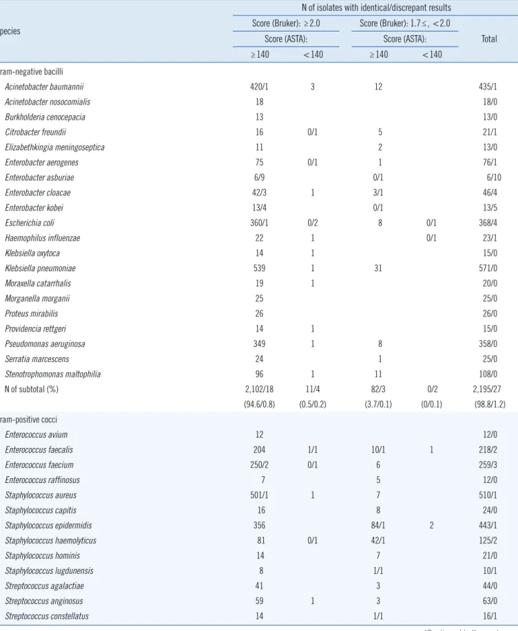
From the 4,919 isolates studied, identical results with confi- dence scores (≥2.0 for the Bruker Biotyper MS system) and ac- ceptable scores (≥140 for the ASTA MicroIDSys system) were obtained for 4,234 (86.1%) isolates (Table 1). For the bacteria that are frequently isolated in clinical microbiology laboratories, the high agreement rates were as follows: K. pneumonia (100%), E. coli (98.9%), P. aeruginosa (100%), A. baumannii (99.8%), S. aureus (99.8%), S. epidermidis (99.8%), E. faecium (98.9%), and E. faecalis (99.1%). In addition, 4,841 (98.4%) isolates had a Bruker Biotyper score ≥1.7 and an ASTA MicroIDSys score
≥140. Only 78 (1.6%) isolates showed discrepant results be- tween the two systems. For these isolates, we performed 16S rRNA gene sequencing. However, some species in the isolates were not accurately identified by either of the two methods; the 16S rRNA gene sequence similarity was very high for Enterobac- ter and Streptococcus mitis groups [7].
From the observed discrepant results between the two MALDI- TOF MS systems, we suspected that a known limitation of other MALDI-TOF MS systems might also be present in the ASTA Mi- croIDSys. Microorganisms are identified by MALDI-TOF MS sys- tems using prerecorded protein spectra that are present in the system library, and these spectra are mostly based on ribosomal proteins. Therefore, MALDI-TOF MS systems are intrinsically limited to differentiate closely related species or strains of Sal- monella spp., Raoutella, Klebsiella, Enterobacter, and Citrobac- ter [4, 8].
Identical results, with scores between 1.7 and 2.0 for the Bruker Biotyper MS system and acceptable scores ≥140 for the ASTA MicroIDSys system, were obtained for 581 (11.8%) isolates;
these included 242 (58.6%) Candida spp., 205 (10.6%) gram- positive cocci, and 82 (3.7%) gram-negative bacilli. Only two isolates of C. albicans showed discrepant results with an ASTA MicroIDSys score <140. This result suggested that the thresh- old score for identification with the Bruker Biotyper should be 1.7, instead of the usual 2.0, in order to compensate for the spectrum quality of Candida spp. In contrast, the cutoff score used for identification by the ASTA MicroIDSys was the typical
Table 1. Comparison of the results for frequently isolated bacteria from the Bruker Biotyper and ASTA MicroIDSys systems
Species
N of isolates with identical/discrepant results Score (Bruker): ≥2.0 Score (Bruker): 1.7≤, <2.0
Total Score (ASTA): Score (ASTA):
≥140 <140 ≥140 <140
Gram-negative bacilli
Acinetobacter baumannii 420/1 3 12 435/1
Acinetobacter nosocomialis 18 18/0
Burkholderia cenocepacia 13 13/0
Citrobacter freundii 16 0/1 5 21/1
Elizabethkingia meningoseptica 11 2 13/0
Enterobacter aerogenes 75 0/1 1 76/1
Enterobacter asburiae 6/9 0/1 6/10
Enterobacter cloacae 42/3 1 3/1 46/4
Enterobacter kobei 13/4 0/1 13/5
Escherichia coli 360/1 0/2 8 0/1 368/4
Haemophilus influenzae 22 1 0/1 23/1
Klebsiella oxytoca 14 1 15/0
Klebsiella pneumoniae 539 1 31 571/0
Moraxella catarrhalis 19 1 20/0
Morganella morganii 25 25/0
Proteus mirabilis 26 26/0
Providencia rettgeri 14 1 15/0
Pseudomonas aeruginosa 349 1 8 358/0
Serratia marcescens 24 1 25/0
Stenotrophomonas maltophilia 96 1 11 108/0
N of subtotal (%) 2,102/18 11/4 82/3 0/2 2,195/27
(94.6/0.8) (0.5/0.2) (3.7/0.1) (0/0.1) (98.8/1.2) Gram-positive cocci
Enterococcus avium 12 12/0
Enterococcus faecalis 204 1/1 10/1 1 218/2
Enterococcus faecium 250/2 0/1 6 259/3
Enterococcus raffinosus 7 5 12/0
Staphylococcus aureus 501/1 1 7 510/1
Staphylococcus capitis 16 8 24/0
Staphylococcus epidermidis 356 84/1 2 443/1
Staphylococcus haemolyticus 81 0/1 42/1 125/2
Staphylococcus hominis 14 7 21/0
Staphylococcus lugdunensis 8 1/1 10/1
Streptococcus agalactiae 41 3 44/0
Streptococcus anginosus 59 1 3 63/0
Streptococcus constellatus 14 1/1 16/1
(Continued to the next page)
Species
N of isolates with identical/discrepant results Score (Bruker): ≥2.0 Score (Bruker): 1.7≤, <2.0
Total Score (ASTA): Score (ASTA):
≥140 <140 ≥140 <140
Streptococcus mitis 20/1 0/1 3/1 0/1 27/4
Streptococcus oralis 30 1/2 4/3 2/4 46/9
Streptococcus parasanguinis 21 7 28/0
Streptococcus pneumoniae 14/10 2/2 4/3 35/15
Streptococcus salivarius 23 10 33/0
N of subtotal (%) 1,671/14 6/8 205/12 5/5 1,887/39
(86.8/0.7) (0.3/0.4) (10.6/0.6) (0.3/0.3) (98.0/2.0) Other bacteria
Clostridium difficile 30 3 1 34
Clostridium hathewayi 13 1 14
Corynebacterium amycolatum 18 18
Corynebacterium striatum 190/1 0/4 6 196/5
Lactobacillus crispatus 8 1 13 22
Neisseria flavescens 11 2 13
Rothia mucilaginosa 28 27 1 56
N of subtotal (%) 298/1 1/4 52/0 2/0 353/5
(83.2/0.3) (0.3/1.1) (14.5/0) (0.6/0) (98.6/1.4)
Candida spp.
Candida albicans 93 0/1 149/3 0/2 242/6
Candida glabrata 27 21 48
Candida krusei 11 3 1 15
Candida parapsilosis 4 19 23
Candida tropicalis 28 50 0/1 79
N of subtotal (%) 163/0 0/1 242/3 1/3 406/7
(39.5/0) (0/0.2) (58.6/0.7) (0.2/0.7) (98.3/1.7)
Total (%) 4,234/33 18/17 581/18 8/10 4,841/78
(86.1/0.7) (0.4/0.3) (11.8/0.4) (0.2/0.2) (98.4/1.6) Table 1. Continued
140 value itself, indicating that the power to discriminate be- tween Candida species was higher in the ASTA MicroIDSys sys- tem than in the Bruker Biotyper system. The ASTA MicroIDSys MS system also showed high accuracy rates for overall identifi- cation of bacteria and Candida spp. from isolates.
In this study, we identified clinically relevant bacteria and Can- dida species from clinical specimens using the ASTA MicroID- Sys system. Our findings on ASTA MicroIDSys system performance in the identification of bacteria and Candida species are in high agreement with findings from the Bruker Biotyper system. Espe- cially for frequently isolated bacteria, such as K. pneumoniae, E.
coli, P. aeruginosa, A. baumannii, S. aureus, S. epidermidis, E.
faecium, and E. faecalis, high agreement rates (98.9–100%) were shown. In conclusion, the ASTA MicroIDSys has comparable iden- tification capability to the Bruker Biotyper system. The ASTA Mi- croIDSys system can reliably identify microorganisms that are commonly isolated in clinical microbiological laboratories.
Authors’ Disclosures of Potential Conflicts of Interest
No potential conflicts of interest relevant to this article were reported.
Acknowledgments
This work was supported by the National Research Foundation of Korea (2014M3A9E5073818), the BioNano Health-Guard Research Center funded by the Ministry of Science, ICT & Fu- ture Planning (MSIP) of Korea as a Global Frontier Project (H- GUARD_2014M3A6B2060509), and the Ministry of Health &
Welfare, Republic of Korea (HI14C1324).
We are deeply grateful to Yong Ha In, Kyu Hwan Park, and Hyung Soon Park (ASTA Inc.), as well as to Hyungsun Kim and Sori Jong (The Research Institute of Antimicrobial Resistance, Yonsei University College of Medicine) for their help.
REFERENCES
1. Seng P, Drancourt M, Gouriet F, La Scola B, Fournier PE, Rolain JM, et al. Ongoing revolution in bacteriology: routine identification of bacteria by matrix-assisted laser desorption ionization time-of-flight mass spec- trometry. Clin Infect Dis 2009;49:543-51.
2. Claydon MA, Davey SN, Edwards-Jones V, Gordon DB. The rapid iden- tification of intact microorganisms using mass spectrometry. Nat Bio-
technol 1996;14:1584-6.
3. Martiny D, Busson L, Wybo I, El Haj RA, Dediste A, Vandenberg O. Com- parison of the Microflex LT and Vitek MS systems for routine identifica- tion of bacteria by matrix-assisted laser desorption ionization-time of flight mass spectrometry. J Clin Microbiol 2012;50:1313-25.
4. Bilecen K, Yaman G, Ciftci U, Laleli YR. Performances and reliability of Bruker Microflex LT and VITEK MS MALDI-TOF mass spectrometry sys- tems for the identification of clinical microorganisms. Biomed Res Int 2015;2015:516410.
5. Richter SS, Sercia L, Branda JA, Burnham CA, Bythrow M, Ferraro MJ, et al. Identification of Enterobacteriaceae by matrix-assisted laser de- sorption/ionization time-of-flight mass spectrometry using the VITEK MS system. Eur J Clin Microbiol Infect Dis 2013;32:1571-8.
6. Bessède E, Angla-Gre M, Delagarde Y, Sep Hieng S, Ménard A, Mégraud F. Matrix-assisted laser-desorption/ionization biotyper: experience in the routine of a university hospital. Clin Microbiol Infect 2011;17:533-8.
7. Janda JM and Abbott SL. 16S rRNA gene sequencing for bacterial iden- tification in the diagnostic laboratory: pluses, perils, and pitfalls. J Clin Microbiol 2007;45:2761-4.
8. Wang H, Fan YY, Kudinha T, Xu ZP, Xiao M, Zhang L, et al. A compre- hensive evaluation of the Bruker Biotyper MS and Vitek MS matrix-as- sisted laser desorption ionization-time of flight mass spectrometry sys- tems for identification of yeasts, part of the national china hospital inva- sive fungal surveillance net (CHIF-NET) study, 2012 to 2013. J Clin Mi- crobiol 2016;54:1376-80.
Species
N of isolates with:
Agreement rate (%) Identical
results Discrepant results Gram-negative bacilli
Acinetobacter baumannii 420 1 99.8
Acinetobacter nosocomialis 18 100.0
Burkholderia cenocepacia 13 100.0
Citrobacter freundii 16 100.0
Elizabethkingia meningoseptica 11 100.0
Enterobacter aerogenes 75 100.0
Enterobacter asburiae 6 9 40.0
Enterobacter cloacae 42 3 93.3
Enterobacter kobei 13 4 76.5
Escherichia coli 360 1 99.7
Haemophilus influenzae 22 100.0
Klebsiella oxytoca 14 100.0
Klebsiella pneumoniae 539 100.0
Moraxella catarrhalis 19 100.0
Morganella morganii 25 100.0
Proteus mirabilis 26 100.0
Providencia rettgeri 14 100.0
Pseudomonas aeruginosa 349 100.0
Serratia marcescens 24 100.0
Stenotrophomonas maltophilia 96 100.0
N of subtotal 2,102 18 99.2
Gram-positive cocci
Enterococcus avium 12 100.0
Enterococcus faecalis 204 100.0
Enterococcus faecium 250 2 99.2
Enterococcus raffinosus 7 100.0
Staphylococcus aureus 501 1 99.8
Staphylococcus capitis 16 100.0
Species
N of isolates with:
Agreement rate (%) Identical
results Discrepant results
Staphylococcus epidermidis 356 100.0
Staphylococcus haemolyticus 81 100.0
Staphylococcus hominis 14 100.0
Staphylococcus lugdunensis 8 100.0
Streptococcus agalactiae 41 100.0
Streptococcus anginosus 59 100.0
Streptococcus constellatus 14 100.0
Streptococcus mitis 20 1 95.2
Streptococcus oralis 30 100.0
Streptococcus parasanguinis 21 100.0
Streptococcus pneumoniae 14 10 58.3
Streptococcus salivarius 23 100.0
N of subtotal 1,671 14 99.2
Other bacteria
Clostridium difficile 30 100.0
Clostridium hathewayi 13 100.0
Corynebacterium amycolatum 18 100.0
Corynebacterium striatum 190 1 99.5
Lactobacillus crispatus 8 100.0
Neisseria flavescens 11 100.0
Rothia mucilaginosa 28 100.0
N of subtotal 298 1 99.7
Candida spp.
Candida albicans 93 100
Candida glabrata 27 100
Candida krusei 11 100
Candida parapsilosis 4 100
Candida tropicalis 28 100
N of subtotal 163 100
Total 4,234 33 99.2
Supplemental Data Table S1. Comparison of the identified bacteria and yeast with score values ≥2.0 for the Bruker Biotyper system and
≥140 for the ASTA MicroIDSys system