←
人 합 放g‘f 짧 짧 챙 會~.: ~21 환 ;:P 3 YJJi pp. 419-423 , 1985 Journal of Korean Radiological Society, Vo1.21 , No.3, 1985
開放性氣管技 徵候를 보이 는 細氣管技師脫處
( bronchioloalveolar carcinoma ) 의 CT
서 울大學俊 醫科大學 放射線科學敎室
任 廷 基·金 鍾 哲 ·韓 萬 좁 - Abstract -
Computed Tomography of Bronchioloalveolar Carcinoma Showing Open Bronchus Sign
Chung Kie 1m
,
Jong Chul Kim,
Man Chung HanDepartment of Radi%gy, College of Medicine, 5eou/ Nationa/ University
Open brochus with diffuse narrowing
,
stretching,
and leafless tree appearance of the bronchi is the well know bronchographic criterior of bronchioloalveolar carcinoma. Though similar findings are expected in CT, authors could find no report concerning the open bronchus sign of bronchioloalveolar carcinoma demonstrated by CTAuthors presents CT 01 bronchioloalveolar carcinoma showing lobar or segmental distribution and patent bronchus within the tumor mass
1.
績 論대해서 注意를 할 必要가 있다.
著者等은 CT 스캔 상 睡塊內에 開放性氣管技를 보이 는 細氣管技뼈뼈찮 3 例를 보고하며 감별진단에 있어서 뼈에 睡場이 있을시 原發性이거나 或은 總發性이거나 매우 特異度가 높은 所見임을 강조하고자 한다.
관계없이 睡場內의 氣管技가 막히거냐 수위로 빌리거나 하기 때문에 睡塊內에는 초氣로 찬 가관지를 볼수가 없 는 것이 상례이냐, 細氣管技8市뼈痛( bronchioloalveolar
carcinoma) 나 럼프睡 等은 예외가 될 수 있음은 잘 알
려진 사실이 다 1
,
2,
3)이 와같은 소위 開放性氣管校 徵候는 特異度가 높은 所
II.
효 例f숱例 75 세 男子
1. 臨똥所見
見이기는 하나 단순뼈部 X線에서는 거의 보이는 경우가 3 年前부터 I!M莫*의 진단하에 내과적 치료를 받아왔 없고, 기관지조영술에서 잘 나타날 수 있으나 睡塊性病 으며, 近來에는 柱被性 객담과 呼댔困難을 主訴로 入院 變에서는 이러한 검사를 잘 시행하지 않는 등의 이유로
별 관심을 끌지 못하고 있다.
그러나 CT 스캔에서는 睡塊內 開放{生氣管技가있다면, 이플 잘 表出해 내므로 이와같은 特異度가 높은 所見에
하여 시행한 객담 결핵균검사 및 細@힘談에 陰性이였으며,
늑막전.Ä)에서 흉出被이 배출되지 않았다.
2.
放射線學的 所見單純뼈部 X線上 右測 하부에 균질성음영도 증가가 있 이 논문은 1985년 4월 15 일에 접수하여 1985 년 6 월 어 助模용出 및 무기펴l를 의심하게 하는 소견이 있으며 3 일에 채택되었음 CFig. 1-A) CT 스캔상 右中葉 및 下葉 전반에 결쳐 뼈
-大합}jj( q‘tl'~짧쟁싹표 : 第 21 卷 第3械 1985
實質의 硬化( consolidation) 의 소견이 있는데, 中葉의 전 기있는 %쨌性을 띠는 비교적 軟한 睡塊로 되어 있으며,
방부에는 分葉性邊緣Cl
obulated margin)
을 보이고 있 어 단순한 硬化와는 다른 양상을 나타내고 있다. 中葉氣 管技의 내측 및 외측 分技角이 정 상보다 커져서 各分技 가 넓게 퍼져있£며, 각각의 氣管技는 내경은 작아져 있 으연서 1$張된 양상을 보여주고 있다. 下葉에는 여려개 의 훌睡樣陰影을 보이 고 있으며,lI:IJ體홍出의 所見은 보 이지 않고있다 CFig.l-B , C).3.
病理組織學的 所見手術時 硬化된 右뼈ij 中葉 및 下棄에서 탁한 %波性짧 體가 특히 압박시 흘러 나왔으며, 육안적 병리소견은 중
이 내부의 기관지는 開通되어 있으나 압박된 양상을 보 였다 현미경적 소견은 싫被質을 함유하는 細氣管技師 8힘?휩
(bronchioloalveolar
carcinoma ) .2.로 냐타났다.jiË例2 57 세 女子 1. 臨똥所見
1 年前 객 담 및 발열증상으로 입원 우하엽 !li1ï~ 듭?斷 요로 치료받은적 있으며, 최근 같은 증상으로 입원하였 다.
2.
放射線學的 所見엽 및 하엽 전체가 硬化되어 있으며 절단연은 회색의 윤 單純뼈部 X線上 우하엽의 後基底分節 부위에 경계가
A
Case 1 , 75 year old male
A. Chest P A view shows homogenous increased density in right lower lungsuperiorly bound by rather bulging minor fissure.
B. CT scan at the level of carina
shows∞ llapsed
right lower lobe with round hypodense area in lateral side.
C. CT scan 3cm below B shows entirely patent but stretched and diffusely narrowed lobar and segmental bron- chi of middle lobe. Medial and
‘lateral segmental bronchi of middle
10be are spread a part. S ee also lobulated medial border of middle
10be suggesting a mass lesion rather than simple consolidation.
B
c
A
-ff廷基 外:00放↑生氣管技 微候를 보이는 細氣풍技l!ilîn힐짧의
CT-
불분영한 睡塊樣陰影이 觀察되며
(Fig.
2 -A) CT스캔에 서 는 分葉樣邊緣(lobulated margin)
의 내부에 작은 開 放性 기관지을 包含하고 있는 睡塊樣陰影으로 보이는데 이의 分布는 分節性으로 後基底分節의 말초부까지, 즉기침 및 뼈部 불쾌감이 1 年前 부터 있었으며, 過去歷 에 폐결핵에 대한 治癡를 받은적이 있었다.
2.
放射線學的 所見tl1J骨橫隔題角
(Costophrenic Sulcus)
까지 뼈張되어 마치 單純뼈部 X線上 左下葉에7x7x7cm
정도 크기의 비뼈賢質의 硬化와 같은 所見을
나타내코있으며, 이의
내교적 경제가
분명한睡塊로
보이며 (Fig.3-A), CT스 부에는여러개의 작은 底陰影度의 훌性部位를 함유하고 캔에서는 역시
睡塊내에 開放性氣管技와 分葉狀 경계를있 다
(Fig.
2-B, C).
3.
細뼈病理學的 所見견g 皮的뼈生檢時 약
lcc
정도의 햄被 및 類R훗狀의 被體 가 흡인되었으며, 吸引細뼈등? 및 찍홍%찢細R힘응?j즈로細氣管 技8市6힘짧j즈로 진단되었다.1ft.例 3 : 49 세 男子 1. 臨뚱所見
보이는 睡塊로 나타나는데, 이의 外測으로는 分節樣 分 布의 所見도 보이고 있다 Gallium 을 이 용한 睡傷숱훌 像에 서는 睡塊에 同位元素의 흉수를 보이고 있 다. 超音 波 擺影像에서는 賢皮質과 비슷한 에코도을 냐타내는역 시 分棄狀의 睡塊로 보이고 있으며, 睡塊의 내부에 開 放性氣管技내의 공기에 의한 것£로 생각되는 강한 선 상의 에코도를 나타내고 있다 (Fig. 3-B,
C
,D).
B
Fig.2. Case
2,
57 year old fema1e.
A. Chest P A shows 피
defined mass
Iike density in
r핑htlower lung field.
B.
CT scan
showsirregular marginated mass in posterolateral basal segment area of right lower lobe with small patent bronchi in the peripheral portion of the mass
C.
CT
scanbelow B shows consolida- tion
파ceappearance of the mass draping the surface of costophrenic
sulcus.
C
-大햄放射線뺨쩔會誌 : 第21卷 第3號
1985-
B
A
D
C
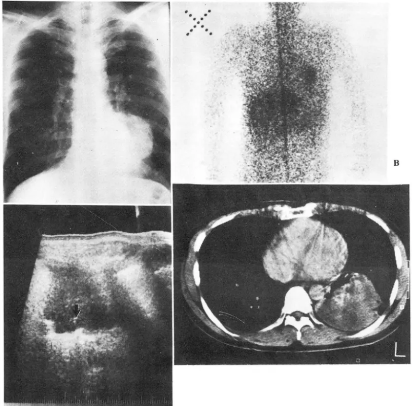
Fig. 3. Case 3 , 49 year old male
A. Chest PA shows mass density in right lower lobe.
B. Gallium scan shows po
sitive uptake of radioisotope within the tumor.C.
Ultrasonogram shows lobulated relative hypoechoic mass with dense tubular
echogenic area within the mass(arrow) suggesting patent air-filled bronchus.
D. CT scan shows homogenous lobulated mass in posterolateral basal segment area of left lower lobe with tiny air-filled bronchi in the anterior portion of the mass.
ßI.
考 察細氣管技師6힘}홈은 原發師僅의 1.5%~ 6.5%의 頻度를 나타내 며 3
’”
單-性 師睡塊, "힘硬化 ( conso1ida tion)
및 鋼漫性小結節(Diffuse nodule)
등의 다양한 뼈部 X線 所見을 냐타내 는 것으로 臨 VR的요로나 放射線學的으로 特 異한 原發性H市}끊이며 주로 말초부의 細氣管技 或은 8市뼈 에서 發生하여 이들의 뚫 및 內흩을 통해서 주위로 퍼 지게 되므로 광범위한 睡塊에도 불구하고 氣管技의 閒銷 가 없다는 점이 特徵의 하나로 지적되어 왔다 5-9)
따라서 이와같은 氣뾰 (A
ir
-space) 을 통한 주위로의-任廷꿇 外: f바IiJc l生氣깝技 ~候를 보이는 뼈I氣管技R빼包?힘으1 CT-
파급이 기존의 싫構( fissure) 에 의하여 경계지워 질때 는 평性葉性硬化 (chronic lobar cons oli dation)의 양상을 띠게 뇌므로 治爾에 反應이 적은뼈갖, 폐쇄성폐렴. H읍 Il15 j生 j뼈씻 및 8市結核 等으로 등2斷되기 쉽고 lo,ll,l2), 실 제로 본
iiEf71 J
1, 2 에서도 이러한 所見을 보이고 있다이와같이 細氣管技뼈뼈않이 師寶質의 漫性硬化의 所 見으로 나타날때 우선 염증성질환을 생각하게 되겠으나,
효例
1에 서와 같이 病 짧部 氣용技가 漫性*효性 훌愚에
서 보이는 織維化 및 용적감소에 의한 不規則한 變形없 이 各各의 氣管技가 內쩔이 均一하게 약간 졸어들어 있 으면서 f$張된 所見은 細氣管技師뼈평의 特異한 點으로 생각된다.
이러한 病짧部氣管技의 閒銷가 없는 均-한 俠쩔 및 f申張의 所見은 氣管技造影術에서의 細氣管技 R市R힘}훨의 特徵的인 所見으로 잘 알려져 있으나 1 ,,>, CT스캔상
이와같은 所見을 나타내 는 효例에 대 한 報告는 著者等 이 아는한 국내외로 발표된 바가 없다.
Metzger등 11>은 細氣管技뼈뼈鎬에서 CT 스캔을 함 으로써 單純B關lX線에 서 발견되 지 않는 bI漫性 病쩔를 찾을 수 있다는 점에서 CT 의 治爾方針 결정 및 예후 판정에서의 역할을 강조한 바 있는데 본 증례 1 에서도 左測 뼈門주위의 結節들이 CT 상 잘 나타냐고 있어 轉移 或은 通氣性擬散( Aeorogenous spread) 을 의 심 케 하고 %다.
IV.
結 論CT 상 開放性氣管技 및 大葉性 分布의 所見을 보이
는 3 f71J의 細氣管技師뼈햄의 iiEØ1J 를 報告하며, 뼈의 睡 塊樣 病變의 감별진단에 있어서 감안되어야 할特異度가 높은 所見으로 생 각된 다.
REFERENCES
1. Zheutlin N
,
Lasser EC,
Rigler LG: 8ronchographic abnor malities in alveolar cell carcinoma of the lung: A new diagnostic sign. Dis. Chest 25:542, 19542. Felson B: Chest roentgenology. Philadelphia
,
Saunders’124-128
,
19133. Storey C, Knudtson K, Lawrence B: 8ronchiolo ("alveolar cell') carcinoma of the lung. } Thorac Surg 26:331-406
,
19534. Watson W
,
Farpour A: Terminal bronchiolar or alveolar cell cancer of the lung: 265 cases: Cancer 19:166-180, 19665. Berkman YM: The many facets of alveolar-cell carcinoma of the lung. Radiology 92:193-198
,
19696. Theros EG, Hlghman B: 8ronchioloalveolar-cell carcinoma:
RPC of the Month from the AFIP. Radiology 91:661- 668, 1910
7. Ludington LG, Verska
JJ
, Howard T, Kypridakis ι Brewer LA: 8ronchilar carcinoma 껴Iveolar cel끼 another great Imltatoη a review of 41 cases. Chest 61:622-628, 1912.8. Shapiro R, Wilson GL, Yesner R, Shuman H‘ A useful roen tgen sign in the diagnosis of localized bronchioloalveolar carcinoma. A}R 114:516-524, 1912
9. Berkman YM: The many faces of bronchiolo-alveolar car- cinoma. Semin Roentgenol 12:201-214, 1911.
10. Miller WT, Husted J. Freiman 0, Atkinson B, Pietra GG 8ronchioloalveolar carcinoma: two clinical entities with one pathologic diagnosis. A}R 130:905-912, 1918 11. Metzger RA, Mulhern CB, Arger PH, Coleman BG, Eps-
tein DM
,
Gefter WM: CT differentiation of solitary from diffuse bronchioloalveolar cell carcinoma. } Comput Assist Tomogr 5:830-833, 198112. Epstein DM, Gefter WB, Miller WT: Lobar bron chioloalveolar cell carcinoma. A}R 139:463-468, 1982.