http://dx.doi.org/10.4078/jrd.2012.19.3.125
125
<Received:May 30, 2012, Revised:June 17, 2012, Accepted:June 18, 2012>
Corresponding to:Seok-Jung Kim, Department of Orthopedic Surgery, Uijeongbu St. Mary's Hospital, The Catholic University of Korea, 65-1, Geumo-dong, Uijeongbu 480-130, Korea. E-mail : [email protected]
pISSN: 2093-940X, eISSN: 2233-4718
Copyright ⓒ 2012 by The Korean College of Rheumatology
This is a Free Access article, which permits unrestricted non-commerical use, distribution, and reproduction in any medium, provided the original work is properly cited.
줄기 세포를 이용한 관절 연골 손상의 치료
윤형문2ㆍ김석중1ㆍ김태균1
가톨릭대학교 의과대학 정형외과학교실1, 건국대학교 의과대학 충주병원 정형외과학교실2
Stem Cell Therapy in Articular Cartilage Injury
Hyung Moon Yoon2, Seok-Jung Kim1, Tae-Gyun Kim1
Department of Orthopedic Surgery, College of Medicine, The Catholic University of Korea1, Seoul, Department of Orthopedic Surgery, Konkuk University Chungju Hospital,
Konkuk University School of Medicine2, Chungju, Korea
The natural history after articular cartilage injury is unclear. However, it is generally accepted that once artic- ular cartilage is injured, its ability to regenerate is limited and that injury progresses to arthritis with time. Over the years various treatments have been developed and are used, such as arthroscopic debridement, microfracture, multiple drilling, osteochondral transfer, and Autologous Chondrocyte Implantation (ACI). These can be divided in- to treatment methods which apply cells and those which
apply tissue. The former include abrasion chondroplasty, microfracture, multiple drilling, and ACI. The latter in- clude osteochondral transfer and allograft. Combination treatments using both cells and tissues are new-generation ACI and microfracture with biomaterials. The clinical ap- plications of stem cell therapy is still at an early stage, but shows much promise, particularly in the management of cartilage defects.
Key Words. Cartilage, Stem cell, ACI, Microfracture, Knee
서 론
관절연골 손상에 대한 자연력(natural history)은 아직까지 도 명확히 밝혀진 바는 없지만, 시간이 지나면서 관절염이 발생할 것이라는 가정이 통념으로 받아들여지고 있다 (1).
골관절염(osteoarthritis)은 65세 이상의 인구 중 약 70%에서 발생할 정도로 가장 흔한 관절의 질환이며 관절연골의 파 괴로 인한 관절기능의 상실을 특징으로 한다 (2).
특히 관절연골의 손상은 매우 심한 통증을 유발할 수 있 으며, 이로 인해 여러 가지 활동을 제한하여 추가적인 문 제(medical problem)들이 발생할 수 있다 (2).
관절 연골은 연골 세포(chondrocyte)의 밀도가 낮아 상대적
으로 세포 외 기질(extracellular matrix, ECM)이 차지하는 부 분이 많고 (1), 관절 연골이 손상된 후 그 주변에 있는 연골 세포(chondrocytes)의 이동(migration)이 제한적이어서 관절 연골의 회복 능력은 다른 조직에 비해서 상대적으로 낮다 (3). 따라서 관절연골의 손상이 작은 경우에는 주변에서 이 동해온 연골세포에 의해서 손상부위가 어느 정도 치유가 될 수 있지만 손상이 큰 경우에는 재생능력이 제한적이고, 재생되는 조직이 정상 초자연골(hyaline cartilage)이 아닌 기 계적 성질이 약한 섬유 연골(fibrocartilage)로 재생된다.
연골 손상 시 치유를 가능하게 할 수 있는 세포들로는 손 상 주변의 연골 세포와 골수(bone marrow), 활막(synovium)
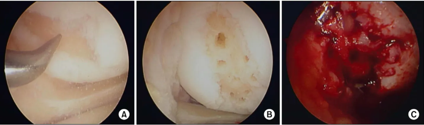
Figure 1. (A) Microfracture using metal awl. (B) Arthroscopic finding of microfracture. (C) Bone marrow bleeding through holes of microfracture.
및 지방 조직에 존재하는 줄기 세포 등이 있다 (4). 그러나 이러한 세포들 중에서 골수에 존재하는 줄기세포는 연골 하 골(subchondral bone)을 침범하는 전층(full thickness) 연 골 손상의 경우에만 연골 재생에 관여할 수 있으며 활막 및 지방 조직의 줄기세포는 연골 손상 시 자발적으로 회복 에 관여하는지 알려져 있지 않다 (4).
오래 전부터 관절 연골 손상이나 초기 관절염의 치료로 는 약물이나 물리치료 등을 통한 통증 조절이 일차 치료였 으며, 증상의 호전이 없는 경우에는 관절경 수술(arthrosco- pic surgery), 인공관절 치환술(arthroplasty)과 같은 수술적 치료가 이루어졌다.
1994년 Brittberg 등 (5)에 의해서 최초로 자기유래 연골세 포 이식술(autologous chondrocyte implantation, ACI)이 시행 된 이후에 세포를 이용한 관절 연골 손상의 치료 방법이 급속도로 발전하기는 하였으나, 세포적 측면에서만 본다 면 이미 오래 전부터 미세 골절술(microfracture) 등 골수줄 기세포를 이용한 관절연골 재생술이 시행되고 있었다.
여기서는 관절 연골 재생을 위해 골수줄기세포를 이용하 는 전통적 방법인 미세 골절술, 생체적합 물질을 사용한 차세대 미세 골절술(2nd generation microfracture), 환자 본인 의 연골세포를 배양한 후 이식하는 자기유래 연골세포 이 식술, 이외에 줄기세포를 비롯한 각종 세포 이식을 통한 연골재생술에 대해서 간단히 살펴보기로 한다.
본 론 미세 골절술
관절 연골 손상의 대표적인 수술적 치료법들로는 관절경 하 변연 절제술(arthroscopic debridement), 미세 골절술, 다 발성 천공술(multiple drilling) 등이 있다. 이 방법들 중에서 관절경하 변연절제술의 효과는 제한적이어서 최근에는 주 수술법으로 사용되고 있지는 않다.
조직 공학의 개념이 발전하기 이전에는 연골손상이 있으 면 단순히 골수를 자극해서 연골 손상을 치료하는 것이 기 본적 치료였다. 비교적 작은 결손에서는 좋은 결과들이 발
표되어 왔지만, 큰 결손에 있어서는 한계가 있다는 것이 오래 전부터 인지되어 왔다.
1959년 Pridie (6)가 처음으로 연골 하 골을 천공(drilling) 하여 여기서 흘러나온 골수의 줄기세포에 의해 새로운 연 골이 형성되는 것을 기술한 이후, Rodrigo 등 (7)은 천공 시 열에 의한 골괴사를 피하기 위해서 뾰족한 정(awls)을 사용한 관절경하 미세 골절술을 소개하였다. 이러한 술기 는 1990년대부터 연골 손상을 치료하는 일차적인 시술 방 법으로 사용되었다.
미세 골절술은 연골 하 골이 손상되면 골수줄기세포 (bone marrow mesenchymal stem cells)를 포함하는 골수성 분이 새어 나오고, 이 세포들이 분화해서 연골을 형성하는 원리를 이용한 치료이다(Figure 1). 하지만 대부분 생성되 는 연골은 정상적인 관절 연골인 초자 연골이 아닌 섬유연 골이 형성되게 된다. 이러한 섬유연골은 제1형 콜라겐이 풍부하고 proteoglycan의 함량은 적어 마모에 견디는 성질 이 떨어진다. 따라서 손상된 연골조직이 섬유연골로 재생 이 되면 수술 후 2년 정도까지는 60∼70%의 증상 호전을 보이나 그 이후에는 구조적인 와해가 일어나 증상의 악화 가 일어나는 경우도 있고 결손 부위가 클수록 증상의 악화 가 더 심해지게 된다고 알려져 있다 (8-11). 현재까지는 관 절 연골 손상 부위의 크기가 2 cm2 보다 큰 경우에는 환자 들의 임상 결과가 좋지 못하기 때문에, 이 결손의 크기가 미세 골절술의 허용한계로 여겨져 왔고 그 외에도 나이가 35세 이상이나 체질량지수(BMI)가 25 kg/m2 이상인 경우 에도 예후가 좋지 않다고 보고되고 있다 (12).
그럼에도 불구하고 경제적 측면이나 시술의 간편성 때문 에 많은 의사들은 비교적 큰 결손부위에서도 미세 골절술 을 하는 경우가 많으며 슬관절의 대퇴골 연골의 손상에 대 해서는 현재까지 일차적 치료법으로 자리잡고 있다.
차세대 미세 골절술(2ND generation microfracture using bioscaffold)
비록 미세골절술을 통해서 재생된 연골이 기계적 성질이
Figure 2. (A) Cutting collagen membrane during AMIC proce- dure. (B) Attaching collagen membrane onto the cartilage defect.
Figure 3. ACI: autologous cho- ndrocyte implantation.
약한 섬유연골이라 할지라도 좋은 단기임상결과, 경제적 측면, 술기적 측면 등을 고려한다면, 미세골절술은 그 나 름의 장점을 지녀 앞으로도 오랫동안 사용될 수 있는 술기 이다. 따라서 연골손상을 치료하기 위해서 새로운 치료법 을 개발하기 보다는 이를 좀 더 나은 방법으로 발전시켜야 한다는 필요성이 증가되고 있다.
Autologous matrix-induced chondrogenesis (AMIC)는 이러한 필요성과 더불어 개발된 대표적인 예로서 손상된 연골을 변 연 절제한 뒤 노출된 연골하골에 미세 골절술을 시행하여 골수 줄기세포가 손상부위로 나오도록 한 뒤 제1/3형 콜라 겐 막으로 손상 부위를 봉합하는 방법이다 (8,9,11,13,14) (Figure 2). 미세골절술 후 덮어진 콜라겐 막은 골수에서 흘
러나온 줄기세포들이 손상부위에 집중되도록 하여 연골형 성을 촉진하게 한다고 알려져 있으며 (15), 때로는 연골세포 로 분화를 촉진하는 인자들이 들어있는 생체적합물질(biosc- affold) 등을 막 안에 주입하기도 한다. 이 방법의 장점은 배 양 후 재수술이 필요하지 않으며, 체외 세포 배양 방법에 비 하여 비용이 적게 든다는 장점이 있다. 결손 부위에 콜라겐 막을 봉합하기 위하여 수술창(incision)이 커지는 단점이 있 음에도 불구하고, Gille 등 (14)은 AMIC의 5년 중기 추시 보 고에서 87%의 환자가 만족했다고 보고하기도 하였다.
최근에는 막으로 고정하는 방법이 아닌 Gel형태의 이식 물을 이용한 방법도 발표되었다. Shetty와 Kim 등 (16)은 연골이 손상된 부위에 미세골절술을 시행한 뒤 콜라겐 겔
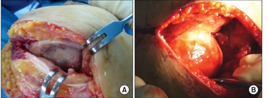
Figure 4. (A) Severe arthritic findings. (B) Operation findings after gel type ACI.
(gel)을 이용하여 골수 줄기세포를 고정하는 Autologous Collagen Induced Chondrogenesis (ACIC)를 보고하였는데, 모든 수술 과정이 관절경하에서 이루어지기 때문에 따로 절개가 필요 없고 회복이 빠른 장점이 있다.
자기유래 연골 세포 이식술
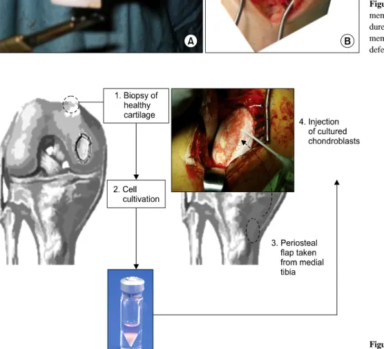
자기유래 연골 세포 이식술은 현재 세포를 이용한 치료 중 가장 널리 사용되고 있는 방법이다. 이는 연골 결손 부 위에 체외에서 배양된 연골 세포을 이식하여 연골의 재생 을 유도하는 방법으로 중, 장기 추시에서 80% 이상의 높은 성공률이 보고되고 있다 (17-19). 체중 부하를 하지 않는 관절연골의 일부를 소량(200∼300 mg) 채취한 뒤 연골 세 포를 분리, 배양 한 후 이식 수술을 시행하는 것으로 (Figure 3), 이러한 자가 연골 세포 이식술은 세포 배양 기 술과 수술 방법에 따라서 세대(generation)를 구분할 수 있 다 (20). 1세대 자가 연골 세포 이식술은 Brittberg 등 (5)이 처음 개발하여 사용한 방법으로 관절 연골 결손 부위에 경 골 근위부에서 채취한 골막을 봉합한 뒤 액상의 연골 세포 를 주입하는 방법으로 여러 연구를 통하여 좋은 결과가 증 명되었다. 그러나 골막을 채취해야 하고 액상의 연골 세포 액이 흐르지 않도록 단단히 봉합해야 하는 기술적 어려움, 이를 위해 절개창이 커지는 문제, 그리고 배양된 연골 세 포가 탈분화 및 석회화 하는 문제들이 제기되고 있다.
2세대 자가 연골 세포 이식술은 collagen membrane (21), hyalograft C (22), fibrin glue (23) 등의 생체적합물질에 연 골 세포를 심어 배양한 후 이를 결손 부위에 고정하는 방 법이다. 이러한 방법들은 골막 채취를 위한 추가의 절개가 필요 없고, 수술창이 작거나 관절경으로도 시술이 가능한 경우도 있어, 술 후 재활 및 회복이 빠른 장점이 있다. 이 중 특히 Kim 등은 국내 제품(2세대 연골세포 이식술, 세원 셀론텍, 한국)을 이용하여 관절염을 치료한 증례를 보고하 기도 하였다(Figure 4) (24).
차세대 자가 연골 세포 이식술은 유전 치료(gene therapy) 기술 및 여러 성장 인자(growth factor)의 도입으로 연골 형 성 능력을 더욱 향상시키려는 시도라 할 수 있다. 활막, 지 방 등 다양한 조직에서 얻어진 줄기세포를 이용하여 연골
세포를 대체하려는 시도가 있으며, 생체 재료(biomaterials) 공학의 발달로 보다 안정적이고 세포 친화적인 환경이 만 들어졌다고 평가되기도 한다 (25). 하지만 유전 치료 기술 및 성장 인자의 세포에 대한 안정성이 명확히 확인된 것이 아니며 모든 연구가 동물 또는 in vitro 연구라는 한계가 있다. 또한, 최근에 동종 줄기세포를 이용한 연골세포 이 식술이 제품으로 사용되고 있지만, 장기 추시의 부재, 윤 리적인 문제, 분화 능력이 뛰어난 줄기 세포의 사용한계 등이 문제점으로 제기되고 있다.
줄기세포를 이용한 연골의 재생
자가 연골세포 이식술은 연골조직채취를 위한 추가적인 수술이 필요하고, 사용되는 세포가 이미 분화가 진행된 세 포이기 때문에 체외 배양 시 탈분화가 일어나 표현형 (phenotype)이 변할 수 있다는 제한점이 있다 (26-28). 또한 동종의 연골 세포를 이용한 세포 이식의 경우 면역 거부 반응이 발생할 가능성이 높아서 시행되고 있지 않다. 이러 한 상황에서 간엽 줄기 세포(mesenchymal stem cells, MSCs)는 좋은 대안이 될 수 있다. 1970년대 골수에서 간엽 줄기세포의 존재를 발견하여 보고한 이후 (29), 다양한 분 화능력과 함께 면역 억제력, 혈관 형성 능력, 세포 사멸 (apoptosis) 및 섬유화 방지, 측분비 유도체(paracrine media- tors) 분비 능력까지 있는 것으로 알려져 왔다 (30). 이러한 다양한 능력으로 인하여 간엽 줄기 세포는 퇴행성관절염, 류마티스질환 등의 퇴행성 및 염증 질환의 치료뿐 만 아니 라 유전적 골-연골 질환의 치료에도 적용하고자 하는 많은 연구가 이루어지고 있다.
골수 유래 간엽 줄기 세포(bone marrow stem cells, BMSCs)는 주로 골반의 장골능(iliac crest)에서 채취하며 다른 부위 간엽 세포에 비해 연골세포, 골형성 세포, 지방 세포, 섬유세포 등 다양한 세포로의 분화가 용이하여 많은 임상 시험이 시행되었다 (31-33). 하지만 골수 줄기세포는 채취할 수 있는 양이 제한적이고, 분리과정이 힘들다는 단 점이 있어 골수 이외에 다른 곳에서 간엽줄기세포를 찾으 려는 연구가 계속되고 있다.
지방 조직은 인체의 여러 곳에 분포하고 있고 많은 양을
간단한 지방 흡입을 통하여 얻을 수 있고 반복적으로 채취 가 가능하다는 장점이 있으며, 골수 유래 간엽 세포와 비 교하여 골-연골세포 형성 능력이 비슷하다는 보고도 있다 (34). 하지만 아직 지방 줄기세포의 분화 조절은 초기단계 에 있어 더 많은 연구가 필요하다.
탯줄(umblical cord)은 와튼제대 교질(Whartone’s jelly)에 간엽줄기세포가 다수 존재하는 것이 확인되면서 세포 치 료에 이용이 늘고 있다 (35-37). 탯줄의 줄기세포는 배아줄 기세포에 비하여는 분화능력이 제한적이지만 성인 줄기세 포에 비하여 분화능력이 뛰어나다. 또한 채취과정이 안전 하고 배아 줄기세포에 비하여 윤리적인 문제가 적고, 면역 거부 반응도 다른 조직에 비하여 낮기 때문에 동종 줄기세 포 치료의 가능성이 높아 여러 기업에서 개발 중에 있으며 최근에는 상용화된 제품도 개발되었다.
활막은 관절을 싸고 있는 관절낭(joint capsule)과 관절 내 공간 사이에 있는 얇은 연부조직으로 macrophage-like cell인 A형 세포(type A cell)와 fibroblast-like cells인 B형 세 포(type B cells)가 존재한다. 이중 B형 세포가 활막유도줄 기세포(synovium-derived stem cells, SDSCs)의 공급원 (source)이 된다고 알려져 있다 (38,39). 특히 SDSCs에서 hyaluronan 생성 시 필수 enzyme인 UDP-glucose de- hydrogenase (UDPGD)의 활성도가 높고 (40,41), 연골 생성 능력의 표지인자라 여겨지는 CD44 발현이 높아 (42-44), 연골 손상 시 새로운 간엽줄기세포의 공급원으로 기대되 고 있다 (45).
동물 실험을 통해서 퇴행성관절염을 지방유래 또는 골수 유래 간엽줄기세포의 관절 내 주입으로 치료했다는 보고 (46)는 있지만, 사람을 대상으로 하는 실제 관절염의 치료 시도는 매우 제한적으로 이루어져 왔고 그 보고 또한 매우 적다. 최근, 간엽줄기세포를 이용한 치료 가능성에 대한 보고들이 시작되었고 (31,32,47,48), 슬관절 퇴행성관절염 을 가진 4명의 환자에게 골수유래 줄기세포치료를 한 뒤 1년 추시에 대한 결과가 보고되는 등 (49) 줄기세포 치료 시도가 점차 늘어가는 추세이다.
줄기세포를 이용하여 치료한 결과 거의 대부분의 환자에 서 보행 능력이 향상되고, 보행시의 통증이 감소되었으며, 이러한 임상적인 증상 호전 외에 좀 더 객관적인 자료로써 자기 공명 영상 검사 등을 통하여 손상 부위에서 조직재생 을 확인하기도 하였다 (50). 또한, 전층 연골 손상에서 간 엽줄기세포를 platelet-rich fibrin glue와 함께 이식하여 성 공적인 결과가 보고되기도 하였으며 (51), 최근에는 자기 유래 연골세포 이식술과 간엽세포를 이용한 치료 결과를 비교하여 두 방법간의 결과에 차이가 없지만 경제적 비용 과 수술의 크기 및 횟수를 고려하면 간엽줄기세포치료가 더 좋다는 보고도 있다 (52).
결 론
관절 연골은 한번 손상이 되면 자연적으로 치유되는 것
에는 한계가 있어 현재까지도 많은 치료법들이 개발되어 오고 있다. 이전의 치료법들은 그 효과나 방법 면에서 다 소 수동적이었으나, 최근 세포 및 조직공학의 발전으로 이 두 분야를 접목한 치료가 소개되면서 그 결과가 매우 고무 적이다. 줄기세포를 이용한 관절 연골 손상 치료는 여러 실험 및 임상을 통해서 매우 유용한 치료법으로 소개되고 있다. 줄기세포를 이식 한 후, 그 분화의 조절은 아직 연구 의 초기 단계이므로 이에 대하여 많은 연구가 필요하다.
그러나, 줄기세포 배양 및 분화에 대한 기술이 점점 발달 되고 있어 가까운 미래에는 줄기세포치료가 관절 연골 손 상 및 관절염의 주된 치료법이 될 것으로 기대된다.
참고문헌
1. Buckwalter JA, Mankin HJ. Articular cartilage: degener- ation and osteoarthritis, repair, regeneration, and transplantation. Instr Course Lect 1998;47:487-504.
2. Reginster JY, Khaltaev NG. Introduction and WHO per- spective on the global burden of musculoskeletal conditions. Rheumatology (Oxford) 2002;41 Supp 1:1-2.
3. Brown TD, Johnston RC, Saltzman CL, Marsh JL, Buckwalter JA. Posttraumatic osteoarthritis: a first esti- mate of incidence, prevalence, and burden of disease. J Orthop Trauma 2006;20:739-44.
4. Min BH, Lee HJ, Kim YJ. Cartilage repair using mesen- chymal stem cells. J Korean Med Assoc 2009;52:1077-89.
5. Brittberg M, Lindahl A, Nilsson A, Ohlsson C, Isaksson O, Peterson L. Treatment of deep cartilage defects in the knee with autologous chondrocyte transplantation. N Engl J Med 1994;331:889-95.
6. Pridie KH. A method of resurfacing osteoarthritic knee joints. In: Proceedings of the British Orthopaedic Association. J Bone Joint Surg Br 1959;41:618-9.
7. Rodrigo J, Steadman J, Silliman J, Fulston H. Improve- ment of full thickness chondral defect healing in the hu- man knee after debridement and microfracture using con- tinuous passive motion. Am J Knee Surg 1994;7:109-16.
8. Breinan HA, Martin SD, Hsu HP, Spector M. Healing of canine articular cartilage defects treated with micro- fracture, a type-II collagen matrix, or cultured autologous chondrocytes. J Orthop Res 2000;18:781-9.
9. Dorotka R, Bindreiter U, Macfelda K, Windberger U, Nehrer S. Marrow stimulation and chondrocyte trans- plantation using a collagen matrix for cartilage repair.
Osteoarthritis Cartilage 2005;13:655-64.
10. Kang SW, Bada LP, Kang CS, Lee JS, Kim CH, Park JH, et al. Articular cartilage regeneration with micro- fracture and hyaluronic acid. Biotechnol Lett 2008;30:
435-9.
11. Kramer J, Böhrnsen F, Lindner U, Behrens P, Schlenke P, Rohwedel J. In vivo matrix-guided human mesen- chymal stem cells. Cell Mol Life Sci 2006;63:616-26.
12. Asik M, Ciftci F, Sen C, Erdil M, Atalar A. The micro- fracture technique for the treatment of full-thickness artic- ular cartilage lesions of the knee: midterm results.
Arthroscopy 2008;24:1214-20.
13. Benthien JP, Behrens P. Autologous matrix-induced chon- drogenesis (AMIC). A one-step procedure for retropatellar articular resurfacing. Acta Orthop Belg 2010;76:260-3.
14. Gille J, Schuseil E, Wimmer J, Gellissen J, Schulz AP, Behrens P. Mid-term results of Autologous Matrix- Induced Chondrogenesis for treatment of focal cartilage defects in the knee. Knee Surg Sports Traumatol Arthrosc 2010;18:1456-64.
15. Steinwachs MR, Guggi T, Kreuz PC. Marrow stimulation techniques. Injury 2008;39 Suppl 1:S26-31.
16. Shetty AA, Kim SJ, Stelzeneder DBP. Surgical Treatment of Chondral Defects of Knee Using Microdrilling and Atelocollagen Gel as One Stage Arthroscopic Procedure.
International Cartilage Repair Society - ICRS World Con- gress Montreal, 2012.
17. Peterson L, Minas T, Brittberg M, Lindahl A. Treatment of osteochondritis dissecans of the knee with autologous chondrocyte transplantation: results at two to ten years.
J Bone Joint Surg Am 2003;85-A Suppl 2:17-24.
18. Peterson L, Minas T, Brittberg M, Nilsson A, Sjögren- Jansson E, Lindahl A. Two- to 9-year outcome after au- tologous chondrocyte transplantation of the knee. Clin Orthop Relat Res 2000;374:212-34.
19. Marlovits S, Zeller P, Singer P, Resinger C, Vécsei V.
Cartilage repair: generations of autologous chondrocyte transplantation. Eur J Radiol 2006;57:24-31.
20. Chiang H, Jiang CC. Repair of articular cartilage defects:
review and perspectives. J Formos Med Assoc 2009;
108:87-101.
21. Behrens P, Bitter T, Kurz B, Russlies M. Matrix-asso- ciated autologous chondrocyte transplantation/ im- plantation (MACT/MACI)--5-year follow-up. Knee 2006;
13:194-202.
22. Aigner J, Tegeler J, Hutzler P, Campoccia D, Pavesio A, Hammer C, et al. Cartilage tissue engineering with novel nonwoven structured biomaterial based on hyaluronic acid benzyl ester. J Biomed Mater Res 1998;42:172-81.
23. Visna P, Pasa L, Cizmár I, Hart R, Hoch J. Treatment of deep cartilage defects of the knee using autologous chondrograft transplantation and by abrasive techniques--a randomized controlled study. Acta Chir Belg 2004;104:
709-14.
24. Kim SJ, Chang CH, Suh DS, Ha HK, Suhl KH.
Autologous chondrocyte implantation for rheumatoid ar- thritis of the knee: a case report. J Med Case Rep 2009;3:6619.
25. Campoccia D, Doherty P, Radice M, Brun P, Abatangelo G, Williams DF. Semisynthetic resorbable materials from hyaluronan esterification. Biomaterials 1998;19:2101-27.
26. Min BH, Kim HJ, Lim H, Park CS, Park SR. Effects of ageing and arthritic disease on nitric oxide production by human articular chondrocytes. Exp Mol Med 2001;
33:299-302.
27. Kim HJ, Park SR, Park HJ, Choi BH, Min BH. Potential predictive markers for proliferative capacity of cultured human articular chondrocytes: PCNA and p21. Artif Organs 2005;29:393-8.
28. Khang G, Kim SH, Kim MS, Rhee JM, Lee HB. Recent and future directions of stem cells for the application of re- generative medicine. Tissue Eng Regen Med 2007;4:441-70.
29. Friedenstein AJ, Chailakhjan RK, Lalykina KS. The de- velopment of fibroblast colonies in monolayer cultures of guinea-pig bone marrow and spleen cells. Cell Tissue Kinet 1970;3:393-403.
30. Vinatier C, Mrugala D, Jorgensen C, Guicheux J, Noël D. Cartilage engineering: a crucial combination of cells, biomaterials and biofactors. Trends Biotechnol 2009;27:
307-14.
31. Wakitani S, Mitsuoka T, Nakamura N, Toritsuka Y, Nakamura Y, Horibe S. Autologous bone marrow stromal cell transplantation for repair of full-thickness articular cartilage defects in human patellae: two case reports. Cell Transplant 2004;13:595-600.
32. Wakitani S, Nawata M, Tensho K, Okabe T, Machida H, Ohgushi H. Repair of articular cartilage defects in the pa- tello-femoral joint with autologous bone marrow mesen- chymal cell transplantation: three case reports involving nine defects in five knees. J Tissue Eng Regen Med 2007;1:74-9.
33. Yan H, Yu C. Repair of full-thickness cartilage defects with cells of different origin in a rabbit model.
Arthroscopy 2007;23:178-87.
34. Im GI, Shin YW, Lee KB. Do adipose tissue-derived mesenchymal stem cells have the same osteogenic and chondrogenic potential as bone marrow-derived cells?
Osteoarthritis Cartilage 2005;13:845-53.
35. Wang HS, Hung SC, Peng ST, Huang CC, Wei HM, Guo YJ, et al. Mesenchymal stem cells in the Wharton's jelly of the human umbilical cord. Stem Cells 2004;22:1330-7.
36. Mitchell KE, Weiss ML, Mitchell BM, Martin P, Davis D, Morales L, et al. Matrix cells from Wharton's jelly form neurons and glia. Stem Cells 2003;21:50-60.
37. Fu YS, Cheng YC, Lin MY, Cheng H, Chu PM, Chou SC, et al. Conversion of human umbilical cord mesen- chymal stem cells in Wharton's jelly to dopaminergic neurons in vitro: potential therapeutic application for Parkinsonism. Stem Cells 2006;24:115-24.
38. Vandenabeele F, De Bari C, Moreels M, Lambrichts I, Dell'Accio F, Lippens PL, et al. Morphological and im- munocytochemical characterization of cultured fibro- blast-like cells derived from adult human synovial membrane. Arch Histol Cytol 2003;66:145-53.
39. Kurth TB, Dell'accio F, Crouch V, Augello A, Sharpe PT, De Bari C. Functional mesenchymal stem cell niches in adult mouse knee joint synovium in vivo. Arthritis Rheum 2011;63:1289-300.
40. Edwards JC. The nature and origins of synovium: ex- perimental approaches to the study of synoviocyte differentiation. J Anat 1994;184:493-501.
41. Edwards JC. Fibroblast biology. Development and differ- entiation of synovial fibroblasts in arthritis. Arthritis Res 2000;2:344-7.
42. Grogan SP, Barbero A, Diaz-Romero J, Cleton-Jansen AM, Soeder S, Whiteside R, et al. Identification of mark-
ers to characterize and sort human articular chondrocytes with enhanced in vitro chondrogenic capacity. Arthritis Rheum 2007;56:586-95.
43. Jones E, Churchman SM, English A, Buch MH, Horner EA, Burgoyne CH, et al. Mesenchymal stem cells in rheu- matoid synovium: enumeration and functional assessment in relation to synovial inflammation level. Ann Rheum Dis 2010;69:450-7.
44. Jones EA, Crawford A, English A, Henshaw K, Mundy J, Corscadden D, et al. Synovial fluid mesenchymal stem cells in health and early osteoarthritis: detection and func- tional evaluation at the single-cell level. Arthritis Rheum 2008;58:1731-40.
45. Jones BA, Pei M. Synovium-derived stem cells: a tis- sue-specific stem cell for cartilage engineering and regeneration. Tissue Eng Part B Rev 2012. [Epub ahead of print]
46. Murphy JM, Fink DJ, Hunziker EB, Barry FP. Stem cell therapy in a caprine model of osteoarthritis. Arthritis Rheum 2003;48:3464-74.
47. Kuroda R, Ishida K, Matsumoto T, Akisue T, Fujioka H, Mizuno K, et al. Treatment of a full-thickness articular cartilage defect in the femoral condyle of an athlete with
autologous bone-marrow stromal cells. Osteoarthritis Cartilage 2007;15:226-31.
48. Wakitani S, Imoto K, Yamamoto T, Saito M, Murata N, Yoneda M. Human autologous culture expanded bone marrow mesenchymal cell transplantation for repair of cartilage defects in osteoarthritic knees. Osteoarthritis Cartilage 2002;10:199-206.
49. Davatchi F, Abdollahi BS, Mohyeddin M, Shahram F, Nikbin B. Mesenchymal stem cell therapy for knee osteoarthritis. Preliminary report of four patients. Int J Rheum Dis 2011;14:211-5.
50. Jorgensen C, Noël D. Mesenchymal stem cells in osteo- articular diseases. Regen Med 2011;6 Suppl 6:44-51.
51. Haleem AM, Singergy AA, Sabry D, Atta HM, Rashed LA, Chu CR, et al. The clinical use of human culture-ex- panded autologous bone marrow mesenchymal stem cells transplanted on platelet-rich fibrin glue in the treatment of articular cartilage defects: A pilot study and prelimi- nary results. Cartilage 2010;1:253-61.
52. Nejadnik H, Hui JH, Feng Choong EP, Tai BC, Lee EH.
Autologous bone marrow-derived mesenchymal stem cells versus autologous chondrocyte implantation: an ob- servational cohort study. Am J Sports Med 2010;38:1110-6.