ABSTRACT
Objective: To determine factors influencing overall survival following recurrence (OSFR) in women with low-risk endometrial cancer (EC) treated with surgery alone.
Methods: A multicenter, retrospective department database review was performed to identify patients with recurrent “low-risk EC” (patients having less than 50% myometrial invasion [MMI] with grade 1 or 2 endometrioid EC) at 10 gynecologic oncology centers in Turkey.
Demographic, clinicopathological, and survival data were collected.
Results: We identified 67 patients who developed recurrence of their EC after initially being diagnosed and treated for low-risk EC. For the entire study cohort, the median time to recurrence (TTR) was 23 months (95% confidence interval [CI]=11.5–34.5; standard error [SE]=5.8) and the median OSFR was 59 months (95% CI=12.7–105.2; SE=23.5). We observed 32 (47.8%) isolated vaginal recurrences, 6 (9%) nodal failures, 19 (28.4%) peritoneal failures,
Original Article
Nazli Topfedaisi Ozkan ,1 Mehmet Mutlu Meydanlı ,1 Mustafa Erkan Sarı ,1 Fuat Demirkiran ,2 Ilker Kahramanoglu ,2 Tugan Bese ,2 Macit Arvas ,2 Hanifi Şahin ,1 Ali Haberal ,3 Husnu Celik ,3 Gonca Coban ,3 Tufan Oge ,4 Omer Tarik Yalcin ,4 Özgür Akbayır ,5 Baki Erdem ,5 Ceyhun Numanoğlu ,5 Nejat Özgül ,6 Gökhan Boyraz ,6 Mehmet Coşkun Salman ,6 Kunter Yüce ,6 Murat Dede ,7 Mufit Cemal Yenen ,7 Salih Taşkın ,8 Duygu Altın ,8
Uğur Fırat Ortaç ,8 Hülya Aydın Ayık ,9 Tayup Şimşek ,9 Tayfun Güngör ,1 Kemal Güngördük ,10 Muzaffer Sancı ,10 Ali Ayhan 3
1 Department of Gynecologic Oncology, Zekai Tahir Burak Women's Health Training and Research Hospital, Faculty of Medicine, University of Health Sciences, Ankara, Turkey
2 Division of Gynecologic Oncology, Department of Obstetrics and Gynecology, Cerrahpasa Faculty of Medicine, Istanbul University, Istanbul, Turkey
3 Division of Gynecologic Oncology, Department of Obstetrics and Gynecology, Faculty of Medicine, Baskent University, Ankara, Turkey
4 Division of Gynecologic Oncology, Department of Obstetrics and Gynecology, Faculty of Medicine, Osmangazi University, Eskisehir, Turkey
5 Department of Gynecologic Oncology, Kanuni Sultan Suleyman Teaching and Research Hospital, Istanbul, Turkey
6 Division of Gynecologic Oncology, Department of Obstetrics and Gynecology, Faculty of Medicine, Hacettepe University, Ankara, Turkey
7 Division of Gynecologic Oncology, Department of Obstetrics and Gynecology, Gulhane Training and Researh Hospital, Ankara, Turkey
8 Division of Gynecologic Oncology, Department of Obstetrics and Gynecology, School of Medicine, Ankara University, Ankara, Turkey
9 Division of Gynecologic Oncology, Department of Obstetrics and Gynecology, Faculty of Medicine, Akdeniz University, Antalya, Turkey
10 Department of Gynecologic Oncology, Tepecik Education and Research Hospital, Faculty of Medicine, University of Health Sciences, Izmir, Turkey
Factors associated with survival after relapse in patients with low- risk endometrial cancer treated with surgery alone
Received: Mar 26, 2017 Revised: May 11, 2017 Accepted: May 18, 2017 Correspondence to Mustafa Erkan Sarı
Department of Gynecologic Oncology, Zekai Tahir Burak Women's Health Training and Research Hospital, Faculty of Medicine, University of Health Sciences, Talatpasa Bulvarı, Hamamonu, Altindag/Ankara 06230, Turkey.
E-mail: [email protected] Copyright © 2017. Asian Society of Gynecologic Oncology, Korean Society of Gynecologic Oncology
This is an Open Access article distributed under the terms of the Creative Commons Attribution Non-Commercial License (https://
creativecommons.org/licenses/by-nc/4.0/) which permits unrestricted non-commercial use, distribution, and reproduction in any medium, provided the original work is properly cited.
ORCID iDs
Nazli Topfedaisi Ozkan
https://orcid.org/0000-0001-9399-0637 Mehmet Mutlu Meydanlı
https://orcid.org/0000-0001-6763-9720 Mustafa Erkan Sarı
https://orcid.org/0000-0003-1202-9823 Fuat Demirkiran
https://orcid.org/0000-0002-0439-2502 Ilker Kahramanoglu
https://orcid.org/0000-0003-0956-1050 Tugan Bese
https://orcid.org/0000-0003-4866-1695 Macit Arvas
https://orcid.org/0000-0003-2548-1667 Hanifi Şahin
https://orcid.org/0000-0001-8522-9119
and 10 (14.9%) hematogenous disseminations. Overall, 45 relapses (67.2%) were loco- regional whereas 22 (32.8%) were extrapelvic. According to the Gynecologic Oncology Group (GOG) Trial-99, 7 (10.4%) out of 67 women with recurrent low-risk EC were qualified as high- intermediate risk (HIR). The 5-year OSFR rate was significantly higher for patients with TTR
≥36 months compared to those with TTR <36 months (74.3% compared to 33%, p=0.001).
On multivariate analysis for OSFR, TTR <36 months (hazard ratio [HR]=8.46; 95% CI=1.65–
43.36; p=0.010) and presence of HIR criteria (HR=4.62; 95% CI=1.69–12.58; p=0.003) were significant predictors.
Conclusion: Low-risk EC patients recurring earlier than 36 months and those carrying HIR criteria seem more likely to succumb to their tumors after recurrence.
Keywords: Endometrial Neoplasms; Recurrence; Local Neoplasm Recurrence; Survival Analysis
INTRODUCTION
Various investigators have defined low-risk endometrial cancer (EC) as patients with disease confined to the uterine corpus, histologic grade 1 or 2, endometrioid histologic subtype and less than 50% myometrial invasion (MMI) [1-3]. Low-risk EC has a minimal risk for pelvic lymph node (LN) metastasis (≤5%) [3,4] and vaginal recurrence (1% to 3%) [2,5-7], with a high disease-free survival (DFS) rate of 95% [8]. Among patients not treated with adjuvant vaginal brachytherapy, the recurrence rate has been reported to range from 4% to 7%
[2,5-7]. However, in routine practice, it appears that a subgroup of women with early-stage endometrioid EC are at increased risk of recurrence and death [9-11].
European Society for Medical Oncology (ESMO) Clinical Practice Guidelines has also defined low-risk EC as women having less than 50% MMI with grade 1 or 2 endometrioid EC [12].
Jeppesen et al. [13,14] have recently used the same “low-risk” terminology for management and follow-up strategies. According to the Surveillance, Epidemiology, and End Results (SEER) data, this subgroup of patients represents approximately 61% of endometrioid ECs grossly confined to the uterus [15,16]. Straughn et al. [17] reported this prevalence as 78%
among women with surgically-staged stage I endometrioid EC. Although the recurrence rate is very low in this subgroup of patients, the absolute number of recurrences in low-risk EC seems to be actually considerable as approximately 70% of women with EC are given a diagnosis while the disease is confined to the uterus [18]. In addition, the incidence of EC is rapidly increasing worldwide, with the highest disease burden reported in North America and Western Europe [19].
Multiple factors have been identified for relative high risk of recurrence among women with early-stage EC such as age, depth of MMI, lymphovascular space invasion (LVSI), grade, and tumor size [3,11,12]. However, relapse patterns and predictive factors for survival following recurrence in low-risk EC have not been thoroughly delineated. Therefore, we conducted a retrospective multicenter study in order to involve an increased number of women with recurrent low-risk EC.
The purpose of this study was to determine factors influencing overall survival following recurrence (OSFR) in women with low-risk EC treated with surgery alone.
Ali Haberal
https://orcid.org/0000-0002-1486-7209 Husnu Celik
https://orcid.org/0000-0003-1185-9227 Gonca Coban
https://orcid.org/0000-0002-3285-5519 Tufan Oge
https://orcid.org/0000-0002-1951-9713 Omer Tarik Yalcin
https://orcid.org/0000-0001-5626-1881 Özgür Akbayır
https://orcid.org/0000-0002-2699-4969 Baki Erdem
https://orcid.org/0000-0002-6407-8718 Ceyhun Numanoğlu
https://orcid.org/0000-0003-4164-1763 Nejat Özgül
https://orcid.org/0000-0002-4257-9431 Gökhan Boyraz
https://orcid.org/0000-0001-6165-1497 Mehmet Coşkun Salman
https://orcid.org/0000-0003-1504-1756 Kunter Yüce
https://orcid.org/0000-0001-7768-8278 Murat Dede
https://orcid.org/0000-0002-6361-3256 Mufit Cemal Yenen
https://orcid.org/0000-0002-0368-0836 Salih Taşkın
https://orcid.org/0000-0002-9651-2224 Duygu Altın
https://orcid.org/0000-0002-9072-9393 Uğur Fırat Ortaç
https://orcid.org/0000-0001-6735-5760 Hülya Aydın Ayık
https://orcid.org/0000-0002-3028-7247 Tayup Şimşek
https://orcid.org/0000-0003-1088-3970 Tayfun Güngör
https://orcid.org/0000-0002-3261-1186 Kemal Güngördük
https://orcid.org/0000-0002-2325-1756 Muzaffer Sancı
https://orcid.org/0000-0002-8494-4302 Ali Ayhan
https://orcid.org/0000-0001-7155-9096 Conflict of Interest
No potential conflict of interest relevant to this article was reported.
Author Contributions
Conceptualization: T.O.N., M.M.M.; Data curation: T.O.N., M.M.M., S.M.E., D.F., K.I., B.T., A.T., Ş.H., H.A., C.H., C.G., O.T., Y.O.T., A.Ö., E.B., N.C., Ö.N., B.G., S.M.C., Y.K., D.M., Y.M.C., T.S., A.D., O.U.F., A.A.H., Ş.T., G.T., G.K., S.M.,
A.A.; Formal analysis: S.M.E.; Investigation:
M.M.M., S.M.E.; Methodology: T.O.N., M.M.M.;
Project administration: M.M.M.; Resources:
T.O.N., D.F., K.I., B.T., A.T., Ş.H., H.A., C.H., C.G., O.T., Y.O.T., A.Ö., E.B., N.C., Ö.N., B.G., S.M.C., Y.K., D.M., Y.M.C., T.S., A.D., O.U.F., A.A.H., Ş.T., G.T., G.K., S.M., A.A.; Software:
T.O.N., S.M.E.; Supervision: M.M.M.; Validation:
S.M.E.; Visualization: M.M.M.; Writing - original draft: M.M.M.; Writing - review & editing:
M.M.M., D.F., B.T., A.T., H.A., C.H., O.T., Y.O.T., A.Ö., N.C., Ö.N., S.M.C., Y.K., D.M., Y.M.C., T.S., O.U.F., Ş.T., G.T., G.K., S.M., A.A.
MATERIALS AND METHODS
1. Study design and eligibility
Medical records of women who underwent primary surgical treatment for EC between January 2000 and December 2016 at 10 gynecologic oncology centers from Turkey were retrospectively reviewed. The study protocol was approved by the local Institutional Review Boards. All patients provided an informed consent regarding research use of their medical information.
The study population included women who developed recurrence of their endometrioid EC after initially being diagnosed and treated for low-risk EC. Women were included if they had undergone primary surgical treatment, including at least total hysterectomy with bilateral salpingo-oopherectomy, with or without pelvic±para-aortic lymphadenectomy.
Since this study focused only on women having <50% MMI with grade 1 or 2 endometrioid EC; women with grade 3 tumors, those with ≥50% MMI were excluded as well as patients with non-endometrioid and mixed histologies. We also excluded patients who received adjuvant radiotherapy (RT), women with synchronous malignancies, and those with incomplete medical records.
2. Clinical information
Patient data were extracted from 10 institutions with maintained EC databases. With the eligible cases, the following information was abstracted from medical records: age at diagnosis, menopausal status, body mass index (BMI), date of diagnosis, surgical procedure applied, time to recurrence (TTR) (as a continuous variable or dichotomous;
<18 months or ≥18 months, <24 months or ≥24 months, and <36 months or ≥36 months) [20], site of recurrence, type of salvage treatment received (surgical resection only, RT only, chemotherapy [CT] only, or a combination of one or more of these therapies), length of follow-up, and survival. Data were collected from centers with an online standardized form.
Tumor characteristics were abstracted from original pathology reports, and the following data were recorded: primary tumor diameter (PTD) (as a continuous variable or dichotomous; <20 mm or ≥20 mm), presence of LVSI, lower uterine segment involvement (LUSI), and the status of peritoneal cytology examination (negative, positive, or not performed).
Data on the extent of surgery included status of lymphadenectomy (yes or no), pelvic lymphadenectomy only (yes or no), pelvic and para-aortic lymphadenectomy (yes or no), number of total LNs harvested (if applicable), number of pelvic LNs removed (if applicable), and number of para-aortic LNs removed (if applicable). All operations were performed by gynecologic oncologists. Lymphadenectomy was performed according to the results of frozen section analysis.
All surgical specimens were examined and interpreted by gynecologic pathologists. Low- risk EC (grade 1 or 2 endometrioid EC with <50% MMI) was diagnosed after examination of permanent sections. Architectural grading was defined by standard International Federation of Gynecology and Obstetrics (FIGO) criteria. Tumor size was macroscopically measured on fresh tissue by gynecologic pathologists who noted size in 3 largest dimensions. The largest of 3 dimensions of the tumor was defined as PTD [3]. LVSI was defined as the presence of adenocarcinoma of any extent, in endothelium lined channels of uterine specimens extracted at the time of surgery [11].
After diagnosis of recurrence, all the hematoxylin and eosin-stained slides of the primary tumor were reviewed by a gynecologic pathologist at each participating institution before initiating treatment for recurrence and the primary diagnosis of low-risk EC was confirmed.
All tumors were staged according to the FIGO staging system [21]. In patients treated before 2009, stage was determined retrospectively on the basis of surgical and pathologic assessment. None of the patients included in the study received adjuvant hormonal therapy after initial diagnosis.
Postoperative cancer surveillance included follow-up visits quarterly for the first 2 years, and biannually thereafter. A chest radiograph and vaginal smears were obtained once a year. The visits included a gynecologic medical history and a gynecologic examination that was further supplemented with biopsies in case of suspicious findings and imaging studies in case of suspicion of distant metastases. If an isolated recurrence was diagnosed, treatment with curative intent was initiated unless precluded by the patient or disease factors. Salvage radiation therapy was defined as the use of any type of RT to any relapse site whether loco-regional or distant. After treatment for relapse, patients were again evaluated every 3 months for the first 2 years and every 6 months thereafter.
All women included in the study were followed until death or to the end of study period (31st December 2016). The survival status of the patients was determined as alive with no evidence of disease (NED), alive with disease (AWD), dead of intercurrent disease (DOID), and dead of disease (DOD) at the time of the last follow-up. For all study subjects with a recorded death, this was confirmed by performing a social security death index search.
3. Definitions
Within the time frame of 5 years after surgery, recurrence was defined as documentation of metastasis either by biopsy or imaging techniques after a DFS ≥3 months.
Site of recurrence was grouped into 4 categories: 1) vaginal relapse: recurrence within vaginal walls or vaginal cuff [22], 2) nodal failure: recurrence in pelvic, para-aortic node regions, or other node-bearing area (i.e., groin, axilla, supraclavicular, or mediastinal) as the primary site of failure [23], 3) peritoneal (abdominal) failure: disease recurring in the upper abdomen or involving the pelvic peritoneum (or both) generally manifested by ascites, peritonitis carcinomatosa, or intestinal obstruction [24], 4) hematogenous dissemination:
lung, liver, or other sites (i.e., adrenals, breast, brain, bone, or skin-via hematogenous spread) [25]. In case of several concomitant recurrence localizations, the patient was involved in the group with the most advanced disease.
For statistical purposes, we also classified tumor relapses at the surgical vaginal cuff, vagina, pelvic sidewall, or pelvic LNs as loco-regional and all other recurrences (peritoneal, hematogenous, and LN recurrences outside the pelvis) as extrapelvic.
According to the Gynecologic Oncology Group (GOG) Trial-99, women in the high- intermediate risk (HIR) group were defined as patients; 1) 50–70 years of age with grade 2 histology and presence of LVSI, or 2) over 70 years of age with either grade 2 histology or presence of LVSI [11]. The primary outcome of this study was to determine independent predictors of OSFR in recurrent low-risk EC patients treated with surgery alone. The secondary outcome was to identify the recurrence rate and failure patterns in low-risk EC patients.
4. Statistical analysis
Statistical analyses were performed using the statistical software package SPSS version 23.0 (SPSS Inc., Chicago, IL, USA). The data were expressed as median and range for continuous variables. Binary variables were reported as counts and percentages. Categorical variables were evaluated using the χ2 test or Fisher's exact test as appropriate for the group size.
DFS was calculated from the date of EC diagnosis to the date of recurrence. We defined OSFR as the period from the diagnosis of recurrence to the patient's death or to the date of last follow-up. Survival curves were generated using the Kaplan-Meier method, and the differences between survival curves were calculated using the log-rank test. In order to evaluate the prognostic factors for OSFR, a Cox regression model was used. A p-value <0.05 was considered to indicate statistical significance.
RESULTS
During the study period, a total of 8,769 endometrioid EC were treated at 10 participating centers. Of these women, 3,545 (40.5%) had low-risk EC (FIGO grade 1 or 2 disease with less than 50% MMI). We identified 78 women who developed recurrent low-risk EC.
The prevalence of recurrent low-risk EC was 2.2%. We excluded 7 patients who received postoperative RT, 1 with synchronous breast cancer and 3 women with incomplete medical records. Therefore, the present analysis addresses the remaining 67 women with recurrent low-risk EC.
The median age of the patients was 58 years (range, 37–83), and their median BMI was 30.8 kg/m2 (range, 20.6–49.1 kg/m2). Histologic grade was determined as grade 1 in 35 women (52.2%) whereas 32 patients had grade 2 histology (47.8%). LVSI was present in 6 women (9%). PTD was ≤20 mm in 25 patients (37.3%) and >20 mm in 33 (49.3%); in patients for whom this information was available (all but 9). LUSI was identified in 7 patients (10.4%).
Fifteen women (22.4%) were treated only with total hysterectomy and bilateral salpingo- oopherectomy. Pelvic LN dissection only was performed in 19 patients (28.4%). However, pelvic and para-aortic lymphadenectomy was performed in 33 women (49.3%). The mean number of total LNs removed was 22.5±14.7 (range, 5–74). The mean number of pelvic and para-aortic LNs removed was 17.2±9.4 (range, 4–41) and 8.5±7.5 (range, 1–33), respectively.
None of the patients in our cohort received postoperative adjuvant RT. According to the GOG Trial-99 [11], 7 (10.4%) out of 67 women with recurrent low-risk EC were qualified as HIR.
All patients with HIR qualification were older than 70 years. One of them had grade 2 disease with LVSI whereas the remaining 6 had only grade 2 tumors. Table 1 demonstrates baseline characteristics of women with recurrent low-risk EC.
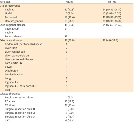
Median TTR was 23 months (95% confidence interval [CI]=11.5–34.5; standard error [SE]=5.8). The diagnosis of recurrence was biopsy proven in 36 women (53.7%) whereas 31 patients (46.3%) had their recurrences diagnosed by only imaging studies. In our study, 34 of recurrences (50.7%) occurred within 2 years after primary surgery whereas 44 of recurrences (65.7%) occurred within 3 years of initial diagnosis. We observed 32 (47.8%) isolated vaginal recurrences, 6 (9%) nodal failures, 19 (28.4%) peritoneal failures, and 10 (14.9%) hematogenous disseminations. Overall, 45 relapses (67.2%) were loco-regional while 22 (32.8%) were extrapelvic. The median TTR was not statistically significant between loco-
regional and extrapelvic recurrences (24 months [95% CI=12.5–35.5; SE=5.8] vs. 19 months [95% CI=4.0–33.9; SE=7.6]; p=0.516). Site of recurrences, median TTR, and type of salvage therapies are summarized in Table 2.
Treatment of the recurrences was applied according to the institutional practices at that time, and consisted of surgical resection (n=4, 6%), radiation (n=12, 17.9%), CT (n=17, 25.4%), surgical resection plus radiation (n=6, 9%), surgical resection plus CT (n=6, 9%), chemoradiaton (n=13, 19.4%), and surgical resection plus chemoradiaton (n=9, 13.4%).
The most frequent site of recurrence was the vaginal vault (47.8%) which was associated with a 5-year OSFR rate of 53.6%. The median OSFR has not been reached yet in women with isolated vaginal recurrence. Complete remission after salvage treatment for isolated vaginal Table 1. Baseline characteristics of women with recurrent low-risk EC
Characteristics Values
Age (yr) 58 (37–83)
≤60 40 (59.7)
>60 27 (40.3)
BMI (kg/m2) 30.8 (20.6–49.1)
<30 26.0 (38.8)
≥30 41.0 (61.2)
Menopausal status
Premenopausal 11 (16.4)
Postmenopausal 56 (83.6)
Postoperative histologic grade
Grade 1 35 (52.2)
Grade 2 32 (47.8)
LVSI
Absent 59 (88)
Present 6 (9)
Unknown 2 (3)
Tumor size (postoperative, mm) 24.5±12.9
≤20 25 (37.3)
>20 33 (49.3)
Unknown 9 (13.4)
LUSI
Yes 7 (10.4)
No 60 (89.6)
Site of lymphadenectomy
Pelvic only 19 (28.4)
Pelvic and para-aortic 33 (49.3)
Not-performed 15 (22.4)
No. of LNs removed 22.5±14.7
No. of pelvic LNs removed 17.2±9.5
No. of para-aortic LNs removed 8.5±7.5
Peritoneal cytology
Positive 0 (0)
Negative 63 (94)
Not-performed 4 (6)
HIR*
Yes 7 (10.5)
No 60 (89.5)
Values are presented as median (range), number (%), or mean.
BMI, body mass index; EC, endometrial cancer; GOG, Gynecologic Oncology Group; HIR, high-intermediate risk;
LN, lymph node; LUSI, lower uterine segment involvement; LVSI, lymphovascular space invasion.
*HIR criteria according to GOG Trial-99 [11]. Women in the HIR group were defined as patients; 1) 50–70 years of age with grade 2 histology and presence of LVSI, or 2) over 70 years of age with either grade 2 histology or presence of LVSI.
relapse was 20/32 (62.5%). The 5-year OSFR rate for nodal, peritoneal and hematogenous failures was 37.5%, 35.3%, and 42%, respectively. No statistically significant difference was observed in terms of 5-year OSFR with regard to the site of recurrence (p=0.760) (Fig.
1). According to the site of recurrence (loco-regional vs. extrapelvic), the 5-year OSFR rate was 51.2% for women with loco-regional recurrences compared to 35.9% for women with extrapelvic recurrences (p=0.065).
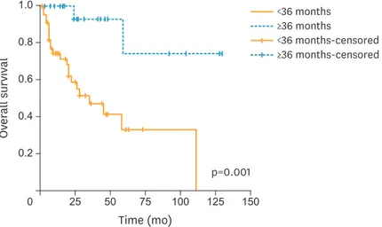
Overall, for the entire study cohort, the median OSFR was 59 months (95% CI=12.7–105.2;
SE=23.5). The 5-year OSFR rate was significantly higher for patients with TTR ≥36 months compared to those with TTR <36 months (74.3% vs. 33%, respectively; p=0.001). Fig. 2 shows the Kaplan-Meier plots for OSFR comparing those with TTR ≥36 months to TTR <36 months.
After further stratification, according to the site of recurrence (loco-regional vs. extrapelvic), patients with loco-regional recurrence had no difference in the 5-year OSFR rate regardless of when they recurred (<24 months or ≥24 months) (49.8% vs. 52.6%, respectively; p=0.640).
However, patients with extrapelvic recurrence and TTR <24 months had a significantly worse 5-year OSFR rate compared to those who had extrapelvic recurrences later than 24 months. For patients who had extrapelvic recurrences earlier than 24 months, the 5-year OSFR rate was 0%
compared to 87.5% for those with extrapelvic recurrences and TTR ≥24 months (p=0.009).
Table 2. Recurrence characteristics of patients
Variables Values TTR (mo)
Site of recurrence
Vaginal 32 (47.8) 24 (10.89–25.11)
Nodal 6 (9.0) 10 (3.99–16.00)
Peritoneal 19 (28.4) 18 (10.89–25.11)
Hematogenous 10 (14.9) 28 (10.95–45.04)
Loco-regional disease 45 (67.2) 24 (12.50–34.50)
Vaginal cuff 31
Vagina 1
Pelvic sidewall 13
Extrapelvic disease 22 (32.8) 19 (4.0–33.9)
Abdominal (peritoneal) disease 4
Liver-lung 2
Liver-vaginal cuff 3
Liver-para-aortic LN 1
Liver-peritoneal disease 1
Para-aortic LN 3
Bowel 1
Diaphragm 1
Mediastinal LN 1
Lung 2
Inguinal LN 1
Inguinal LN-para-aortic LN 1
Skin 1
Salvage therapies -
Surgical resection alone 4 (6.0)
RT alone 12 (17.9)
CT alone 17 (25.4)
Surgical resection plus RT 6 (9.0)
Surgical resection plus CT 6 (9.0)
Surgical resection plus CRT 9 (13.4)
CRT 13 (19.4)
Values are presented as number (%) or median (95% CI).
CI, confidence interval; CT, chemotherapy; CRT, chemoradiotherapy; LN, lymph node; RT, radiotherapy; TTR, time to recurrence.
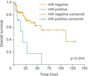
We compared the OSFR rates of 7 patients in the HIR group with 60 women in the non-HIR group. As expected, the 5-year OSFR rate was significantly higher for patients in the non-HIR group compared to those with HIR criteria (55.7% compared to 0%, respectively; p=0.004).
However, the 3-year OSFR rate for the HIR group was 17.9% while the corresponding rate was 67.5% for the non-HIR group. Fig. 3 shows the Kaplan-Meier plots for OSFR comparing those with HIR criteria to the non-HIR group.
On multivariate analysis for OSFR, TTR <36 months (hazard ratio [HR]=8.46; 95% CI=1.65–
43.36; p=0.010) and presence of HIR criteria (HR=4.62; 95% CI=1.69–12.58; p=0.003) were significant predictors. Table 3 shows univariate and multivariate analyses of factors associated with OSFR in women with recurrent low-risk EC.
At the time of reporting, of the 67 patients with recurrent low-risk EC, 35 (52.2%) were alive with NED, 8 (11.9%) were AWD, 1 (1.5%) died of a cause not related to the disease and was disease free at death, and 23 (34.3%) were DOD.
p=0.760
Time (mo)
0.6 0.4 0.2
0 50 75 150
Overall survival
0.8 1.0
100 125 25
Lymphatic Hematogenous Vaginal Peritoneal
Lymphatic-censored Hematogenous-censored Vaginal-censored Peritoneal-censored
Fig. 1. Overall survival from recurrence with regard to the site of recurrence (n=67).
p=0.001
Time (mo)
0.6 0.4 0.2
0 50 75 150
Overall survival
0.8 1.0
100 125 25
≥36 months
<36 months
≥36 months-censored
<36 months-censored
Fig. 2. Kaplan-Meier plots for OSFR comparing those with TTR ≥36 months (n=23) to TTR <36 months (n=44).
OSFR, overall survival following recurrence; TTR, time to recurrence.
DISCUSSION
Our results indicate that all types of relapse patterns (vaginal, nodal, peritoneal, and hematogenous) may be observed in women with low-risk EC; vaginal vault being the most frequent site of recurrence. Recurrent low-risk EC patients with TTR <36 months were 8 times more likely to succumb to their tumors following recurrence whereas patients carrying HIR criteria were 4 times more likely to be DOD after relapse.
However, we should underline some limitations of the current study. First, the retrospective nature of the study cannot exclude any bias. Second, lack of central pathology review seems to be an important limitation. However, as mentioned before, pathology review was performed at each participating institution individually before initiating treatment for relapse. Finally, data interpretation is constrained by variation in institutional practices.
We culled, treated, and observed patients from 10 gynecologic cancer centers; possibly analyzing clinical outcomes among patients with recurrence may not be equally balanced and objectively represented. However, given the low rate of recurrence in low-risk EC, the study design used here was necessary to achieve a satisfactory sample size. Despite above limitations, our study contributes to the limited body of knowledge associated with OSFR in low-risk recurrent EC.
Patients having <50% MMI with grade 1 or 2 disease represent the largest group of those with endometrioid EC [26]. We found the rate of low-risk EC as 40.5% among all endometrioid ECs at 10 gynecologic cancer centers in Turkey. Given the high rate of low- risk EC and the rising incidence of EC worldwide, clinicians will have to deal with a large number of recurrent low-risk EC patients although the recurrence rate is very low in this clinical setting.
The recurrence rate of low-risk EC has been reported to range from 1% to 7% [3,9,11,17,27- 33]. The recurrence rate in low-risk EC was 2.2% in the current study; similar to those
p=0.004
Time (mo)
0.6
0.4
0.2
0 50 75 150
Overall survival
0.8 1.0
100 125
25
HIR positive HIR negative
HIR positive-censored HIR negative-censored
Fig. 3. Kaplan-Meier plots for OSFR comparing those with HIR criteria (n=7) to the non-HIR group (n=60). Women in the HIR group were defined as patients; 1) 50–70 years of age with grade 2 histology and presence of LVSI, or 2) over 70 years of age with either grade 2 histology or presence of LVSI.
HIR, high-intermediate risk; LVSI, lymphovascular space invasion; OSFR, overall survival following recurrence;
TTR, time to recurrence.
reported in the literature. However, because of the retrospective design of the current study, the true overall rate of recurrence in patients with low-risk EC may have been underestimated.
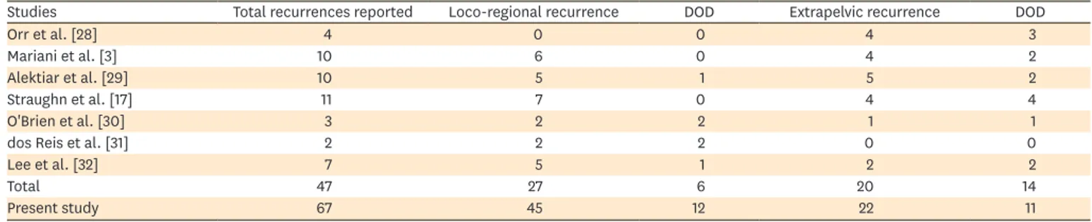
The anatomic locations of recurrences have been reported to be roughly equivalent between loco-regional and extrapelvic disease [11,34] with the most common sites being vaginal vault, pelvis, intraabdominal region, and lungs [35]. Our results implied that 67% of recurrences was loco-regional whereas 33% was extrapelvic. However, all types of relapse patterns described by Mariani et al. [22-25] were observed in our low-risk cohort. It should be emphasized that in only a limited number of studies in the literature, patients with low-risk EC have been accurately described for sites of recurrence and outcome (Table 4).
Improved survival after isolated vaginal recurrence is a well-known feature of early-stage EC. Complete remission after salvage treatment for isolated vaginal relapse has been Table 3. Univariate and multivariate prognostic factors for overall survival from recurrence
Characteristics No. of events (%) Univariate analyses
p-value Multivariate analyses
HR 95% CI p-value
Age (yr) 0.216 - - -
≤60 11/40 (27.5)
>60 13/27 (48.1)
BMI (kg/m2) 0.878 - - -
<30 8/26 (30.7)
≥30 16/41 (39.0)
Histologic grade 0.320 - - -
Grade 1 9/35 (25.7)
Grade 2 15/32 (46.8)
Tumor size (mm) 0.928 - - -
≤20 9/25 (36.0)
>20 13/33 (39.4)
LVSI 0.319 - - -
Absent 20/59 (33.9)
Present 4/6 (66.6)
LUSI 0.369 - - -
Yes 1/7 (14.3)
No 23/60 (38.3)
Lymphadenectomy 0.761 - - -
Yes 20/52 (38.5)
No 4/15 (26.6)
Site of recurrence 0.065 2.320 0.988–5.486 0.053
Loco-regional 13/45 (28.8)
Extrapelvic 11/22 (50.0)
TTR (mo) 0.019 0.389 0.418–9.436 0.390
≥18 10/39 (25.6)
<18 14/28 (50.0)
TTR (mo) 0.033 0.400 0.092–2.617 0.410
≥24 8/33 (24.2)
<24 16/34 (47.0)
TTR (mo) 0.001 8.460 1.664–43.367 0.010
≥36 2/23 (8.7)
<36 22/44 (50.0)
HIR criteria 0.004 4.620 1.697–12.585 0.003
Yes 6/7 (85.7)
No 18/60 (30.0)
Women in the HIR group were defined as patients; 1) 50–70 years of age with grade 2 histology and presence of LVSI, or 2) over 70 years of age with either grade 2 histology or presence of LVSI.
BMI, body mass index; CI, confidence interval; HIR, high-intermediate risk; HR, hazard ratio; LUSI, lower uterine segment involvement; LVSI, lymphovascular space invasion; TTR, time to recurrence.
reported to range from 40% to 80% in previously unirradiated patients [36]. In our study, the corresponding rate was 62.5% which was associated with a 5-year OSFR rate of 53.6%.
Colombo et al. [12] reported the standard treatment for isolated vaginal recurrence as radiation therapy (external beam plus vaginal brachytherapy); with high rates of local control, complete response, and a 5-year survival rate of 50%. However, we were unable to demonstrate a significant difference in terms of 5-year OSFR with regard to the site of recurrence. The 5-year OSFR rate was 51.2% for women with loco-regional recurrences compared to 35.9% for women with extrapelvic recurrences (p=0.065). According to our findings, it does not seem reasonable to recommend a distinction between loco-regional and extrapelvic recurrences while evaluating the prognosis following recurrence in low-risk EC.
Various factors have been suggested to be prognostic for OSFR in recurrent EC including site of recurrence (loco-regional or distant), endometrioid histology, use of adjuvant radiation therapy, initial tumor grade, age at the time of recurrence, and TTR [12,20,37].
However, predictive factors for OSFR in recurrent low-risk EC have not been specifically reported to date. Presence of HIR criteria and TTR <36 months were found to be
independent risk factors for decreased OSFR in low-risk EC in the current study. It should be kept in mind that low-risk EC patients may be qualified as HIR with the contribution of age (>70 years), histologic grade (grade 2), and presence of LVSI. Our data showed that patients with HIR criteria had a statistically significant OSFR disadvantage when compared to women in the non-HIR group.
Robbins et al. [20] reported that the prognostic impact of TTR was less important than the site of recurrence in a series of 57 women with recurrent early-stage endometrioid EC. The authors suggested the uniform endometrioid histology and the fairly high success rate of salvage therapy as probable reasons why TTR was not statistically significant in their cohort [20]. However, TTR <36 months appeared as an independent risk factor for decreased OSFR in our low-risk study population. This may be due to the difference between the study populations (recurrent early-stage endometrioid EC vs. recurrent low-risk EC) as well as different institutional practices for salvage therapies.
Because this study was limited only to patients having less than 50% MMI with grade 1 or 2 histology, it is not surprising that some of the prognostic factors such as grade, LVSI and LUSI were not significant predictors of OSFR. However, to the best our knowledge, this study reports on the largest series of recurrent low-risk EC patients and is the first study evaluating OSFR specifically in those patients. The strengths of the current study lie in its multicenter nature with a large number of patients with recurrent low-risk EC, and the uniformity of applying no adjuvant treatment.
Table 4. The absolute number of recurrences, site of recurrence, and outcome of patients with recurrent low-risk EC reported in the English literature
Studies Total recurrences reported Loco-regional recurrence DOD Extrapelvic recurrence DOD
Orr et al. [28] 4 0 0 4 3
Mariani et al. [3] 10 6 0 4 2
Alektiar et al. [29] 10 5 1 5 2
Straughn et al. [17] 11 7 0 4 4
O'Brien et al. [30] 3 2 2 1 1
dos Reis et al. [31] 2 2 2 0 0
Lee et al. [32] 7 5 1 2 2
Total 47 27 6 20 14
Present study 67 45 12 22 11
DOD, dead of disease; EC, endometrial cancer.
Since low-risk EC generally shows an excellent prognosis, its potential risk of lethal outcome is frequently underestimated. However, 34% of women with recurrent low-risk EC were DOD in the present study. It can be speculated that among women with low-risk EC, there seems to be subsets of patients with unknown additional negative prognostic factors leading them into a dead end.
According to our results, TTR <36 months and presence of HIR criteria seem to be independent predictors of decreased OSFR in low-risk EC patients. We conclude that clinicians should be cautious about the possibility of presence of HIR criteria among women with low-risk EC. Low-risk EC patients carrying HIR criteria should receive adjuvant treatment rather than surgery alone.
REFERENCES
1. Morrow CP, Bundy BN, Kurman RJ, Creasman WT, Heller P, Homesley HD, et al. Relationship between surgical-pathological risk factors and outcome in clinical stage I and II carcinoma of the endometrium: a Gynecologic Oncology Group study. Gynecol Oncol 1991;40:55-65.
PUBMED | CROSSREF
2. Carey MS, O'Connell GJ, Johanson CR, Goodyear MD, Murphy KJ, Daya DM, et al. Good outcome associated with a standardized treatment protocol using selective postoperative radiation in patients with clinical stage I adenocarcinoma of the endometrium. Gynecol Oncol 1995;57:138-44.
PUBMED | CROSSREF
3. Mariani A, Webb MJ, Keeney GL, Haddock MG, Calori G, Podratz KC. Low-risk corpus cancer: is lymphadenectomy or radiotherapy necessary? Am J Obstet Gynecol 2000;182:1506-19.
PUBMED | CROSSREF
4. Schink JC, Rademaker AW, Miller DS, Lurain JR. Tumor size in endometrial cancer. Cancer 1991;67:2791-4.
PUBMED | CROSSREF
5. Elliott P, Green D, Coates A, Krieger M, Russell P, Coppleson M, et al. The efficacy of postoperative vaginal irradiation in preventing vaginal recurrence in endometrial cancer. Int J Gynecol Cancer 1994;4:84-93.
PUBMED | CROSSREF
6. Leijon T, Rosenberg P, Boeryd B. Total abdominal hysterectomy and bilateral salpingo-oophorectomy. A sufficient treatment for patients with low risk endometrial carcinoma. Int J Gynecol Cancer 1997;7:376-80.
CROSSREF
7. Poulsen HK, Jacobsen M, Bertelsen K, Andersen JE, Ahrons S, Bock J, et al. Adjuvant radiation therapy is not necessary in the management of endometrial carcinoma stage I, low-risk cases. Int J Gynecol Cancer 1996;6:38-43.
CROSSREF
8. Kong A, Johnson N, Kitchener HC, Lawrie TA. Adjuvant radiotherapy for stage I endometrial cancer: an updated Cochrane systematic review and meta-analysis. J Natl Cancer Inst 2012;104:1625-34.
PUBMED | CROSSREF
9. Scholten AN, van Putten WL, Beerman H, Smit VT, Koper PC, Lybeert ML, et al. Postoperative radiotherapy for stage 1 endometrial carcinoma: long-term outcome of the randomized PORTEC trial with central pathology review. Int J Radiat Oncol Biol Phys 2005;63:834-8.
PUBMED | CROSSREF
10. Nugent EK, Bishop EA, Mathews CA, Moxley KM, Tenney M, Mannel RS, et al. Do uterine risk factors or lymph node metastasis more significantly affect recurrence in patients with endometrioid adenocarcinoma? Gynecol Oncol 2012;125:94-8.
PUBMED | CROSSREF
11. Keys HM, Roberts JA, Brunetto VL, Zaino RJ, Spirtos NM, Bloss JD, et al. A phase III trial of surgery with or without adjunctive external pelvic radiation therapy in intermediate risk endometrial adenocarcinoma:
a Gynecologic Oncology Group study. Gynecol Oncol 2004;92:744-51.
PUBMED | CROSSREF
12. Colombo N, Preti E, Landoni F, Carinelli S, Colombo A, Marini C, et al. Endometrial cancer: ESMO Clinical Practice Guidelines for diagnosis, treatment and follow-up. Ann Oncol 2013;24 Suppl 6:vi33-8.
PUBMED | CROSSREF
13. Jeppesen MM, Jensen PT, Gilså Hansen D, Iachina M, Mogensen O. The nature of early-stage endometrial cancer recurrence-a national cohort study. Eur J Cancer 2016;69:51-60.
PUBMED | CROSSREF
14. Jeppesen MM, Mogensen O, Hansen DG, Iachina M, Korsholm M, Jensen PT. Detection of recurrence in early stage endometrial cancer - the role of symptoms and routine follow-up. Acta Oncol 2017;56:262-9.
PUBMED | CROSSREF
15. Mahdi H, Munkarah AR, Ali-Fehmi R, Woessner J, Shah SN, Moslemi-Kebria M. Tumor size is an independent predictor of lymph node metastasis and survival in early stage endometrioid endometrial cancer. Arch Gynecol Obstet 2015;292:183-90.
PUBMED | CROSSREF
16. Vargas R, Rauh-Hain JA, Clemmer J, Clark RM, Goodman A, Growdon WB, et al. Tumor size, depth of invasion, and histologic grade as prognostic factors of lymph node involvement in endometrial cancer: a SEER analysis. Gynecol Oncol 2014;133:216-20.
PUBMED | CROSSREF
17. Straughn JM Jr, Huh WK, Kelly FJ, Leath CA 3rd, Kleinberg MJ, Hyde J Jr, et al. Conservative management of stage I endometrial carcinoma after surgical staging. Gynecol Oncol 2002;84:194-200.
PUBMED | CROSSREF
18. Siegel R, Ma J, Zou Z, Jemal A. Cancer statistics, 2014. CA Cancer J Clin 2014;64:9-29.
PUBMED | CROSSREF
19. McAlpine JN, Temkin SM, Mackay HJ. Endometrial cancer: not your grandmother's cancer. Cancer 2016;122:2787-98.
PUBMED | CROSSREF
20. Robbins JR, Yechieli R, Laser B, Mahan M, Rasool N, Elshaikh MA. Is time to recurrence after hysterectomy predictive of survival in patients with early stage endometrial carcinoma? Gynecol Oncol 2012;127:38-42.
PUBMED | CROSSREF
21. Pecorelli S. Revised FIGO staging for carcinoma of the vulva, cervix, and endometrium. Int J Gynaecol Obstet 2009;105:103-4.
PUBMED | CROSSREF
22. Mariani A, Dowdy SC, Keeney GL, Haddock MG, Lesnick TG, Podratz KC. Predictors of vaginal relapse in stage I endometrial cancer. Gynecol Oncol 2005;97:820-7.
PUBMED | CROSSREF
23. Mariani A, Webb MJ, Keeney GL, Aletti G, Podratz KC. Predictors of lymphatic failure in endometrial cancer. Gynecol Oncol 2002;84:437-42.
PUBMED | CROSSREF
24. Mariani A, Webb MJ, Keeney GL, Aletti G, Podratz KC. Endometrial cancer: predictors of peritoneal failure. Gynecol Oncol 2003;89:236-42.
PUBMED | CROSSREF
25. Mariani A, Webb MJ, Keeney GL, Calori G, Podratz KC. Hematogenous dissemination in corpus cancer.
Gynecol Oncol 2001;80:233-8.
PUBMED | CROSSREF
26. Abeler VM, Kjørstad KE. Endometrial adenocarcinoma in Norway. A study of a total population. Cancer 1991;67:3093-103.
PUBMED | CROSSREF
27. Fung-Kee-Fung M, Dodge J, Elit L, Lukka H, Chambers A, Oliver TCancer Care Ontario Program in Evidence-based Care Gynecology Cancer Disease Site Group. Follow-up after primary therapy for endometrial cancer: a systematic review. Gynecol Oncol 2006;101:520-9.
PUBMED | CROSSREF
28. Orr JW Jr, Holimon JL, Orr PF. Stage I corpus cancer: is teletherapy necessary? Am J Obstet Gynecol 1997;176:777-88.
PUBMED | CROSSREF
29. Alektiar KM, McKee A, Venkatraman E, McKee B, Zelefsky MJ, Mychalczak BR, et al. Intravaginal high- dose-rate brachytherapy for Stage IB (FIGO Grade 1, 2) endometrial cancer. Int J Radiat Oncol Biol Phys 2002;53:707-13.
PUBMED | CROSSREF
30. O'Brien DJ, Flannelly G, Mooney EE, Foley M. Lymphovascular space involvement in early stage well- differentiated endometrial cancer is associated with increased mortality. BJOG 2009;116:991-4.
PUBMED | CROSSREF
31. dos Reis R, Burzawa JK, Tsunoda AT, Hosaka M, Frumovitz M, Westin SN, et al. Lymphovascular space invasion portends poor prognosis in low-risk endometrial cancer. Int J Gynecol Cancer 2015;25:1292-9.
PUBMED | CROSSREF
32. Lee JY, Kim JH, Seo JW, Kim HS, Kim JW, Park NH, et al. Detecting asymptomatic recurrence in early- stage endometrial cancer: the value of vaginal cytology, imaging studies, and CA-125. Int J Gynecol Cancer 2016;26:1434-9.
PUBMED | CROSSREF
33. Therasakvichya S, Kuljarusnont S, Petsuksiri J, Chaopotong P, Achariyapota V, Srichaikul P, et al.
Clinical outcomes of stage I endometrial carcinoma patients treated with surgery alone: Siriraj Hospital experiences. J Gynecol Oncol 2016;27:e48.
PUBMED | CROSSREF
34. Creutzberg CL, van Putten WL, Koper PC, Lybeert ML, Jobsen JJ, Wárlám-Rodenhuis CC, et al. Surgery and postoperative radiotherapy versus surgery alone for patients with stage-1 endometrial carcinoma:
multicentre randomised trial. PORTEC Study Group. Post Operative Radiation Therapy in Endometrial Carcinoma. Lancet 2000;355:1404-11.
PUBMED | CROSSREF
35. Menczer J. Endometrial carcinoma. Is routine intensive periodic follow-up of value? Eur J Gynaecol Oncol 2000;21:461-5.
PUBMED
36. Creutzberg CL, van Putten WL, Koper PC, Lybeert ML, Jobsen JJ, Wárlám-Rodenhuis CC, et al. Survival after relapse in patients with endometrial cancer: results from a randomized trial. Gynecol Oncol 2003;89:201-9.
PUBMED | CROSSREF
37. Xu Y, Burmeister C, Hanna RK, Munkarah A, Elshaikh MA. Predictors of survival after recurrence in women with early-stage endometrial carcinoma. Int J Gynecol Cancer 2016;26:1137-42.
PUBMED | CROSSREF