Original Article
The effect of combined treatment with cisplatin and histone deacetylase inhibitors on HeLa cells
Ke Long Jin1,*, Jeong-Yeol Park1, Eun Joo Noh2, Kwang Lae Hoe2, Joo Hak Lee3, Jong-Hyeok Kim1, Joo-Hyun Nam1
1Department of Obstetrics and Gynecology, Asan Medical Center, University of Ulsan College of Medicine, Seoul,
2Intergrative Omics Research Center, Korea Research Institute for Biology and Biotechnology, Daejeon, Korea,
3Department of Western Medicine, Life University School of Oriental Medicine, Gardena, CA , USA
Objective: To investigate the combined effects of cisplatin and the histone deacetylase (HDAC) inhibitors suberoylanilide hydroxamic acid (SAHA) or sirtinol on HeLa cells and assess the mechanism underlying HDAC inhibitor-cisplatin synergy.
Methods: The antineoplastic actions of cisplatin, SAHA and sirtinol, alone and in combination, were evaluated using the tetrazolium dye-based MTT cell proliferation assay, DAPI nuclear staining and cytotoxicity analysis.
Results: Exposure to cisplatin, SAHA or sirtinol alone induced a dose-dependent reduction in HeLa cell viability.
Combined treatment with cisplatin and SAHA or sirtinol was significantly more cytotoxic than cisplatin alone.
Individually, cisplatin, SAHA and sirtinol activated caspase-3 and induced apoptosis, but the effects of combined treatment were greater. Importantly, both HDAC inhibitors dose-dependently inhibited the expression of the antiapoptotic proteins Bcl-2 and x-linked inhibitor of apoptosis protein (XIAP).
Conclusion: The combination of cisplatin and SAHA or sirtinol had synergistic effect on the HeLa cell viability. This potentiation of cisplatin activity was associated with HDAC inhibitor-mediated down-regulation of Bcl-2 and XIAP. These may result from the relaxation of chromatin by these HDAC inhibitors that increase cisplatin sensitivity by enhancing the accessibility of DNA to cisplatin and transcriptional regulators.
Key Words: Cervical cancer, Apoptosis, Cisplatin, Suberoylanilide hydroxamic acid, Sirtinol
Received October 7, 2010, Revised December 6, 2010, Accepted December 6, 2010
Correspondence to Joo-Hyun Nam
Department of Obstetrics and Gynecology, Asan Medical Center, University of Ulsan College of Medicine, 388-1 Poongnap-2dong, Songpa-gu, Seoul 138-736, Korea
Tel: 82-2-3010-3633, Fax: 82-2-476-7331 E-mail: [email protected]
*Current address: Department of Gynecology, Huai An Women and Children's Healthcare Hospital, Huai An, China
INTRODUCTION
Modulation of chromatin structure through histone acetyla- tion/deacetylation is one of the major mechanisms involved in the regulation of gene expression.1-3 Increased histone ace- tylation causes chromatin to decondense and is associated with increased transcription.4-7 Increased activity of histone deacetylase (HDAC), which down-regulates histone acetyla- tion, has been observed in various cancer types.8 Through ace- tylation of histones, HDAC inhibitors induce chromatin re- laxation and favor transcription.
Previous studies have demonstrated that treatment with
HDAC inhibitors alters gene transcription, resulting in a pro- apoptotic expression profile characterized by increased ex- pression of pro-apoptotic genes/proteins, and decreased ex- pression of anti-apoptotic genes/proteins, including Bcl-2, Bcl-Xl, MCL1 and x-linked inhibitor of apoptosis protein (XIAP).9-14 Treatment with HDAC inhibitors also promotes ac- cumulation of ROS in tumor cells.10,15,16 These inhibitors in- duce growth arrest and apoptosis in various cancer cell lines in vitro and suppress tumor growth in animal xenograft mo- dels.17-19 Several HDAC inhibitors, including suberoylanilide hydroxamic acid (SAHA), tributyrin, and depsipeptide, are cur- rently under investigation in clinical trials as antineoplastic agents for use in patients with hematologic and solid malig- nancies.20-22
SAHA, which inhibits class I and class II HDAC enzymes, has been shown to overcome multidrug resistance in different cancer cells in vitro, and to induce p53-independent apoptosis via the mitochondrial pathway.23 Sirtinol belongs to the class III histone deacetylase family and is an inhibitor of SIRT1 (silent mating type information regulation 2 homolog 1), the closest mammalian homolog of yeast Sir2. It has been shown that the SIRT1 levels are up-regulated in various drug-re- sistant cell lines and SIRT1 knockdown by siRNA transfection
covalent platinum-DNA adducts. It is known that a tight chro- matin structure prevents cisplatin from accessing DNA, and relaxation of chromatin by HDAC inhibitors may increase the accessibility of DNA to chemotherapeutic agents.25 Cisplatin is effective against many types of cancer, including cervical cancer, but its use is often restricted due to side effects and the development of drug-resistance. Cisplatin resistance is often thought to arise due to the induction of DNA repair enzymes, overexpression of Bcl-2, and increased levels of reduced gluta- thione (GSH) and associated enzymes. XIAP, a direct in- hibitor of caspase-3 and caspase-7, modulates the Bax/cyto- chrome c pathway by inhibiting caspase-9.26,27 Cisplatin sen- sitivity in ovarian and uterine cancer cells has been shown to involve XIAP.28,29
Increasing the effectiveness of cisplatin-based chemotherapy will require a means to overcome the side effects of, and resist- ance to, cisplatin. Given the biological effects of HDAC in- hibitors on cancer cells and the known mechanisms of cisplatin resistance noted above, we postulated that the combined treat- ment with cisplatin and an HDAC inhibitor would increase the anticancer efficacy of cisplatin. Here, we tested this hypothesis by investigating the potentiation of cisplatin activity by HDAC inhibitors SAHA and sirtinol in HeLa cervical cancer cells, and sought to determine the mechanism underlying the synergistic effect of combined treatment.
MATERIALS AND METHODS 1. Cell culture and reagents
HeLa cells, obtained from the American Type Culture Collec- tion, were cultured in DMEM medium supplemented with 10%
fetal bovine serum, 100 IU/mL penicillin and 100 μg/mL strep- tomycin in a humidified atmosphere of 5% CO2 in air at 37oC.
Cisplatin and sirtinol were purchased from Sigma Chemical Co.
(St. Louis, MO, USA), SAHA was purchased from Alexis Bio- chemicals (San Diego, CA, USA).
2. MTT cell viability assay
The viability of HeLa cells following treatment with cisplatin, SAHA or sirtinol alone, and in the indicated combinations, was measured using an MTT (3-[4,5-dimethylthiazol-2-yl]-2,5-di- phenyl tetrazolium bromide) assay (Sigma). Cells were plated in triplicate wells (1.5×10⁴ cells/well) of 96-well flat-bottomed plates and incubated overnight prior to drug exposure. Cells were then incubated with different concentrations of cisplatin, SAHA, sirtinol or a combination of cisplatin and one of the HDAC inhibitors. After exposure to the indicated concen- trations/drug combinations for 48 hours, 10 μL of MTT reagent was added to each well. Cells were then incubated for 3 hours with MTT, after which 100 μL of stop mix solution (20% SDS in 50% dimethyl formamide) was added and cells were in- cubated for an additional 1 hour. Absorbance was measured at
3. Nuclear staining with DAPI
After treatment with HDAC inhibitor and/or cisplatin, the cells were fixed with 3.7% paraformaldehyde (Sigma) in PBS for 8 minutes at room temperature. Fixed cells were washed with PBS and stained with a 4,6-diamidino-2-phenylindole (DAPI; Sigma) solution for 5 minutes at room temperature.
The cells were then washed three more times with PBS and analyzed using a fluorescence microscope.
4. Western blot analysis
The cells were harvested and lysed, and the protein concen- tration in lysates was quantified using the Bradford method. For Western blot analysis, an equal amount of protein (40 μg) was separated by electrophoresis on SDS-polyacrylamide gels and transferred to nitrocellulose membranes by electroblotting.
Blots were probed with the desired antibodies for 1 hour, in- cubated with diluted enzyme-linked secondary antibody, and then visualized by enhanced chemiluminescence as recom- mended by the manufacturer (Amersham).
5. Cytotoxicity analysis
We used the combination index method of Chou and Talalay30 to determine whether the observed interactions between cis- platin and SAHA or sirtinol were additive or synergistic. If the interaction is additive, the sum of the effects of the two drugs should be equal to the product of their fractional activities. The representative function is f(u)1,2=f(u)1 ×f(u)2, where f(u)1 is the fraction unaffected by drug 1, f(u)2 is the fraction unaffected by drug 2 and f(u)1,2 is the fraction unaffected by drugs 1 and 2. The expected (presumed to be additive) and observed surviv- al rates of HeLa cells obtained from the three independent drug-combination treatments were analyzed by Student’s t-test. p<0.05 was considered significant.
RESULTS
1. Cytotoxicity of cisplatin, SAHA or sirtinol, alone and in combination
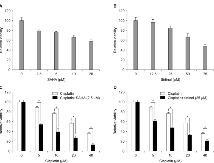
We first exposed HeLa cells to the conventional anticancer drug cisplatin or to SAHA or sirtinol, two HDAC inhibitors be- longing to different structural classes, to investigate the possi- ble cytotoxic effects of these three drugs alone. MTT assays re- vealed that, after incubating with HeLa cells for 48 hours, each of these drugs had a dose-dependent inhibitory effect on HeLa cell viability (Fig. 1A and B, and the black portion of C). Of the three compounds, cisplatin reduced cell viability to the greatest extent. We then tested the responses of HeLa cells to combina- tions of different concentrations of cisplatin (5, 10, 20, and 40 μm) and SAHA (2.5 μm) or sirtinol (25 μm). When tested alone, both SAHA (2.5 μm) and sirtinol (25 μm) reduced cell viability by about 20% (21% for SAHA and 16% for sirtinol). As shown in Fig. 1, the observed viability in HeLa cells treated with combi-
Fig. 1. Inhibition of HeLa cell growth by suberoylanilide hydroxamic acid (SAHA), sirtinol or cisplatin, alone and in combination. Cells were assayed for viability using MTT assays after treating for 48 hr with increasing doses of SAHA (A), sirtinol (B), or cisplatin (C or D black col- umn) alone, or with a combination of cisplatin and SAHA (C) or cisplatin and sirtinol (D). Results are expressed as the means±SEs of three independent experiments. *p<0.05.
nations of cisplatin and SAHA (2.5 μm) or sirtinol (25 μm) was less than that expected for an additive model at each concen- tration of cisplatin tested, indicating synergy. Although both HDAC inhibitors synergized with cisplatin to inhibit cell via- bility, the combination of cisplatin and SAHA was more effec- tive than the cisplatin/sirtinol combination (Fig. 1C and D).
2. Morphological features of apoptosis in HeLa cells treated with HDAC inhibitors and/or cisplatin
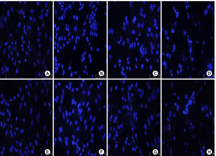
To determine whether the growth inhibition by cisplatin and/or HDAC inhibitors was associated with apoptotic cell death, we treated cells for 48 hours with cisplatin, SAHA or sirti- nol, alone, or with HDAC inhibitor/cisplatin combinations, and then examined nuclear morphology in DAPI- stained cells.
With DAPI nuclear staining, cells with condensed and frag- mented nuclei are judged to be apoptotic. Treatment of HeLa cells with SAHA (2.5 μm), sirtinol (25 μm) or cisplatin (10 μm) alone induced a modest level of apoptosis. However, the combi- nation of cisplatin (10 μm) and SAHA (2.5 μm) or sirtinol (25
μm) induced a significantly greater degree apoptosis than that seen with cisplatin alone (Fig. 2). These results indicate a good correspondence between the extent of apoptosis and growth in- hibition, and suggest that the cell death induced in HeLa cells by cisplatin, SAHA and sirtinol, alone or in combination, is largely apoptotic in nature.
3. Inhibition of Bcl-2 and XIAP expression by HDAC inhibitors
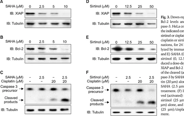
Apoptotic cell death is highly regulated by pro-apoptotic and anti-apoptotic modulators. Bcl-2 and XIAP are anti-apoptotic proteins that are relevant to chemoresistance to cisplatin. To determine whether SAHA or sirtinol down-regulated the ex- pression of Bcl-2 and/or XIAP in HeLa cells, we isolated cellular proteins after exposure to different concentrations of SAHA and sirtinol for 24 hours, and performed immunoblotting. The results of Western-blot analyses indicated that treatment with either SAHA (0, 2.5, 5, and 10 μm) or sirtinol (0, 12.5, 25, and 50 μm) induced a dose-dependent decrease in the levels of Bcl-2
Fig. 2. Induction of apoptosis in HeLa cells by suberoylanilide hydroxamic acid (SAHA), sirtinol or cisplatin, alone and in combination. Cells were incubated for 48 hr with 2.5 μm SAHA (B), 25 μm sirtinol (F) or 10 μm cisplatin (C and G) alone, or with 2.5 μm SAHA/10 μm cisplatin (D) or 25 μm sirtinol/10 μm cisplatin (H) combinations. (A) and (E) are controls. The cells were sampled, fixed and stained with DAPI;
stained nuclei were then observed under a fluorescence microscope using a blue filter.
and XIAP (Fig. 3A, B, D, and E).
4. Increased levels of activated caspase-3 by combined cisplatin/HDAC inhibitor treatment
Caspases are thought to be vital in mediating various apoptotic responses, including those induced by cisplatin. Caspase-3, which is cleaved proteolytically from its inactive precursor form into active fragments by various apoptotic stimuli, is an im- portant apoptotic effector in both the intrinsic and extrinsic pathways of apoptotic cell death. To assess the effects of HDAC inhibitors on the caspase pathway, we measured caspase-3 acti- vation in HeLa cells after a 24-hour treatment with cisplatin, SAHA or sirtinol, alone or in combination, using an immuno- blotting method. We found that SAHA (2.5 μm) and sirtinol (25 μm) alone induced minimal activation of caspase-3, where- as the level of caspase-3 activation induced by cisplatin alone (20 μm) was readily apparent. Notably the cisplatin (20 μm)/
SAHA (2.5 μm) and cisplatin (20 μm)/sirtinol (25 μm) combi- nations strongly induced caspase-3 activation (Fig. 3C and F),
demonstrating that the combinations of cisplatin with SAHA or sirtinol were more effective apoptosis-inducing stimuli than were cisplatin or HDAC inhibitors alone.
DISCUSSION
Cisplatin is effective in neoadjuvant and conventional che- motherapy in the treatment of cervical cancer. Despite the im- pressive anti-neoplastic activity of cisplatin, severe side ef- fects and drug resistance restricts its use in cancer therapy.
High doses of cisplatin are more effective than low doses in cancer chemotherapy. Unfortunately, several severe side ef- fects, notably nephrotoxicity and ototoxicity, limit the dose that can be given to patients. Moreover, the repeated use of cisplatin promotes resistance to cisplatin-induced apoptosis in cancer cells. Thus, drugs that sensitize cancer cells to cis- platin could increase the clinical efficacy of cisplatin. Unlike conventional chemotherapeutic agents, which often cause DNA damage in both cancer and normal tissues, HDAC in-
Fig. 3. Down-regulation of XIAP and Bcl-2 levels and activation of cas- pase-3. HeLa cells were treated with the indicated concentrations of SAHA, sirtinol or cisplatin, alone or as SAHA/
cisplatin or sirtinol/cisplatin combi- nations, for 24 hours and then ana- lyzed by immunoblotting. (A, B, D, and E) SAHA (0, 2.5, 5, or 10 μm) or sirtinol (0, 12.5, 25, or 50 μm) in- duced a dose-dependent reduction in XIAP and Bcl-2 levels. (C) Induction of the cleaved (activated) form of cas- pase-3 by SAHA (2.5 μm) and cispla- tin (20 μm) alone, and by combined SAHA (2.5 μm)/cisplatin (20 μm) treatment. (F) Induction of the clea- ved (activated) form of caspase-3 by sirtinol (25 μm) and cisplatin (20 μm) alone, and by combined sirtinol (25 μm)/cisplatin (20 μm) treat- ment.
hibitors display strong cancer selectivity and cause less tox- icity to normal tissues.31
In this study, we found that both SAHA and sirtinol exerted a dose-dependent cytotoxic effect on HeLa cells. Importantly, low concentrations of SAHA or sirtinol synergized with cis- platin in combination therapy to induce a level of cytotoxicity greater than that mediated by either agent alone, or that pre- dicted by an additive model. Of the two combinations, the cis- platin/SAHA combination produced a more marked reduc- tion in cell viability. DAPI staining and Western-blot analyses revealed that combined treatment with cisplatin and a low concentration of either of the two HDAC inhibitors markedly increased caspase-3 activation and apoptosis in HeLa cells compared with cisplatin alone.
Cisplatin cytotoxicity appears to correlate with DNA adduct formation.32 Cisplatin, along with a number of other exogenous toxins, is detoxified by glutathione, the most abundant intra- cellular thiol and a critical cellular antioxidant,33,34 through the formation of glutathione adducts.35,36 Among the mechanisms that contribute to cellular resistance to cisplatin are those that involve up-regulation of the anti-apoptotic proteins Bcl-2 and XIAP. Bcl-2 overexpression has been shown to increase cispla- tin resistance in a number of experimental models.37-41 Bcl-2 overexpression in the mitochondrial outer membrane inhibits the characteristic increase in reactive oxygen species observed in cells exposed to a number of apoptotic triggers.42,43 More- over, Bcl-2-mediated cisplatin resistance is associated with an increase in glutathione levels, and glutathione synthesis is re- quired for Bcl-2-mediated cisplatin resistance.44 Similarly, XIAP levels are known to regulate cisplatin sensitivity in some
uterine cell lines.29
Through acetylation of histones, SAHA and sirtinol relax chromatin, increasing the accessibility of DNA to transcription factors and thereby regulating the expression of a number of genes, including those for Bcl-2 and XIAP. Thus, the sig- nificantly increased cell death induced in cells by the combina- tion of cisplatin and low concentrations of SAHA or sirtinol compared with cisplatin alone is likely due, at least in part, to down-regulation of Bcl-2 and XIAP by these HDAC inhibitors.
By promoting chromatin decondensation, SAHA and sirtinol also provides greater DNA access to cisplatin. These actions sensitize HeLa cells to cisplatin, and thus are also likely to con- tribute to the synergistic cytotoxicity of cisplatin and HDAC in- hibitors toward HeLa cells. Collectively, these results may in- dicate that relaxation of chromatin by SAHA or sirtinol in- creases the effectiveness of cisplatin by enhancing the accessi- bility of DNA to both cisplatin and transcriptional regulators.
The first action directly increases the sensitivity of cells to cis- platin; the second contributes to increased sensitivity (and po- tentially to decreased resistance) to cisplatin by down-regulat- ing anti-apoptosis regulators.
CONFLICT OF INTEREST
No potential conflict of interest relevant to this article was reported.
REFERENCES
1. Khochbin S, Verdel A, Lemercier C, Seigneurin-Berny D. Functional
2. Marmorstein R, Roth SY. Histone acetyltransferases: function, structure, and catalysis. Curr Opin Genet Dev 2001; 11: 155-61.
3. Roth SY, Denu JM, Allis CD. Histone acetyltransferases. Annu Rev Biochem 2001; 70: 81-120.
4. Lee DY, Hayes JJ, Pruss D, Wolffe AP. A positive role for histone acetylation in transcription factor access to nucleosomal DNA.
Cell 1993; 72: 73-84.
5. Grunstein M. Histone acetylation in chromatin structure and transcription. Nature 1997; 389: 349-52.
6. Struhl K. Histone acetylation and transcriptional regulatory mechanisms. Genes Dev 1998; 12: 599-606.
7. Strahl BD, Allis CD. The language of covalent histone modifica- tions. Nature 2000; 403: 41-5.
8. Marks PA, Rifkind RA, Richon VM, Breslow R, Miller T, Kelly WK. Histone deacetylases and cancer: causes and therapies. Nat Rev Cancer 2001; 1: 194-202.
9. Zhang XD, Gillespie SK, Borrow JM, Hersey P. The histone de- acetylase inhibitor suberic bishydroxamate regulates the ex- pression of multiple apoptotic mediators and induces mi- tochondria-dependent apoptosis of melanoma cells. Mol Cancer Ther 2004; 3: 425-35.
10. Rosato RR, Maggio SC, Almenara JA, Payne SG, Atadja P, Spiegel S, et al. The histone deacetylase inhibitor LAQ824 induces human leukemia cell death through a process involving XIAP down-regu- lation, oxidative injury, and the acid sphingomyelinase-dependent generation of ceramide. Mol Pharmacol 2006; 69: 216-25.
11. Mitsiades CS, Mitsiades NS, McMullan CJ, Poulaki V, Shringarpure R, Hideshima T, et al. Transcriptional signature of histone deacety- lase inhibition in multiple myeloma: biological and clinical implications. Proc Natl Acad Sci USA 2004; 101: 540-5.
12. Peart MJ, Smyth GK, van Laar RK, Bowtell DD, Richon VM, Marks PA, et al. Identification and functional significance of genes regulated by structurally different histone deacetylase inhibitors. Proc Natl Acad Sci U S A 2005; 102: 3697-702.
13. Moore PS, Barbi S, Donadelli M, Costanzo C, Bassi C, Palmieri M, et al. Gene expression profiling after treatment with the his- tone deacetylase inhibitor trichostatin A reveals altered ex- pression of both pro- and anti-apoptotic genes in pancreatic ad- enocarcinoma cells. Biochim Biophys Acta 2004; 1693: 167-76.
14. Duan H, Heckman CA, Boxer LM. Histone deacetylase in- hibitors down-regulate bcl-2 expression and induce apoptosis in t(14;18) lymphomas. Mol Cell Biol 2005; 25: 1608-19.
15. Ruefli AA, Ausserlechner MJ, Bernhard D, Sutton VR, Tainton KM, Kofler R, et al. The histone deacetylase inhibitor and che- motherapeutic agent suberoylanilide hydroxamic acid (SAHA) induces a cell-death pathway characterized by cleavage of Bid and production of reactive oxygen species. Proc Natl Acad Sci U S A 2001; 98: 10833-8.
16. Rosato RR, Almenara JA, Dai Y, Grant S. Simultaneous activa- tion of the intrinsic and extrinsic pathways by histone deacety- lase (HDAC) inhibitors and tumor necrosis factor-related apop- tosis-inducing ligand (TRAIL) synergistically induces mitochon- drial damage and apoptosis in human leukemia cells. Mol Cancer Ther 2003; 2: 1273-84.
17. Marks PA, Richon VM, Rifkind RA. Histone deacetylase in- hibitors: inducers of differentiation or apoptosis of transformed cells. J Natl Cancer Inst 2000; 92: 1210-6.
18. Bolden JE, Peart MJ, Johnstone RW. Anticancer activities of his- tone deacetylase inhibitors. Nat Rev Drug Discov 2006; 5:
769-84.
19. Butler LM, Agus DB, Scher HI, Higgins B, Rose A, Cordon-Cardo C, et al. Suberoylanilide hydroxamic acid, an inhibitor of histone deacetylase, suppresses the growth of prostate cancer cells in vitro
Bender J, et al. Phase I trial of the histone deacetylase inhibitor, depsipeptide (FR901228, NSC 630176), in patients with re- fractory neoplasms. Clin Cancer Res 2002; 8: 718-28.
21. Duvic M, Zhang C. Clinical and laboratory experience of vorino- stat (suberoylanilide hydroxamic acid) in the treatment of cuta- neous T-cell lymphoma. Br J Cancer 2006; 95(S1): S13-9.
22. O’Connor OA. Clinical experience with the novel histone deace- tylase inhibitor vorinostat (suberoylanilide hydroxamic acid) in patients with relapsed lymphoma. Br J Cancer 2006; 95(S1):
S7-12.
23. Ruefli AA, Bernhard D, Tainton KM, Kofler R, Smyth MJ, Johnstone RW. Suberoylanilide hydroxamic acid (SAHA) overcomes multi- drug resistance and induces cell death in P-glycoprotein-expressing cells. Int J Cancer 2002; 99: 292-8.
24. Chu F, Chou PM, Zheng X, Mirkin BL, Rebbaa A. Control of multi- drug resistance gene mdr1 and cancer resistance to chemotherapy by the longevity gene sirt1. Cancer Res 2005; 65: 10183-7.
25. Kim MS, Blake M, Baek JH, Kohlhagen G, Pommier Y, Carrier F.
Inhibition of histone deacetylase increases cytotoxicity to anti- cancer drugs targeting DNA. Cancer Res 2003; 63: 7291-300.
26. Deveraux QL, Roy N, Stennicke HR, Van Arsdale T, Zhou Q, Srinivasula SM, et al. IAPs block apoptotic events induced by caspase-8 and cytochrome c by direct inhibition of distinct caspases. EMBO J 1998; 17: 2215-23.
27. Deveraux QL, Leo E, Stennicke HR, Welsh K, Salvesen GS, Reed JC. Cleavage of human inhibitor of apoptosis protein XIAP results in fragments with distinct specificities for caspases. EMBO J 1999;
18: 5242-51.
28. Asselin E, Mills GB, Tsang BK. XIAP regulates Akt activity and caspase-3-dependent cleavage during cisplatin-induced apopto- sis in human ovarian epithelial cancer cells. Cancer Res 2001;
61: 1862-8.
29. Gagnon V, Van Themsche C, Turner S, Leblanc V, Asselin E. Akt and XIAP regulate the sensitivity of human uterine cancer cells to cisplatin, doxorubicin and taxol. Apoptosis 2008; 13: 259-71.
30. Chou TC, Talalay P. Quantitative analysis of dose-effect relation- ships: the combined effects of multiple drugs or enzyme inhibitors.
Adv Enzyme Regul 1984; 22: 27-55.
31. Papeleu P, Vanhaecke T, Elaut G, Vinken M, Henkens T, Snykers S, et al. Differential effects of histone deacetylase inhibitors in tumor and normal cells-what is the toxicological relevance? Crit Rev Toxicol 2005; 35: 363-78.
32. Reed E, Yuspa SH, Zwelling LA, Ozols RF, Poirier MC. Quantitation of cis-diamminedichloroplatinum II (cisplatin)-DNA-intrastrand adducts in testicular and ovarian cancer patients receiving cisplatin chemotherapy. J Clin Invest 1986; 77: 545-50.
33. Anderson ME. Glutathione: an overview of biosynthesis and modulation. Chem Biol Interact 1998; 111-112: 1-14.
34. Schroder CP, Godwin AK, O'Dwyer PJ, Tew KD, Hamilton TC, Ozols RF. Glutathione and drug resistance. Cancer Invest 1996;
14: 158-68.
35. Ishikawa T, Ali-Osman F. Glutathione-associated cis-diamminedi- chloroplatinum(II) metabolism and ATP-dependent efflux from leukemia cells: molecular characterization of glutathione-plati- num complex and its biological significance. J Biol Chem 1993;
268: 20116-25.
36. Meister A. Glutathione metabolism and its selective modification.
J Biol Chem 1988; 263: 17205-8.
37. Mese H, Sasaki A, Alcalde RE, Nakayama S, Matsumura T.
Regulation of apoptosis reduction in the cisplatin-resistant A431 cell line by Bcl-2 and CPP32. Chemotherapy 2000; 46: 69-76.
38. Miyake H, Hara I, Yamanaka K, Arakawa S, Kamidono S. Synergi- stic enhancement of resistance to cisplatin in human bladder can-
cer cells by overexpression of mutant-type p53 and Bcl-2. J Urol 1999; 162: 2176-81.
39. Miyake H, Hanada N, Nakamura H, Kagawa S, Fujiwara T, Hara I, et al. Overexpression of Bcl-2 in bladder cancer cells inhibits apop- tosis induced by cisplatin and adenoviral-mediated p53 gene transfer. Oncogene 1998; 16: 933-43.
40. Zangemeister-Wittke U, Schenker T, Luedke GH, Stahel RA.
Synergistic cytotoxicity of bcl-2 antisense oligodeoxynucleotides and etoposide, doxorubicin and cisplatin on small-cell lung can- cer cell lines. Br J Cancer 1998; 78: 1035-42.
41. Simonian PL, Grillot DA, Merino R, Nunez G. Bax can antagonize Bcl-XL during etoposide and cisplatin-induced cell death in-
dependently of its heterodimerization with Bcl-XL. J Biol Chem 1996; 271: 22764-72.
42. Hockenbery DM, Oltvai ZN, Yin XM, Milliman CL, Korsmeyer SJ.
Bcl-2 functions in an antioxidant pathway to prevent apoptosis.
Cell 1993; 75: 241-51.
43. Voehringer DW. BCL-2 and glutathione: alterations in cellular redox state that regulate apoptosis sensitivity. Free Radic Biol Med 1999; 27: 945-50.
44. Yao MK, Desilets H, Charles MT, Boulanger R, Tweddell RJ. Effect of mycorrhization on the accumulation of rishitin and solaveti- vone in potato plantlets challenged with Rhizoctonia solani.
Mycorrhiza 2003; 13: 333-6.
Standards for Different Types of Articles
Guidelines for six different types of articles have been adopted by the Journal of Gynecologic Oncology:
1. CONSORT (Consolidated Standards of Reporting Trials) standards for reporting randomized trials 2. PRISMA(Preferred Reporting Items for Systematic Reviews and Meta-analyses) guidelines for repor-
ting systematic reviews and meta-analyses
3. MOOSE (Meta-analysis of Observational Studies in Epidemiology) guidelines for meta-analyses and systematic reviews of observational studies
4. STROBE (Strengthening the Reporting of Observational Studies in Epidemiology) guidelines for the reporting of observational studies
5. STARD(Standards for Reporting of Diagnostic Accuracy) standards for reporting studies of diagnostic accuracy
6. REMARK (Reporting of tumor Markers Studies) guidelines for reporting tumor marker prognostic studies
Investigators who are planning, conducting, or reporting randomized trials, meta-analyses of randomized trials, meta-analyses of observational studies, observational studies, studies of diagnostic accuracy, or tu- mor marker prognostic studies should be familiar with these sets of standards and follow these guidelines in articles submitted for publication.
NOW AVAILABLE ONLINE - http://www.ejgo.org