pISSN 1738-3544 eISSN 2288-1662
The Anti-Inflammatory Effects of Picea wilsonii Mast on HaCaT Cells
Kyung-Jae Cha
Department of Clinical Pathology, Sohae College, Gunsan 54116, Korea
HaCaT 세포주에서 Picea wilsonii Mast의 항염효과
차경재
서해대학교 임상병리과
Atopic dermatitis refers to a chronic, recurrent, skin condition, typically typified by itching, inflamed skin. It precedes other allergic diseases, such as asthma, food allergies, and allergic rhinitis, and is usually accompanied by various other immune disorders and secondary symptoms. In this study, we discovered that when treating TNF- and IFN--stimulated HaCaT cells with various concentrations of Picea wilsonii Mast (PwM) extracts, the cell viability was excellent. In addition, we measured the inflammatory cytokines associated with atopic dermatitis, including IL-6, IL-8, IL-13, and MCP-1. The production of IL-6, IL-13, and MCP-1 decreased in the presence of PwM extracts, whereas there was no significant difference in the production of IL-8. Further studies are necessary to develop an effective cure for atopic dermatitis and inflammation using foreign plant extracts, and PwM efficacy should be determined with an in-depth, objective verification process using protein and mechanism analysis.
Key words: Atopic dermatitis, Keratinocytes, Picea, Tumor necrosis factor-alpha
Corresponding author: Kyung-Jae Cha Department of Clinical Pathology, Sohae College, 6 Seohae-daegil, Gunsan 54116, Korea
Tel: 82-63-460-9252 Fax: 82-63-460-9258 E-mail: [email protected]
This is an Open Access article distributed under the terms of the Creative Commons Attribution Non-Commercial License (http://creativecommons.org/licenses/by-nc/4.0) which permits unrestricted non-commercial use, distribution, and reproduction in any medium, provided the original work is properly cited.
Copyright © 2016 The Korean Society for Clinical Laboratory Science. All rights reserved.
Received: October 28, 2016 Revised 1st: November 19, 2016 Revised 2nd: November 30, 2016 Revised 3rd: December 4, 2016 Revised 4th: December 5, 2016 Accepted: December 5, 2016
Introduction
Atopic dermatitis (AD) is more common in Westernized and industrialized countries, where families are getting smaller; it is apparent in 10∼20% of children and 1∼3% of adults. This prevalence rate is similar in Korea, where the number of patients with AD has been soaring lately [1,2]. AD is a chronic, recurrent, and inflammatory disease that develops before other allergic illnesses such as asthma, food allergies, and allergic rhinitis, and often coexists with a number of other immune disorders and secondary symptoms [3,4]. At the
onset of atopic dermatitis, scratching the skin damages the integrity of the skin barrier, worsening the inflammatory reaction. This is accompanied by erythema, lichenification, sores, and itchiness [5]. The disruption of the skin barrier makes it easier for antigens and stimulants to permeate the skin, resulting in increased production of inflammatory cytokines (IL-1, -2, -3, -4, -5, -6, -11, -13, TNF-, and GM-CSF), and chemokines (IL-8, RANTES [regulated on activation, normal T cell expressed and secreted]), MIP-1
(macrophage inflammatory protein-1), MCP-1 (monocyte
chemotactic protein-1), MCP-3, MCP-4, and eotaxin. These
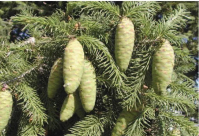
Fig. 1. Picea wilsonii Mast. The tree grows up to 50 m tall, with a trunk up to 1.5 m in diameter. The bark is dark gray and rough, and remains scaly with age. It is found in north-central China, from Qinghai and Sichuan to Nei Mongol, Shanxi, and Hubei, where it is scattered among other spruces and hardwoods in the upper montane forest (1,400∼3,000 m).
cytokines and chemokines then interact with each other to produce atopic dermatitis, which is characterized by an increase in eosinophil, activation of mast cell, and abnormal generation of IgE (a reaction that leads to a vicious circle of inflammation). As a result, one of the potential mechanisms for AD treatment is the regulation of cytokine production [6-8]. Picea wilsonii Mast (PwM) trees grow up to 1.5 m in diameter and 50 m in height, and they have dark gray, rough bark that does not change with age. Their shape is that of a crown cone or cylinder, and their branches spread horizontally; new branches are initially yellowish-gray and free of hair, with sprout-less twigs as long as 6∼8 mm. Once developed, the needles are dark green and 0.5∼2 cm long;
they have a diamond-like appearance with a flat top, and bend forward. The pollen cone is 20∼30 mm long and pink, while the seed cone is 4∼8 cm in length and changes from green (while young) to yellowish-brown (once ripened). The seed itself is 3∼4.5 mm long, resembles an egg, and has wings of 9∼11 mm; it is smooth and thin at the edge, but may also be hard and stiff. These PwM trees grow in north-central China from Qinghai and Sichuan to Nei Mongol, Shanxi, and Hubei, scattered among other spruces and hardwoods in upper montane forest (at 1,400∼3,000 m) [9,10] (Fig. 1). In this study, we aimed to measure cell viability in TNF- and IFN--stimulated HaCaT (human adult low calcium high
temperature) cells, and to observe whether Picea wilsonii Mast. PwM extracts inhibit cytokine secretion in cell culture media, thus identifying whether or not they could be a candidate treatment for AD. We also aimed to raise awareness of the need for research using foreign plant materials in the future.
Materials and Methods
1. Cell culture
Human keratinocyte HaCaT cells were cultured in Dulbecco's modified Eagle's medium (DMEM), supplemented with 10%
heat-inactivated fetal bovine serum, penicillin (100 U/mL), and streptomycin (100 g/mL). These cells were maintained at subconfluence in a 95% air, 5% CO
2humidified atmosphere, at 37
oC.
2. PwM preparation
PwM was purchased from the Foreign Plant Extract Bank (FPEB), and standard extracts have been deposited at the Herbarium of the department of Herbal pharmaceutical development (Korea Institute of Oriental Medicine, Daejeon, Korea) and the division of Life Science (Daejeon University, TUT, Daejeon, Korea) [11]. The whole plants of PwM (33.1 mg) were dried, powdered, and then extracted with dimethyl sulfoxide (DMSO; 0.3, 1, 3, 10, and 30 g/mL) for 1 day at room temperature.
3. 3-(4,5-dimethylthiazol-2-yl)-2,5-diphenyl tetrazollum bromide (MTT) assay
Cell viability was assayed based on the conversion of
3-(4,5-dimethylthiazol-2-yl)-2,5-diphenyltetrazolium
bromide (MTT) by using a cell proliferation kit I (Roche
Diagnostics, Mannheim, Germany). HaCaT cells in 100 L of
culture medium were seeded into a 96-well plate. PwM was
added to the wells at a concentration ranging between 0.3
and 30 g/mL. Following incubation for 24 h at 37
oC, 10 L
MTT solution was added and the plates were incubated again
for 4 h. Solubilization solution (100 L) was subsequently
added to the wells. Following 24 further hours of incubation,
absorbance was measured at 550 nm using an ELx808
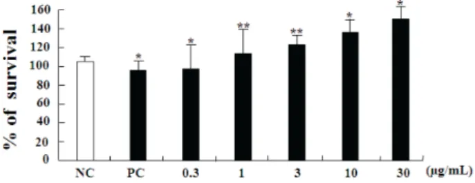
Fig. 2. Cell viability of human keratinocyte cells (HaCaT cells) when treated with PwM extracts. HaCaT cells were treated with various concentrations (0.3, 1, 3, 10, 30 g/mL) of PwM extracts for 24 h and the cytotoxicity was measured by MTT assay colorimetric dye reduction method. Data are expressed as the mean±SD (n=3).
Data are expressed as the mean±SD. *p<0.05 and **p<0.01 were considered to indicate a significant difference between the untreated group and the TNF- and IFN- only treated group or the TNF-
and IFN- only treated group and the PwM treated group.
enzyme-linked immunosorbent assay (ELISA) reader (Bio-Tek Instruments Inc., Winooski, VT, USA).
4. Cell density determination
Cell numbers and viability were assessed by trypan blue (Sigma-Aldrich, MS, USA) dye exclusion. Twenty microliters of trypan blue solution were mixed with 10 L of cell suspension in a microtube to obtain a final density of 1.5×10
5cells/mL, and these microtubes were loaded into a hemocy- tometer.
5. Enzyme-linked immunosorbent assay (ELISA)
Following pre-treatment with PwM for 30 minutes, HaCaT cells were treated with tumor necrosis factor- (TNF-) and interferon- (IFN-). The concentrations of IL-6, IL-8, IL-13, and MCP-1 in the supernatant were measured using sandwich ELISA, by using a BD OptEIA
TMsets (BD Biosciences, San Jose, CA, USA) according to the manufacturer's instructions.
The cytokine concentration was calculated using a linear regression equation obtained from the standard absorbance values. All assays were performed in at least three independent experiments.
6. Statistical analysis
Values are expressed as the mean ± standard deviation.
Data were analyzed with the Student's t-test by using SPSS software, version 18.0 (SPSS Inc., Chicago, IL, USA). * p <0.05 and ** p <0.01 were considered to indicate a statistically significant difference.
Results
1. Cytotoxic effects on HaCaT cells in vitro
Before studying the cytotoxic effects of PwM extracts, we determined the optimal dose of TNF- and IFN- (10 L). The PwM showed a cytotoxic effect on these cells; cell viability was altered after 24 h of PwM treatment using concentrations in the range of 0.3∼30 g/mL (Fig. 2). The results of negative controls and TNF- and IFN--stimulated positive controls revealed excellent cell viability of over 120%. This indicates that the cytotoxicity of PwM extracts is very low.
2. Effect of PwM extracts on cytokine levels in TNF- and IFN--stimulated HaCaT cells
Among the various cytokines and chemokines relating to acute or chronic atopic dermatitis, we observed the effects of PwM upon MCP-1, IL-6, IL-8, and IL-13 production in this study. In HaCaT cells, production of MCP-1 by the TNF- and IFN--stimulated positive control increased remarkably, compared to the negative control. When this positive control was subsequently treated with PwM extracts, MCP-1 production decreased significantly with increasing PwM concentrations, and this was particularly significant at a concentration of 1∼3 g/mL ( p <0.01). The same was true for IL-6; the TNF- and IFN--stimulated positive control HaCaT cells showed a remarkable increase in IL-6 production, compared to the negative control. With PwM treatment under the same conditions, there was a significant reduction in IL-6 production with increasing PwM concentration ( p <0.05). In the case of IL-8, a remarkable increase in IL-8 production occurred in the TNF- and IFN--stimulated positive control HaCaT cells, but there was no significant change with PwM treatment at the various concentrations.
In HaCaT cells, the production of TNF- and IFN--sti-
mulated IL-13 rose noticeably compared to the negative
control, with a remarkable decrease in production at PwM
concentrations of 3 g/mL or more ( p <0.01). In conclusion,
PwM extracts were found to inhibit the production of IL-6,
IL-13, and MCP-1, and to cause no significant change in
IL-8 production.
Fig. 3. PwM extracts inhibited production of IL-6, IL-13, and MCP-1 in TNF- and IFN--stimulated HaCaT cells, but did not affect IL-8 production. Cells were treated with various concen- trations of PwM extracts for 24 h, after being stimulated with 10
L TNF- and IFN-. Data are expressed as the mean±SD. Data are expressed as the mean±SD. *p<0.05 and **p<0.01 indicate a significant difference between the positive control and PwM treated group.
Discussion
The noticeable increase in cases of atopic dermatitis in countries that have been through rapid westernization and
industrialization, like Korea, suggests that environmental factors have a strong effect on the development of atopic dermatitis. Some explain this with the “hygiene hypothesis,”
however this is insufficient explanation. Since it was reported in 2006 that function-loss mutations of filaggrin (FLG) were highly implicated in Caucasian patients with atopic dermatitis in north-west Europe, innate skin barrier damage has surfaced as the primary cause of atopic dermatitis [12].
Recently, views on the pathogenesis of atopic dermatitis have been divided into the outside-in model (that claims skin barrier damage is the primary cause), and the inside-out model (that holds that AD starts from abnormal immune reactions). Meanwhile, the outside-inside-outside model explains that these two mechanisms work together to cause AD [13]. In immunological terms, it is thought that atopic dermatitis is caused by various cells, such as T cells (particularly helper T cells), eosinophil and mast cells. T lymphocytes are involved in the formation of antibodies, and helper T (Th) cells infiltrate atopic dermatitis lesions. In terms of function, helper T cells are divided into Th1 cells and Th2 cells, and the latter are more prevalent in the acute stages of atopic dermatitis. Symptoms like itching, urticaria, erythema, blisters, and psoriasis appear in atopic dermatitis, as well as other dermatological immune reactions. Cytokines are an important 'messenger' protein secreted from cells. They are part of an extracellular signal delivery network that controls innate and adaptive immune reactions, and they are deeply involved in inflammatory reactions caused by viral infections and tissue damage. Therefore, observing changes in their secretion may facilitate an understanding of the development of this inflammatory and immunological reaction [14].
There are many Korean studies concerning the use of natural extracts in the treatment of atopic dermatitis. In particular, traditional medicinal extracts from Korean red ginseng, chestnut, and Chinese herbs [15], Scutellaria baicalensis water [16], Eucommia ulmoides [17], Opuntia humifusa [18], kimchi [19], and angelica [20] have been reported to mitigate the symptoms of atopic dermatitis.
Despite extensive research, however, an innovative cure for
AD has not yet been developed. With this in mind, we
measured in vitro cell viability and inflammatory cytokine
production in TNF- and IFN--stimulated HaCaT cells, with the aim of investigating the potential effect of PwM extracts on the treatment of atopic dermatitis. The HaCaT cells are made up of normal human epithelial keratinocytes. As shown in Fig 2.PwM extract has no cytotoxic effect on HaCaT cells were seeded into 96-well plate at 1.5×10
5cells/well and were treated with PwM extract. In the course of inflam- mation, pro-inflammatory cytokines such as TNF-, IL-1, IL-4, IL-6, and IL-8 increase the number of activated inflammatory and immune-related cells, which cause the inflammatory state to spread. Eosinophil, monocyte, and lymphocyte migrate to the area and cause inflammation after chemokines of IL-8 and MCP-1 are released [21,22]. MCP-1 is one of the CC family chemokines, and it is an influential chemotactic factor for monocytes. MCP-1 is believed to regulate the migration and infiltration of monocytes, macrophages, memory T cells, and NK cells, and it has been implicated in various inflammatory disorders such as asthma and rheumatoid arthritis. Furthermore, in a study of patients with chronic and idiopathic urticaria, a chronic inflammatory skin disease, an increase in MCP-1 was observed. Finally, it has been reported that as MCP-1 increases the reactivity of monocytes and can easily facilitate the production of a pro-inflammatory environment [23-25]. IL-6 is an important inflammatory cytokine secreted from activated mast cells and a multi-functional protein that boosts acute phase protein synthesis in the innate immune response, thus contributing to the systemic inflammatory response [26]. IL-13 inhibits macrophage activation and controls inflammatory reactions such as atopic dermatitis; its dysregulation is known to be a major cause of allergic disorders, and it is generated excessively in the skin of patients with atopic dermatitis as the diseases develops [27]. Finally, IL-8 is a chemokine that guides neutrophils from blood vessels to tissues, and it is released by macrophages for the recruitment of other immune cells [28]. Thus the regulation of cytokine production such as IL-3, IL-4, IL-5, IL-6 caused by allergen is an important process in the pathogenesis and progression of allergic diseases. AD is characterized by elevated number of circulating eosinophils and their infiltration into the skin.
Monocytes produce inflammatory cytokines and differentiate
into inflammatory dendritic epidermal cells that induce the switch from Th2 to Th1/0 [29]. In the present study, inflammatory cytokine secretion was induced in HaCaT cell lines, and then PwM extracts were added at various concentrations. We found that these PwM extracts significantly inhibited the production of IL-6, IL-13, and MCP-1, but there was no significant change in IL-8 production. In Korea, most of the existing studies use indigenous anti-inflammatory plant extracts for the treatment of atopic dermatitis, but research using foreign plant materials has been insufficient. As far as the PwM extracts used in this study are concerned, objective and in-depth verification of their efficacy via protein and mechanism analysis is considered necessary. Furthermore, additional studies should follow using the various foreign plant extracts available, to determine their suitability for the treatment and alleviation of atopic dermatitis.
요 약
아토피피부염은 가려움증을 보이는 만성의, 재발성의 염증성 피 부상태를 말하며 천식, 식품 알레르기, 알레르기 비염 등과 같은 다 른 알레르기 질환보다 먼저 발생하고 다양한 면역학적 이상과 이로 인한 다양한 속발 증상도 갖고 있는 만성 재발성의 습관성 질환이 다. 본 연구는 TNF-와 IFN-로 유도된 HaCaT 세포주를 이용하 여 농도별로 PwM 천연물을 처리하였을 때 세포생존율이 우수한 것을 확인하였으며 아토피피부염과 관련된 염증성 사이토카인 즉, IL-6, IL-8, IL-13, MCP-1을 측정하였다. 그 결과 IL-6, IL-13, MCP-1의 생산량은 PwM 천연물에 의해 감소하였고 IL-8은 유의 한 변화가 없었다. 앞으로의 연구는 해외생물소재를 활용한 아토피 피부염 및 염증에 효과적인 치료제 후보물질을 개발하는 것이며 단 백질 분석과 기전 연구를 통해 보다 객관적인 검증이 더 필요하다 고 사료된다.
Acknowledgements:
This research was supported by Department of Biomedical Laboratory Science, Allergomics Laboratory, School of Medicine, Eulji University, Daejeon.
Funding:
None
Conflict of interest:
None
References
1. Leung DY, Bieber T. Atopic dermatitis. Lancet. 2003;361:
150-160.
2. Yu JS, Lee CJ, Lee HS, Kim J, Han Y, Ahn K, et al. Prevalence of atopic dermatitis in Korea: analysis by using national statistics. J Korea Med Sci. 2012;27:681-685.
3. Sturgil S, Bermard LA. Atopic dermatitis update. Curr Opin Pediatr. 2004;16:396-401.
4. Ozakaya E. Adult-onset atopic dermatitis. J Am Acad Dermatol.
2005;52:579-582.
5. Ou LS, Leung DY. Advances in atopic dermatitis. Chang Gung Med J. 2005; 28(1):1-8.
6. Novak N, Bieber T, Leung DY. Immune mechanisms leading to atopic dermatitis. J Allergy Clin Immunol. 2003;112(6):128-139.
7. Hamid Q, Boguniewicz M, Leung DY. Differential in situ cyto- kine gene expression in acute versus chronic atopic dermatitis.
J Clin Invest. 1994;94(2):870-876.
8. Ono SJ, Nakamura T, Miyazaki D, Ohbayashi M, Dawson M.
Toda M. Chemokines: roles in leukocyte development, traffick- ing, and effector function. J Allergy Clin Immunol. 2003;111 (6):1185-1199.
9. Farjon A. A handbook of the world's conifers. 1st ed. Leiden:
Koninklijke Brill; 2010.
10. Eckenwalder JE. Conifers of the world: The complete reference. 1st ed. Portland: Timber; 2009.
11. Holgate ST, Polosa R. Treatment strategies for allergy and asthma. Nat Rev Immunol. 2008:8:872-892.
12. Palmer CN, Irvine AD, Terron-Kwiatkowski A, Zhao Y, Liao H, Lee SP, et al. Common loss-of-function variants of the epi- dermal barrier protein filaggrin are a major predisposing factor for atopic dermatitis. Nat Genet. 2006;38:441-446.
13. Elias PM, Steinhoff M. “Outside-to-inside” (and now back to
“outside”) pathogenic mechanisms in atopic dermatitis. J Invest Dermatol. 2008;128:1067-1070.
14. Roitt M, Brostoff J, Male D. Immunology. 6th ed. London:
Mosby; 2002. p119-128.
15. Young HT, Choi HJ. Clinical efficacy of functional herbal ex- tracts liquid in atopic dermatitis patients. Kor J Food Nutr.
2005;18:380-384.
16. Kim YH, Park YS. Effect of Scutellaria baicalensis water extract on antioxidative activity and epidermal thickness in DNCB-in- duced allergic contact dermatitis animal model. J Korean Soc
and Food Sci Nutri. 2006;35:543-548.
17. Shon MY, Nam SH. Effect of Eucommia ulmoides extracts on al- lergic contact dermatitis and oxidative damage induced by re- peat elicitation of DNCB. J Korea Soc Food Sci Nutr 2007;36:
1517-1522.
18. Kim KS, Ahn MJ, Kim GS, Park SC, Rhee MH, In JG, et al. Effects of Opuntia humifusa extract on DNCB-induced allergic contact dermatitis in BALB/c mice. Lab Anim Res. 2007;23:169-173.
19. Lee IH, Lee SH, Lee IS, Park YK, Chung DK, Choue R. Effects of probiotic extracts of Kimchi on immue function in NC/Nga mice. Korean J Food Sci Technol. 2008;40:82-87.
20. Kim MA, Son HU, Nam DY, Cha YS, Shin YK, Choi YH, et al.
Inhibitory effect of Angelica keiskei extract in an atopic derma- titis animal model. Korean J Food Presev. 2012;19(5):792-798.
21. Church MK, Levi-Schaffer F. The human mast cell. J Allergy Clin Immunol. 1997;99(2):155-160.
22. Metcalfe DD, Baram D, Mekori YA. Mast cells. Physiol Rev.
1997;77(4):1033-1079.
23. Deshmane SL, Kremlev S, Amini S, Sawaya BE. Monocyte che- moattractant protein-1 (MCP-1): an overview. J Interferon Cytokine Res. 2009;29(6):313-326.
24. Hussain R, Ansari A, Talat N, Hasan Z, Dawood G. CCL2/MCP-I genotype-phenotype relationship in latent tuberculosis infection. PLoS One. 2011;6(10):e25803. doi: 10.1371/journal.
pone.0025803.
25. Santos JC, de Brito CA, Futata EA, Azor MH, Orii NM, Maruta CW, et al. Up-regulation of chemokine C-C ligand 2 (CCL2) and C-X-C chemokine 8 (CXCL8) expression by monocytes in chronic idiopathic urticaria. Clin Exp Immunol. 2012;167(1):
129-136.
26. Baek YM, Choi JY, Lee CW, Jeon YS, Han JT, Jang SI, et al.
Effects of Chinemys reevesii on lipopolysaccharide-indused in- flammatory reactions. J Physiol & Pathol Korean Med. 2012;
26(1):26-34.
27. Simon D, Von Gunten S, Borelli S, Braathen LR, Simon HU. The interleukin-13 production by peripheral blood T cells from atopic dermatitis patients does not require CD2 costimulation.
Int Arch Allergy Immunol. 2003;132(2):148-155.
28. Holgate ST, Polosa R. Treatment strategies for allergy and asthma. Nat Rev Immunol. 2008;8:872-892.
29. Yang EJ, Lee JS, Yun CY, Kim JH, Kim JS. Inhibitory effects of Duchesnea chrysantha extract on ovalbumin-induced lung in- flammation in a mouse model of asthma. J Ethnopharmacol.
2008;118(1):102-107.