Posterior Trans-Dural Repair of Iatrogenic Spinal Cord Herniation after Resection of
Ossification of Posterior Longitudinal Ligament
Seung-Jae Hyun, Hong-Ki Kim, Ki-Jeong Kim, Tae-Ahn Jahng, Hyun-Jib Kim
Department of Neurosurgery, Spine Center, Seoul National University Bundang Hospital, Seoul National University College of Medicine, Seongnam, Korea
Iatrogenic spinal cord herniation is a rare complication following spinal surgery. We introduce a posterior trans-dural repair technique used in a case of thoracic spinal cord herniation through a ventral dural defect following resection of ossification of the posterior longitudinal ligament (OPLL) in the cervicothoracic spine. A 51-year-old female was suffering from paraplegia after laminectomy alone for cervicothoracic OPLL. Magnetic resonance imaging revealed a severely compressed spinal cord with pseudomeningocele identified postoperatively. Cerebrospinal fluid leak and iatrogenic spinal cord herniation persisted despite several operations with duroplasty and sealing agent. Finally, the problems were treated by repair of the ventral dural defect with posterior trans-dural duroplasty. Sev- eral months after surgery, the patient could walk independently. This surgical technique can be applied to treat ventral dural defect and spinal cord herniation.
Keywords: Iatrogenic spinal cord herniation; Duroplasty; Cervicothoracic junction; Ossification of the posterior longitudinal ligament
Copyright Ⓒ 2016 by Korean Society of Spine Surgery
This is an Open Access article distributed under the terms of the Creative Commons Attribution Non-Commercial License (http://creativecommons.org/licenses/by-nc/3.0/) which permits unrestricted non-commercial use, distribution, and reproduction in any medium, provided the original work is properly cited.
Asian Spine Journal • pISSN 1976-1902 eISSN 1976-7846 • www.asianspinejournal.org
Received Aug 11, 2015; Revised Sep 21, 2015; Accepted Sep 24, 2015 Corresponding author: Seung-Jae Hyun
Department of Neurosurgery, Spine Center, Seoul National University Bundang Hospital,
Seoul National University College of Medicine, 82 Gumi-ro 173 Beon-Gil, Bundang, Seongnam 13620, Korea Tel: +82-31-787-7164, Fax: +82-31-787-4097, E-mail: [email protected]
ASJ A SJ
Introduction
Pseudomeningoceles are extravasated collections of extradural cerebrospinal fluid (CSF) that result from a dural tear [1,2]. They may be traumatic, congenital and iatrogenic. Iatrogenic spinal cord herniation occurs after durotomy, usually during cervical or lumbar surgery.
Extradural herniation of the thoracic spinal cord is a rare complication that presents with progressive paraplegia.
After surgical reduction of the herniated spinal cord and dural repair, a patient’s neurological function can be re- covered [3]. In this report, we describe a case of thoracic spinal cord herniation through a ventral dural defect fol- lowing resection of ossification of the posterior longitudi-
nal ligament (OPLL) in the cervicothoracic spine.
Technical Note
A 51-year-old female was referred to our institute for paraplegia and anesthesia below the T2 dermatome. Deep tendon reflex was increased at knee and ankle jerk, with ankle clonus. Cervicothoracic OPLL had been diagnosed and resected with laminectomy at another hospital one week before. CSF leaked out through the surgical wound.
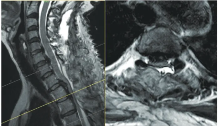
Magnetic resonance imaging (MRI) revealed a severely compressed spinal cord and postoperative pseudomenin- gocele were identified (Fig. 1). We decided to revise the surgical site for neural decompression and stabilization.
1. Operation
Through a posterior approach, we first installed pedicle screws and placed a tentative rod with dekyphotic rod contouring. After further laminectomy of the thoracic spine, severely compressed ventral dura and CSF leak from the ventral dural defect were visualized. Severe adhesion between the OPLL and ventral dura made it impossible to dissect and repair the defect primarily. We were obliged to perform duroplasty using artificial dura, muscle patch and sealant. Postoperatively, the patients’
neurological status improved, but it sequentially deterio- rated after 3 days. Although we performed re-operation using a similar surgical technique on day 5 after the initial operation, CSF leakage and iatrogenic spinal cord hernia- tion with paraparesis (motor grade 2–3) persisted. Finally, the problems were treated by repair of the ventral dural defect with posterior trans-dural duroplasty on day 10 after the initial operation. The spinal cord was exposed by durotomy. To free the spinal cord, the dentate ligament was resected. The ventral OPLL was carefully resected with the attached portion of the ventral dura. The primary repair of the ventral dural defect was not feasible due to severe adhesion between the ventral dura and remained OPLL. An artificial dura was passed on the ventral side of the spinal cord and reinforced to the dura located on the superior aspects of the upper and lower roots, allowing it to function as a check valve (Figs. 2, 3). To prevent anteri- Fig. 1. Magnetic resonance imaging showing severely compressed
spinal cord and postoperative pseudomeningocele.
Fig. 2. Intraoperative photography: posterior trans-dural duro- plasty technique. (A, B, C) An artificial dura was passed on the ventral side of the spinal cord and reinforced to the dura located on superior aspects of the upper and lower roots, allowing func- tion as a check valve.
A
B
C
Fig. 3. Schematic drawing of the posterior trans-dural duro- plasty in the shape of a check valve.
or spinal artery injury or cord damage, careful dissection between the spinal cord and ventral dura and soft artificial dura material are mandatory.
2. Postoperative course
After surgery, the patient gradually recovered from paraplegia. An external orthosis was applied for 3 months to achieve bony fusion. Several months after surgery, the patient could walk independently. Postoperative MRI showed well-decompressed spinal cord without pseudo- meningocele or spinal cord herniation (Fig. 4).
Discussion
Laminectomy without instrumentation has not been rec- ommended for thoracic OPLL because of the kyphotic curvature. Dorsal shift of the spinal cord in the thoracic spine may not occur by laminectomy alone. When decid- ing a posterior approach, neural decompression with in- strumentation should be considered [4-7].
Iatrogenic spinal cord herniation through a dural defect is a rare complication. The first report of iatrogenic spinal cord herniation was published over 40 years ago [3]. A meta-analysis of 126 case reports indicated the several cases with ventral thoracic dural defect [8]. However, no cases of spinal cord herniation following resection of
OPLL in the thoracic spine have been reported.
A previous clinical study involving a series of patinets with idiopathic spinal cord herniation determined that the most common clinical symptom was Brown-Sequard syn- drome [9]. The authors suggested the symptoms might be derived from the involvement of the anterolateral funicu- lus. On the other hand, another investigator explained the symptoms as myeloradiculopathies [10].
In this case, recurrence of iatrogenic spinal cord hernia- tion even after repeat revision surgery is thought to have worsen the neurological symptoms. Despite advance- ments in surgical techniques for thoracic OPLL, favorable results are not always achieved. In patients with thoracic myelopathy resulting from thoracic OPLL, removal of the ossified PLL is the most effective method of relieving pres- sure on the spinal cord [7,11-15]. A surgical technique for OPLL of the thoracic spine which was removed by floating method has been described [16]. For this patient, floating method was not feasible because of extensive adhesion between the dura and OPLL.
Direct primary closure of ventral dural defect is the best way to treat spinal cord herniation [17]. However, primary closure is not always feasible. A previous study reported that a muscle pedicle flap technique to repair a CSF fistula can be used as an alternative to the direct clo- sure of ventral dural defects [18].The authors suggested that the technique can be used as an alternative to the
Fig. 4. (A) Magnetic resonance imaging showing well-decompressed spinal cord without pseudomeningocele or spinal cord her- niation. (B, C) postoperative X-ray shows dekyphosis of cervicothoracic junction by instrumentation.
A B C
direct “water-tight” closure of ventral cervical dural de- fects. A trans-dural duroplasty technique utilizing Gore- Tex through a trans-dural approach has been detailed [8]. However, the method is less effective in preventing the large dural defect. A recent paper reported a case of posterior cord reduction and dural repair after multilevel anterior thoracic vertebrectomy for a giant cell tumor [19].
The authors closed the ventral dural defect using an artifi- cial dura anteriorly to the cord. The edges of the artificial dura were inside the spinal dural edge, entirely covering the defect in an inside-out fashion. However, surgical de- tails or drawings were not presented.
Repair of the ventral dura is an important step of our surgical method with a duroplasty in the shape of a check valve, making it possible to prevent CSF leakage if direct primary closure is not feasible. Sophisticated care should be taken to prevent anterior spinal artery injury or cord damage, when passing the artificial dura beneath the spi- nal cord.
Duroplasty in the shape of a check valve through poste- rior trans-dural approach for iatrogenic spinal cord her- niation is described. This surgical method for dural repair can serve as an option in patients with ventral dural defect and iatrogenic spinal cord herniation if direct primary closure is not feasible.
Conflict of Interest
No potential conflict of interest relevant to this article was reported.
Acknowledgments
We appreciate the assistance of Mijin Jung, the artist who drew Fig. 3 in this manuscript. She worked as a medical illustrator for the Department of Neurosurgery, Baylor College of Medicine from 2009 to 2011, and has worked in the Department of Neurosurgery, Seoul National Uni- versity Bundang Hospital, Seoul National University Col- lege of Medicine since 2013.
References
1. Hawk MW, Kim KD. Review of spinal pseudomenin- goceles and cerebrospinal fluid fistulas. Neurosurg Focus 2000;9:e5.
2. Macki M, Lo SF, Bydon M, Kaloostian P, Bydon A.
Post-surgical thoracic pseudomeningocele causing spinal cord compression. J Clin Neurosci 2014;21:
367-72.
3. Cobb C 3rd, Ehni G. Herniation of the spinal cord into an iatrogenic meningocele. Case report. J Neu- rosurg 1973;39:533-6.
4. Matsumoto M, Toyama Y, Chikuda H, et al. Out- comes of fusion surgery for ossification of the pos- terior longitudinal ligament of the thoracic spine: a multicenter retrospective survey: clinical article. J Neurosurg Spine 2011;15:380-5.
5. Yamazaki M, Okawa A, Fujiyoshi T, Furuya T, Koda M. Posterior decompression with instrumented fu- sion for thoracic myelopathy caused by ossification of the posterior longitudinal ligament. Eur Spine J 2010;
19:691-8.
6. Matsuyama Y, Sakai Y, Katayama Y, et al. Indirect posterior decompression with corrective fusion for ossification of the posterior longitudinal ligament of the thoracic spine: is it possible to predict the surgi- cal results? Eur Spine J 2009;18:943-8.
7. Fujimura Y, Nishi Y, Nakamura M, Toyama Y, Suzuki N. Long-term follow-up study of anterior decom- pression and fusion for thoracic myelopathy resulting from ossification of the posterior longitudinal liga- ment. Spine (Phila Pa 1976) 1997;22:305-11.
8. Groen RJ, Middel B, Meilof JF, et al. Operative treatment of anterior thoracic spinal cord hernia- tion: three new cases and an individual patient data meta-analysis of 126 case reports. Neurosurgery 2009;64:ons145-59.
9. Najjar MW, Baeesa SS, Lingawi SS. Idiopathic spinal cord herniation: a new theory of pathogenesis. Surg Neurol 2004;62:161-70.
10. Watters MR, Stears JC, Osborn AG, et al. Transdural spinal cord herniation: imaging and clinical spectra.
AJNR Am J Neuroradiol 1998;19:1337-44.
11. Hyun SJ, Kim JS, Hong SC. Late occurrence of cervi- cothoracic ossification of posterior longitudinal liga- ments in a surgically treated thoracic OPLL patient. J Korean Neurosurg Soc 2010;47:55-7.
12. Ido K, Shimizu K, Nakayama Y, et al. Anterior de- compression and fusion for ossification of posterior longitudinal ligament in the thoracic spine. J Spinal Disord 1995;8:317-23.
13. Suzuki R, Ohno K, Inaba Y. Fracture of the floor of the anterior cranial fossa and the sella turcica, and
sification of posterior longitudinal ligament in the thoracic spine. J Neurosurg Spine 2012;17:525-9.
17. Aydin AL, Sasani M, Erhan B, Sasani H, Ozcan S, Ozer AF. Idiopathic spinal cord herniation at two separate zones of the thoracic spine: the first reported case and literature review. Spine J 2011;11:e9-e14.
18. Hyun SJ, Rhim SC, Ra YS. Repair of a cerebrospinal fluid fistula using a muscle pedicle flap: technical case report. Neurosurgery 2009;65:E1214-5.
19. Kawsar KA, Bhatia R, Casey AC. Spinal cord hernia- tion as a complication of en bloc, multilevel, anterior thoracic vertebrectomy for a giant cell tumor: success of posterior cord reduction and dural repair. J Neu- rosurg Spine 2014;21:909-12.
successful repair of the cerebrospinal fluid fistula with fibrin glue: case report. Neurol Med Chir (To- kyo) 1986;26:420-5.
14. Tomita K, Kawahara N, Baba H, Kikuchi Y, Nishimu- ra H. Circumspinal decompression for thoracic myelopathy due to combined ossification of the pos- terior longitudinal ligament and ligamentum flavum.
Spine (Phila Pa 1976) 1990;15:1114-20.
15. Tsuzuki N, Hirabayashi S, Abe R, Saiki K. Staged spi- nal cord decompression through posterior approach for thoracic myelopathy caused by ossification of posterior longitudinal ligament. Spine (Phila Pa 1976) 2001;26:1623-30.
16. Kato S, Murakami H, Demura S, Yoshioka K, Hayas- hi H, Tsuchiya H. Novel surgical technique for os-