키토산은 키틴의 탈아세틸화 화합물로 갑각류의 외골 격에 존재하며 뮤코다당류로써 당단백류와 유사한 구조
를 갖고 있다22). 키토산은 독성이 없고 생흡수성이 좋으 며 생체 친화적 물질로 알려져 있고4,13), 체내 항상성 유
332
목 적: Carboxy methyl화된 키토산이 퇴행성 관절염에 미치는 효과를 알아보고자 하였다.
대상 및 방법: 슬관절 전방십자인대 절단을 통해 인위적으로 만들어진 토끼 슬관절의 골관절염에 키토산과 히알루론산을 주기적으로 관절 내에 각각 주입 후 관절 연골과 활액막 조직을 얻어 육안적, 병리 조직학적 검사를 통해 골관절염의 진 행에 미치는 키토산과 히알루론산의 효과를 비교하였다.
결 과: 히알루론산과 키토산 주입군에서는 대조군에 비해 골관절염의 진행이 적게 일어나는 소견을 얻었으며, 키토산군 보다는 히알루론산군에서 이러한 효과가 우수하였으나 통계학적인 차이는 없었다.
결 론: Carboxy methyl화된 키토산이 골관절염 진행은 지연시키는데 긍정적 영향을 미치는 것을 알 수 있었다.
색인 단어: 골관절염, 관절 연골, 키토산, 히알루론산
Purpose: This study evaluates the effect on carboxy methyl chitosan on the articular cartilage of an osteoarthritic knee.
Materials and Methods: The biological effect of chitosan on the osteoarthritic articular cartilage was compared with hyaluronan, which is one of the treatment modalities for an osteoarthritic knee. Chitosan or hyaluronan were injected into the intraarticular space of the experimental osteoarthritic knee of a rab- bit with a transected anterior cruciate ligament at weekly intervals for 5 weeks, and the articular cartilage and synovium were obtained. Nine weeks later, the specimens were analyzed by their gross findings, histological examinations, and histo-morphometric studies.
Results: The hyaluronan and chitosan group showed less severe osteoarthritic changes than the con- trol group. Although the hyaluronan group showed less severe osteoarthiricic changes than the chitosan group, there was no statistical significance.
Conclusion: Carboxy methyl chitosan, which has an excellent affinity to the body, appeared to have an positive effect on delaying the progress of cartilage degeneration.
Key Words: Osteoarthritis, Articular cartilage, Chitosan, Hyaluronan
Depatrment of Orthopaedic Surgery, School of Medicine, Chung-Ang University, Seoul; Department of Biotechnology*, Chung-Ang University, Ahnsung, Korea
Ho-Joong Jung, M.D., Eui Chan Jang, M.D., Eun Woo Lee, M.D., Soo Yong Kang, M.D., and Tae Il Son, Ph.D.*
Effect of Carboxy Methyl Chitosan on Experimental Osteoarthritis in a Rabbit Knee
- Carboxy methyl chitosan vs. Hyaluronan -
332
Carboxy Methyl화된 키토산이 토끼 슬관절의 골관절염에 미치는 효과 - Carboxy Methyl화된 키토산과 히알루론산의 비교 -
정호중ㆍ장의찬ㆍ이은우ㆍ강수용ㆍ손태일*
중앙대학교 의과대학 정형외과학교실, 중앙대학교 생명공학부*
332 332 통신저자 : 강 수 용
서울시 용산구 한강로 3가 65-207 중앙대학교 용산병원 정형외과
TEL: 02-748-9963∙FAX: 02-793-6634 E-mail: [email protected]
Address reprint requests to Soo Yong Kang, M.D.
Department of Orthopaedic Surgery, Yongsan Hospital, Chung-Ang University, 65-207 Hangangro 3-ga, Yongsan-gu, Seoul 140-757, Korea
Tel: +82.2-748-9963, Fax: +82.2-793-6634 E-mail: [email protected]
지와 정균 작용이 있는 것으로 보고되고 있다9,20,21). 또 한 골, 연골 세포의 활성 조절 및 치유에 관여하며7,14-16), 그 유도체들은 관절 연골의 기질이 되는 콘드로이틴, 황 산콘드로이틴, 황산데르마탄, 황산케라탄과 히알루론산 의 생성에도 영향을 주는 것으로 보고되고 있다12,18). 그 러나 퇴행성 관절염이 있는 경우에 키토산이 어떤 효과 가 있는지에 대한 체계적인 연구는 없는 실정이다.
본 실험에서는 키토산의 일부 구조를 변화시켜 히알루 론산의 분자 구조와 좀 더 유사하며(Fig. 1), 수용성 성 질을 가지는 carboxy methyl화된 키토산을 만들어 토 끼 슬관절의 골관절염 진행에 미치는 효과를 히알루론산 과 비교하여 알아보고자 하였다.
대상 및 방법
1. 연구 방법
체중이 4-5 kg이고 생후 12주 이상 되어 성장판이 닫 힌 성숙한 토끼(New Zealand White Rabbit) 30마리 의 우측 슬관절에 Yoshioka 등26)의 방법에 따라 내측 관 절 개방술의 방법을 통해 슬관절 전방십자인대 절단술을 시행하고 특별한 고정 없이 60×60×40 cm크기의 사 육장에서 수술 후 4주까지 사육하였다. 각 토끼를 3군으 로 나누어, 제1 실험군(키토산 주입군, N=10)은 1주에 한번씩 5주간 0.3 mL의 carboxy methyl키토산(OO 대학교 생명공학과 생체 재료 화학 실험실에서 공여 받 음. MW=200,000 10 mg/mL)을 관절 내 주입하였고, 제2 실험군(히알루론산 주입군, N=10)은 같은 방법으 로 0.3 mL의 히알루론산(MW=800,000 10 mg/mL
신풍제약)을 주입하였다. 대조군(무투여군, N=10)은 전 방십자인대 절단 후 아무것도 주입하지 않았다. 수술 후 9주에 토끼를 희생 시켜 실험 동물의 양측 슬관절을 얻 은 후 각 군에서 전방십자인대 제거술을 시행한 우측과 수술을 시행하지 않은 좌측을 비교하였다.
2. 실험 동물 처리 1) 육안적소견
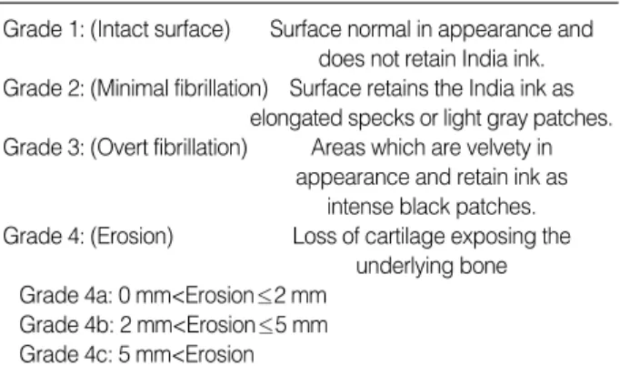
모든 토끼에 대하여 관절 연골의 변화에 대한 육안적 검사를 시행하였다. 대퇴과를 디지털 카메라(Coolpix 5700, Nikon copr, Japan)를 이용하여 근접 촬영 후 India ink를 이용하여 4가지 단계, 즉 정상 연골인 1단 계, 최소한의 섬유성 연축(fibrillation)을 보이는 2단 계, 중등도의 섬유성 연축을 보이는 3단계, 관절 연골의 미란(erosion)이나 전층 결손을 보이는 4단계로 분류하 였다26)(Table 1).
2) 조직학적 검사
전구 실험을 통하여 전방십자인대 절단에 의한 골관절 염 모델에서는 내측 대퇴과가 퇴행성 변화의 진행을 가 장 잘 나타낸다는 것을 알 수 있었기 때문에 내측 대퇴과 의 관절 연골과 골조직을 10% neutral buffered for- malin과 1% cetylpyridinium chloride (CPC)에서 7 일간 고정 후 ethylene-diamine tetra acetic acid (EDTA)로 탈석회화하고 시상면으로 절편을 만들어 파라 핀에 고정하였다. 세포 변화를 보기 위해 H&E염색을 하 고 글리코스아미노글리칸 존재 여부를 보기 위해 safra- nin-O/Fast green염색을 시행하여 광학현미경 하에 서 관찰하였다. 조직학적 변화는 구조적 이상 여부, 세포 배열 및 밀집도, safranin-O stain의 분포, tidemark
Grade 1: (Intact surface) Surface normal in appearance and does not retain India ink.
Grade 2: (Minimal fibrillation) Surface retains the India ink as elongated specks or light gray patches.
Grade 3: (Overt fibrillation) Areas which are velvety in appearance and retain ink as
intense black patches.
Grade 4: (Erosion) Loss of cartilage exposing the underlying bone Grade 4a: 0 mm<Erosion≤2 mm
Grade 4b: 2 mm<Erosion≤5 mm Grade 4c: 5 mm<Erosion
Table 1.Gross morphological assessment
Fig. 1.Molecular structures of hyaluronan and carboxy methyl chitosan.
COOH O
O O
O O OH
OH
NH2
OH
4
4 4
4 1
1 1
1
OH
HO O
O O O
O CH2OH
CH2OH
NHCOCH3
NHCOCH3
OCH2CO2H Hyaluronan
Carboxy methyl chitosan
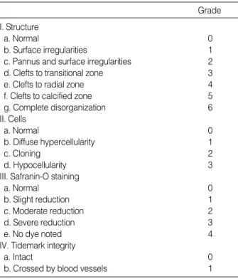
의 유지도 등을 분석하여 각각의 점수를 매겨 병리 조직 화학적 등급을 나누는 Mankin10)의 grading system 을 사용하였다(Table 2). 각 실험 동물의 슬개건 내측 부 위의 활액막을 채취하여 파라핀에 고정 후 H&E염색을 하였다. 조직학적 검사는 준비된 조직을 광학 현미경 하 에서 검사한 후 고해상도 디지털 카메라(Coolpix 5700, Nikon copr, Japan)와 영상 분석 프로그램(Scion image beta ver 4.02, Scion corp.,USA)을 이용하 여 Yoshioka 등26)의 방법으로 관절 연골의 두께와 면적 을 측정하고(Fig. 2), 활액막 세포층의 두께는 100군데 의 표본을 설정하여 평균값을 구하여 측정하였다. 각 측 정치는 개체 변이에 의한 편재(bias)를 제거하기 위해 우측과 좌측의 차이를 계산하고, 이 수치를 각 군별로 비교하였다.
3) 통계처리 방법
각 군에서 양측의 측정치간의 차이를 분석함에 있어서 는 비모수적 방법인 Wilcoxon signed rank test를 이 용하였다. 각 군간의 차이를 비교함에 있어서는, 모수적
방법으로 일원배치 분산분석과 사후 분석으로 Tukey b test를 사용하였고, 비모수적 방법으로 Kruskall-Wal- lis test와 사후 분석으로 Mann-Whiteney U test를 사용하였다. 통계학적 분석은 SPSS 통계프로그램(SPSS for Windows Release 11.5; Chicago, Ilinois)을 이 용하였고, 신뢰구간은 95%로 하였다.
결 과
1. 육안적 소견
전체 30마리 중 전방십자인대를 제거한 우측에서는 26예에서 grade 2 이상의 관절 연골 변성이 나타났고 좌측에서는 3예에서만 grade 2의 관절 연골 변성이 나 타났다. 수술 후 감염은 없었고 내측 관절 개방술 후에 습관성 슬개골 외측 탈구가 1예에서 발생하였다. 히알루 론산이나 키토산의 주입에 대한 특별한 부작용은 없었다.
우측 슬관절의 전방십자인대 제거술을 시행 후 아무것도 주입하지 않은 대조군 중 1예는 원인을 알 수 없이 수술 후 1주에 죽었다. 모든 실험군에서 육안적 소견상 전방 십자인대를 절제한 우측에서는 관절낭의 섬유성 비후와 활액막 세포의 증식이 반대편에 비하여 현저하였다.
1) 대퇴과
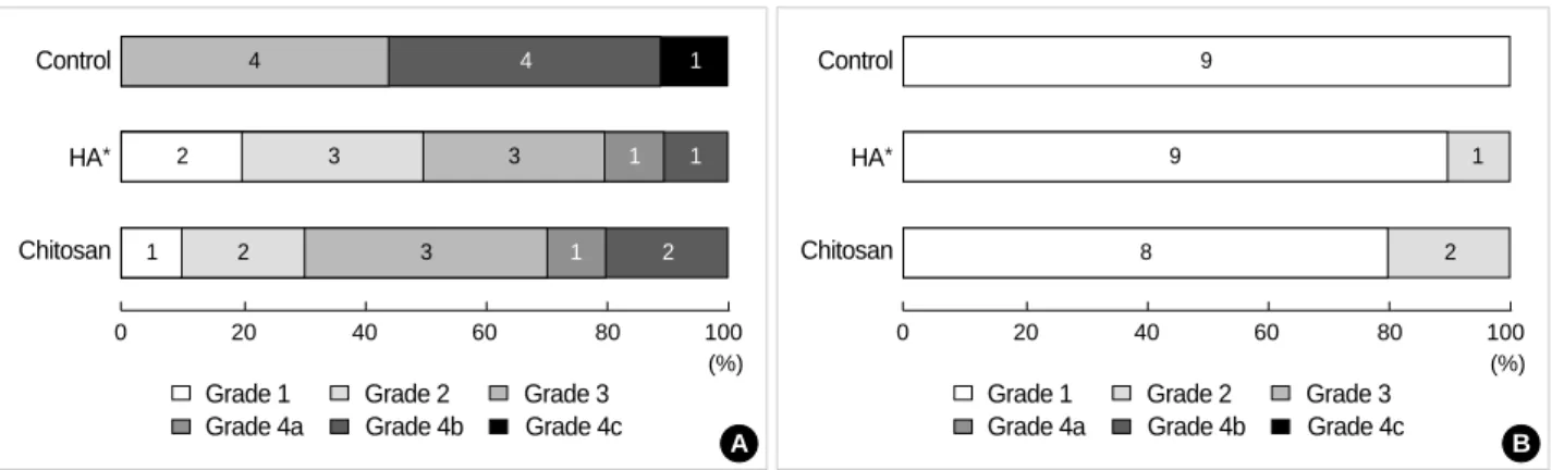
전방십자인대 절단측(우측)을 India ink를 이용해 분 류한 결과, 대조군에서는 수술 후 죽은 1예를 제외하고 9예 모두 grade 3 이상의 관절염을 보였고 이 중 5예에 서 grade 4의 관절염을 보였다. 키토산군과 히알루론산 군에서는 각각 3예와 2예에서만 grade 4의 관절염을 보였으나, 그 정도와 범위가 대조군에 비하여 적은 결과
Grade I. Structure
a. Normal 0
b. Surface irregularities 1
c. Pannus and surface irregularities 2
d. Clefts to transitional zone 3
e. Clefts to radial zone 4
f. Clefts to calcified zone 5
g. Complete disorganization 6
II. Cells
a. Normal 0
b. Diffuse hypercellularity 1
c. Cloning 2
d. Hypocellularity 3
III. Safranin-O staining
a. Normal 0
b. Slight reduction 1
c. Moderate reduction 2
d. Severe reduction 3
e. No dye noted 4
IV. Tidemark integrity
a. Intact 0
b. Crossed by blood vessels 1
*Serial histological sections, cut at five micrometers and stained with hematoxylin and eosin and safranin-O-fast green-iron hema- toxylin, were analyzed for abnormalities in structure, cell popula- tion, safranin-O stain distribution, and tidemark integrity and scores assigned as the histologic-histochemical grade.
Table 2.Histological-Histochemical grading*
Fig. 2.Schematic representation of the typical histology speci- men shown on the image analysis monitor. The geometric para- meters of the cartilage specimens were measured on 5 mm wide condylar weight-bearing areas.
Histolic section
Thickness
5 mm
Area
를 보였다(Fig. 3A, 4A). 키토산군의 1예와 히알루론산 군의 2예에서 grade 1의 정상적인 관절면을 보였다. 골 극은 대퇴 슬개 관절면의 변연에서 보였는데 골극 형성 은 대조군에서 더 심하였고 이 역시 키토산군과 히알루
론산군에서 골극 형성이 적은 양상을 보였다. 전방십자 인대 제거술을 시행하지 않은 반대측(좌측)에서는, 대조 군에서는 9예 모두 정상적인 관절면을 보였고, 키토산 군에서는 8예에서, 히알루론산군은 9예에서 정상적인 Fig. 3.Gross morphological assessment of the femoral condyles (*HA: hyaluronan group). (A) Anterior cruciate ligament transection side. Note that the grade of cartilage damage in the hyaluronan and Chitosan group was generally less severe than in the control group. (B) Intact anterior cruciate ligament side.
Control
HA*
Chitosan
4 4 1
2 3 3 1 1
1 2 3 1 2
0 20 40 60 80 100
Grade 1 Grade 2 Grade 3 Grade 4a Grade 4b Grade 4c
(%)
A
Control
HA*
Chitosan
9
9 1
8 2
0 20 40 60 80 100
Grade 1 Grade 2 Grade 3 Grade 4a Grade 4b Grade 4c
(%)
B
Fig. 5.Histological-histochemical assessment according to the Mankin grade system (*HA: hyaluronan group). (A) Anterior cruciate ligament transection side. Note that the grade of cartilage damage in the hyaluronan and chitosan group was generally less severe than the control group. (B) Intact anterior cruciate ligament side.
Control
HA*
Chitosan
9 10 11 12
2 4 5 6 7 9 10
2 3 4 5 6 7 9
0 10 20 30 40 50 60 70 80 90 100
(%)
A
Control
HA*
Chitosan
0
0 2
0 2
0 20 40 60 80 100
0 2
2 3 4 5 6
7 9 10 11 12
(%)
B
A B
Fig. 4.Gross morphological finding 9 weeks after surgery in the control group. (A) Anterior cruciate ligament transection side. The carti- lage was largely degenerated on the medial femoral condyle. (B) Intact anterior cruciate ligament side. The articular surface was intact.
관절면을 보였고 관절 변성이 있었던 키토산의 2예, 히 알루론산군 1예에서는 grade 2의 최소한의 섬유성 연 축만 보였다(Fig. 3B, 4B).
2) 활액막
활액막의 증식과 갈색으로 변성(brown discoloration) 이 모든 전방십자인대 절단 슬관절에서 관찰되었으며 키
E F
C D
A B
Fig. 6.Histological findings of the femoral condyles. (A) Intact anterior cruciate ligament side (H&E, original magnification ×40). (B) Intact anterior cruciate ligament side (safranin-O/fast green, original magnification ×40), The cartilage matrix is consistently well stained with safranin-O while the cartilage thickness and smoothness is maintained. (C) Anterior cruciate ligament transection side in the control group (H&E, original magnification ×40), Ulceration of the cartilage exposing the surface of the subchondral bone can be seen. (D) Anterior cruciate ligament transection side in the control group (safranin O/fast green, original magnification ×40).
Complete loss of safranin O staining can be seen. (E) A mild degenerative case in the chitosan group (safranin O/fast green, origi- nal magnification ×40). Lower intensity of safranin O staining along with mild degenerative changes in the cartilage can be seen.
(F) A mild degenerative case in the chitosan group (safranin O/fast green, original magnification ×100).
0.5 mm
0.5 mm
0.5 mm 0.1 mm
0.5 mm 0.5 mm
토산군과 히알루론산군에서는 대조군에 비하여 이러한 변성이 심하지 않았다. 각 군의 반대측 슬관절은 이러한 소견이 발견되지 않았다.
2. 조직학적 정성 분석(histology-qualitative observations)
1) 대퇴과
전방십자인대 절단측(우측)의 결과로, 대조군에서는 Mankin 점수의 평균이 10.4±1.1이었고 히알루론산군 은 5.3±2.7, 키토산군은 6.6±2.8로 히알루론산군과 키토산군에서 관절 연골 변성의 정도와 범위가 적었다 (Fig. 5A). 대조군에서는 관절 연골의 결손이나 미란이 심하고 safranin-O 염색의 소실이 많았다(Fig. 6C-F).
전방십자인대 제거술을 시행하지 않은 반대측(좌측)은 키토산군 2예와 히알루론산군 1예를 제외하고는 모두 정 상적인 관절면을 보였고, 관절 변성이 있었던 3예에서 도 Mankin 점수 2의 상태를 보였다(Fig. 5B, 6A, B).
2) 활액막
히알루론산군이나 키토산군 보다는 대조군의 활액막 세포층의 과증식과 단핵성 염증 세포의 침윤이 두드러져 보였고, 각 군의 반대측에서는 이러한 소견이 보이지 않 았다.
3. 조직형태학적 정량 분석(histomorphometry- quantitative analysis)
1) 관절 연골층의 두께
3군 모두에서 반대편과 비교하여 관절 연골층의 두께가
의미있게 감소하였으나(p<0.05) 대조군에서는 히알루론 산군과 키토산군과 비교하여 관절연골층 두께의 감소가 심하였다(p<0.05). 히알루론산군과 키토산군간의 비교 에서는 평균을 비교하였을 때는 키토산군에서의 연골층 두께의 감소가 히알루론산군보다 심하였으나 이러한 두 군간의 차이는 통계학적으로 유의하지 않았다(Fig. 7A).
2) 관절 연골층의 면적
관절 연골층의 두께에서 얻은 결과와 비슷하게, 3군 모 두에서 반대편과 비교하여 관절 연골층의 면적이 감소하 였으나(p<0.05) 대조군에서는 히알루론산군과 키토산군 과 비교하여 관절 연골층 면적의 감소가 심하였다(p<
0.05). 히알루론산군과 키토산군간의 비교에서는 평균 을 비교하였을 때는 키토산군에서의 면적의 감소가 히알 루론산군보다 심하였으나 이러한 두 군간의 차이는 통계 학적으로 유의하지 않았다(Fig. 7B).
3) 활액막 세포층(synovial lining cell layer)의 두께 3군 모두에서 수술을 시행한 우측 슬관절의 활액막 세 포층 두께가 반대측에 비하여 의미있게 증가하였다(p<
0.05). 대조군에서는 반대편과의 차이의 평균이 10.9± 2.1 m로 가장 높은 수치를 보였고 다음으로는 키토산 군으로 평균 5.2±1.9 m, 히알루론산군은 평균 2.9± 1.9 m로 가장 낮은 수치를 보였다. 각 군간의 차이는 통계학적으로 유의한 차이를 보였다(p<0.05, Fig. 7C).
고 찰
본 연구에서 비교 대상으로 선정하였던 히알루론산은 Fig. 7.*HA: hyaluronan group. (A) Side to side differences in the cartilage thickness in the medial femoral condyle (mean±S.D.):
*p<0.05. (B) Side to side differences in the cartilage area in the medial femoral condyle (mean±S.D.): *p<0.05. (C) Side to side dif- ferences in the thickness of the synovial lining cell layer (mean±S.D.): *p<0.05.
A
Difference of cartilage thickness (mm)
0.08
0.06
0.04
0.02
0.00
HA Chitosan Control
*
B Difference of cartilage area (mm2) 0.08
0.06
0.04
0.02
0.00
HA Chitosan Control
*
C
Difference of synovial thickness (microns)
14 12 10 8 6 4 2 0
HA Chitosan Control
*
*
여러 연구에서 퇴행성 관절염에 대한 치료 효과가 보고 되었고 그 기전으로는 양성 되먹임에 의한 내인성 히알 루론산의 생산 증가를 통한 활액막의 과증식 억제6), 관 절액과 연관된 scavenger 성상의 회복1), 관절 연골 퇴 행성 변화에 관여하는 PGE2나 TIMP-1의 생산 억제 등의 기전이 보고되었고24,25) 키토산은 이러한 히알루론 산이나 다른 관절 연골의 기질과 매우 비슷한 구조를 가 지고 있다(Fig. 1).
최근에는 인체에서 연골 결손 치료의 한 방법으로써 배양된 관절 연골 세포의 이식술이 시행되고 있고, 이때 연골 세포의 지지체로써의 키토산의 이용에 관한 연구가 활발하게 진행되고 있으나2,3,17,19) 퇴행성 관절염의 진행 에 키토산이 어떤 작용을 하는지에 대한 체계적인 연구 는 없는 실정이다. 본 연구에서 사용한 carboxy methyl 화된 키토산은 수용성으로서, 히알루론산과 구조가 매 우 비슷하며, 적당한 점도를 가지고 있고 관절 윤활 역 할이 가능하며, 내열성이 있어 소독하기 쉽고, 생체 독 성이 없어 여러 가지 생체 재료 공학에 이용 가능할 것으 로 생각된다.
본 실험에서 사용한 동물 실험 모델에서는 퇴행성 관 절염을 만들기 위하여 전방십자인대를 절단하는 방법을 사용하였는데 이 방법은 Vigonon 등23)이 처음 사용하였 다. 이들은 전방십자인대 절단 후 12주에 연골의 퇴행성 변화가 뚜렷이 나타난다고 보고하였고 Yoshioka 등26) 은 8주에 뚜렷한 변화가 나타난다고 보고하였다. 전방 십자인대 모델을 사용 시에는 반드시 성장판이 닫힌 성 숙한 토끼를 사용하는 것이 중요하며 미성숙 토끼는 관 절 연골의 재생력이 높아 적절치 못한 것으로 보고하였 다. 퇴행성 관절염 모델을 만드는 방법에는 반월상 연골 판을 절단하는 방법11)과 내측 측부 인대와 전방십자인대 와 후방십자인대를 모두 절단하는 방법5)도 있다. 그러나 반월상 연골판 절단 모델에서는 관절 연골의 변성이 빨 리 일어나는 장점이 있으나 관절 연골 전층의 결손 등 골 관절염의 최종 단계까지 일어나지 않는 경우가 많고, 내 측측부인대와 전, 후방십자인대 절단 모델에서는 과도 한 수술적 조작으로 인해 관절 주위 구축이 심하게 일어 나 골극 형성은 빠르게 일어나지만 오히려 관절 연골의 변성은 잘 나타나지 않는 단점이 있어 전방십자인대 절 단 모델이 골관절염 시 관절 연골의 변성을 관찰하는데 적절한 것으로 알려져있다26). 본 실험에서도 전방십자인
대 제거 후 9주에 대부분의 경우에서 슬관절 골관절염이 나타남을 알 수 있었다.
결과에 있어서는 활액막 세포층의 두께를 제외한 나머 지 결과에서는 키토산군과 히알루론산군 간에는 통계적 차이가 없었고, 대조군과는 양 군 모두 뚜렸한 차이를 보 여 키토산도 골관절염의 진행 과정 중에 일어나는 관절 연골의 변성을 막는데 긍정적인 영향이 있다는 것을 알 수 있었다. 이러한 효과를 나타내는 기전으로 예상되는 것은 히알루론산과 비슷한 구조에서 기인된 carboxy methyl화된 키토산의 화학적 작용 뿐만 아니라 관절액 과 비슷한 점도와 마찰계수를 가지고 있기 때문에 기계 적 윤활 역할로 인한 효과도 기여했을 것으로 예상된다.
활액막 세포층 두께 측정의 결과에 있어서 키토산군에 서 비교적 활액막 증식이 많았던 일차적 이유로는 Koyano 등8)이 보고한 바와 같이 수용성 키토산이 활액막 증식을 일으키는 섬유아세포의 증식을 촉진하기 때문일 것으로 사료되며, 일부는 키토산으로부터 carboxy methyl 키 토산으로 전환하는 과정에서 생긴 부산물의 정제 공정과 이미 완제품으로 나와있는 히알루론산의 정제 공정의 차 이에서 기인하였을 가능성도 완전히 배재할 수는 없었다.
앞으로 관절 연골의 기질 형성 등을 생화학적 정량법 으로 평가하여 키토산의 효과를 좀더 정확히 평가할 필 요가 있으며 키토산의 작용 기전을 밝히기 위한 생체 분 석 역학적 기법의 도입도 필요할 것으로 생각된다.
결 론
골관절염 관절 연골에 미치는 키토산의 효과를 알아보 기 위한 기초 자료를 얻기 위한 목적으로, 인위적으로 만들어진 토끼 슬관절의 골관절염에 수용성의 carboxy methyl화된 키토산과 히알루론산을 관절 내에 각각 주 입하여 관절 연골의 상태와 관절의 활액막을 비교 관찰 한 결과, 키토산과 히알루론산 모두 골관절염 진행을 지 연시키는데 긍정적 영향을 미치는 것을 알 수 있었다.
참고문헌
1. Cortivo R, Brun P, Cardarelli L, O’Regan M, Radice M and Abatangelo G:Antioxidant effects of hyaluronan and its alpha- methyl-prednisolone derivative in chondrocyte and cartilage cul- tures. Semin Arthritis Rheum, 26: 492-501, 1996.
2. Frankel SR, Toolan B, Menche D, Pitman M and Pachence
JM:Chondrocyte transplantation using a collagen bilayer matrix for cartilage repair. J Bone Joint Surg, 79-B: 831-836, 1997.
3. Freed LE, Marquis JC, Nohria A, Emmanual J, Mikos AG and Langer R:Neocartilage formation in vitro and in vivo using cells cultured on synthetic biodegradable polymers. J Biomed Mater Res, 27: 11-23, 1993.
4. Hirano S and Noishiki Y: The blood compatibility of chitosan and N-acylchitosans. J Biomed Mater Res, 19: 413-417, 1985.
5. Hulth A, Lindberg L and Telhag H: Experimental osteoarthri- tis in rabbits. Preliminary report. Acta Orthop Scand, 41: 522- 530, 1970.
6. Iwata H: Pharmacologic and clinical aspects of intra-articular injection of hyaluronate. Clin Orthop, 289: 285-292, 1993.
7. Klokkevold PR, Vandemark L, Kenney EB and Bernard GW:Osteogenesis enhanced by chitosan (poly-N-acetyl glucosa- minoglycan) in vitro. J Periodontol, 67: 1170-1175, 1996.
8. Koyano T, Minoura N, Nagura M and Kobayashi KI: Attach- ment and growth of cultured fibroblast cells on PVA/chitosan-blend- ed hydrogels. J Biomed Mater Res, 39: 486-490, 1998.
9. Malette WG, Quigley HJ, Gaines RD, Johnson ND and Rainer WG:Chitosan, a new hemostatic. Ann Thorac Surg, 36:
55-58, 1983.
10. Mankin HJ, Dorfman H, Lippiello L and Zarins A: Bio- chemical and metabolic abnormalities in articular cartilage from osteo-arthritic human hips. II. Correlation of morphology with biochemical and metabolic data. J Bone Joint Surg, 53-A: 523-537, 1971.
11. Moskowitz RW, Davis W, Sammarco J, et al: Experimental- ly induced degenerative joint lesions following partial meniscecto- my in the rabbit. Arthritis Rheum, 16: 397-405, 1973.
12. Moskowitz RW, Schwartz HJ, Michel B, Ratnoff OD and Astrup T:Generation of kinin-like agents by chondroitin sulfate, heparin, chitin sulfate, and human articular cartilage: possible pathophysiologic implications. J Lab Clin Med, 76: 790-798, 1970.
13. Muzzarelli RA, Baldassarre V, Conti F, et al: Biological activi- ty of chitosan: ultrastructural study. Biomaterials, 9: 247-252, 1988.
14. Muzzarelli RA, Biagini G, Bellardini M, Simoelli L, Castal- dini C and Fratto G:Osteoconduction exerted by methylpyrolidi-
none chitosan used in dental surgery. Biomaterials, 14: 39-43, 1993.
15. Muzzarelli RA, Biagini G, Pugnaloni A, et al: Reconstruc- tion of parodontal tissue with chitosan. Biomaterials, 10: 598-603, 1989.
16. Muzzarelli RA, Zucchini C, Ilari P, et al: Osteoconductive properties of methylpyrrolidinone chitosan in an animal model.
Biomaterials, 14: 925-929, 1993.
17. Paige KT and Vacanti CA: Engineering new tissue: Formation of neocartilage. Tissue Eng, 1: 97-106, 1995.
18. Prudden JF, Migel P, Hanson P, Friedrich L and Balassa L:
The discovery of a potent pure chemical wound-healing accelera- tor. Am J Surg, 119: 560-564, 1970.
19. Qi WN and Scully SP: Extracellular collagen modulates the regulation of chondrocytes by transforming growth factor- l. J Orthop Res, 15: 483-490, 1997.
20. Rao SB and Sharma CP: Use of chitosan as a biomaterial: stud- ies on its safety and hemostatic potential. J Biomed Mater Res, 34:
21-28, 1997.
21. Tarsi R, Muzzarelli RA, Guzman CA and Pruzzo C: Inhibi- tion of Streptococcus mutans. Adsorption to hydroxyapatite by low-molecular-weight chitosans. J Dent Res, 76: 665-672, 1997.
22. Varum KM, Holme HK, Izume M, Stokke BT and Smid- srod O.Determination of enzymatic hydrolysis specificity of par- tially N-acetylated chitosans. Biochim Biophys Acta, 1291: 5-15, 1996.
23. Vignon E, Bejui J, Mathieu P, et al: Histological cartilage changes in a rabbit model of osteoarthritis. J Rheumatol, 14: 104-106, 1987.
24. Yasui T, Akatsuka M, Tobetto K, Hayaishi M and Ando T:
The effect of hyaluronan on Interleukin-1 induced prostaglandin- E2 production in human osteoarthritic synovial cells. Agents Actions, 37: 155-156, 1992.
25. Yasui T, Akatsuka M, Tobetto K, et al: Effects of hyaluronan of the production of stromelysin and TIMP-1 in bovine articular chondrocytes. Biomed Res, 13: 343-348, 1992.
26. Yoshioka M, Coutts RD, Amiel D and Hacker SA: Charac- terization of a model of osteoarthritis in the rabbit knee. Osteoarthri- tis Cartilage, 4: 87-98, 1996.