http://dx.doi.org/10.5090/kjtcs.2014.47.3.233 ISSN: 2233-601X (Print) ISSN: 2093-6516 (Online)
Department of Thoracic and Cardiovascular Surgery, Asan Medical Center, University of Ulsan College of Medicine Received: August 27, 2013, Revised: October 17, 2013, Accepted: October 29, 2013, Published online: June 5, 2014
Corresponding author: Joon Bum Kim, Department of Thoracic and Cardiovascular Surgery, Asan Medical Center, University of Ulsan College of Medicine, 88 Olympic-ro 43-gil, Songpa-gu, Seoul 138-736, Korea
(Tel) 82-2-3010-3580 (Fax) 82-2-3010-6966 (E-mail) [email protected]
C
The Korean Society for Thoracic and Cardiovascular Surgery. 2014. All right reserved.
CC
This is an open access article distributed under the terms of the Creative Commons Attribution Non-Commercial License (http://creative- commons.org/licenses/by-nc/3.0) which permits unrestricted non-commercial use, distribution, and reproduction in any medium, provided the original work is properly cited.
Myocardial Injury Following Aortic Valve Replacement for Severe Aortic Stenosis: Risk Factor of Postoperative Myocardial Injury and
Its Impact on Long-Term Outcomes
Chee-Hoon Lee, M.D., Min Ho Ju, M.D., Joon Bum Kim, M.D., Ph.D., Cheol Hyun Chung, M.D., Ph.D., Sung Ho Jung, M.D., Ph.D., Suk Jung Choo, M.D., Ph.D., Jae Won Lee, M.D., Ph.D.
Background: As hypertrophied myocardium predisposes the patient to decreased tolerance to ischemia and in- creased reperfusion injury, myocardial protection is of utmost importance in patients undergoing aortic valve replace- ment (AVR) for severe aortic valve stenosis (AS). Methods: Consecutive 314 patients (mean age, 62.5±10.8 years;
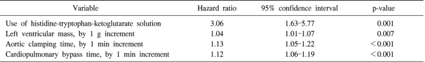
143 females) with severe AS undergoing isolated AVR were included. Postoperative myocardial injury (PMI) was defined as 1) maximum postoperative creatinine kinase isoenzyme MB or troponin-I levels ≥10 times of reference, 2) postoperative low cardiac output syndrome or episodes of ventricular arrhythmia, or 3) left ventricular ejection fraction of less than 55% and decrease in left ventricle (LV) ejection fraction of more than 20% of the baseline value. Results: There were 90 patients (28.7%) who developed PMI. There were five cases of early death (1.6%), all of whom had PMI. On multivariable analysis, the use of histidine-tryptophan-ketoglutarate (HTK) solution instead of blood cardioplegia (odds ratio [OR], 3.06; 95% confidence interval [CI], 1.63 to 5.77; p=0.001), greater LV mass (OR, 1.04; 95% CI, 1.01 to 1.07; p=0.007), and increased cardiac ischemic time (OR, 1.13; 95% CI, 1.05 to 1.22;
p<0.001) were independent predictors for PMI. Patients who had PMI showed significantly inferior long-term surviv- al than those without PMI (p=0.049). Conclusion: PMI occurred in a considerable proportion of patients undergoing AVR for severe AS and was associated with poor long-term survival. HTK cardioplegia, higher LV mass, and lon- ger cardiac ischemic duration were suggested as predictors of myocardial injury.
Key words: 1. Myocardial injury 2. Aortic valve 3. Surgery
INTRODUCTION
Aortic valve stenosis (AS) is the most common valvular disease in the western countries, and approximately 4% of people older than 65 years are reported to have severe AS [1]. For instance, approximately 50,000 cases of AVR for se- vere AS are being performed annually in the United States,
and these are predicted to increase by one and a half times
by the year 2030 [2]. Severe AS can predispose the patient to
symptoms of chest pain, syncope, and dyspnea, and the prog-
nosis is reported to be very disappointing when these symp-
toms accompany. The average survival rate of these patients
has been reported as only two years, and the five-year surviv-
al rate has been reported to be only 12% to 20% if the con-
dition is not treated surgically [3,4]. Surgical aortic valve re- placement (AVR) is a well-proven standard treatment of se- vere AS, and it has shown normalization of survival in se- lected populations [5].
Despite progresses in myocardial protection strategies, car- diopulmonary bypass (CPB), surgical techniques, and peri- operative management, the mortality and morbidity rates fol- lowing AVR for severe AS are still not negligible with the reported mortality rates being 2.6% to 4.0% [2,6]. The left ventricular (LV) wall is usually hypertrophied as the con- sequence of pressure overloading due to valvular obstruction, which develops gradually over several decades. Accordingly, delivery of a cardioplegic solution may be suboptimal in the hypertrophied LV wall, particularly in the endocardial area.
Therefore, myocardial protection of AVR for AS still remains challenging, and hence, significant postoperative myocardial injury (PMI) after AVR for severe AS is the leading cause of early death [7].
In order to improve myocardial protection during AVR for AS, a number of studies have been conducted including those seeking ideal routes of cardioplegic solution delivery or types of cardioplegic solution, and those determining factors related to PMI [8-15]. These studies, however, are limited by ex- perimental study designs and have shown mixed results.
Therefore, there are only few comprehensive clinical studies that assess the risk factors for PMI in the setting of isolated AVR for AS with a reasonably sized cohort. Therefore, we sought to evaluate the incidence of PMI in patients under- going isolated AVR for severe AS, and to determine the risk factors of PMI.
METHODS 1) Study population
Between August 1999 and July 2009, a total of 340 pa- tients underwent elective isolated AVR for severe AS at the Asan Medical Center, Seoul, Korea. Patients were excluded if they had significant (>grade 2) aortic regurgitation (n=16) or significant (>grade 2) mitral regurgitation (n=9) because these factors can affect the analyses as confounding variables.
Finally, 314 patients formed the subject population of this study. The requirement to obtain informed consent was
waived by the institutional review board due to the retro- spective nature of the study.
2) Surgical techniques
Surgery involved a median sternotomy (n=304) or a limited sternotomy including upper sternotomy (n=8) or transverse sternotomy (n=2), depending on the preference of the surgeon. The sternotomy approach involved conventional bi- caval (n=170) or single venous (n=134) combined with distal ascending aorta cannulation, whereas limited sternotomy (n=10) involved femoral and internal jugular venous cannu- lation. For myocardial protection, tepid blood cardioplegia (n=253) or histidine-tryptophan-ketoglutarate (HTK-Custodiol;
Koehler Chemi, Alsbach-Haenlien, Germany) solution (n=61) was administered after aortic clamping under CPB, depending on the preference of the surgeon. Routes of cardioplegic de- livery were ‘antegrade’ via aortic root cannula or directly through coronary ostia in 175 patients (n=175), ‘retrograde’
via coronary sinus in 16 patients (blood cardioplegia in 13 patients, HTK solution in 3 patients), or combined antegrade and retrograde in 123 patients (blood cardioplegia in 104 pa- tients, HTK solution in 19 patients).
3) Postoperative monitoring and measurement of cardiac marker proteins
All patients were transferred to the intensive care unit after the operation, and hemodynamic monitoring was carried out by the measurement of arterial pressure, central venous pres- sure, and pulmonary artery pressure, continuously. Cardiac enzyme levels including those of creatinine kinase isoenzyme MB (CK-MB) and troponin-I were measured immediately af- ter surgery and at postoperative 6, 12, and 24 hours. If any cardiac enzyme increased until 24 hours after the operation, additional measurements of the cardiac enzymes were carried out. The upper normal reference value of CK-MB and tropo- nin I were defined as ‘less than 5 ng/mL’ and ‘less than 1.5 ng/mL,’ respectively.
4) Definition of perioperative significant myocardial injury and follow-up
Significant perioperative myocardial injury was defined if
any of the following criteria were satisfied [11,12,15,16]: (1)
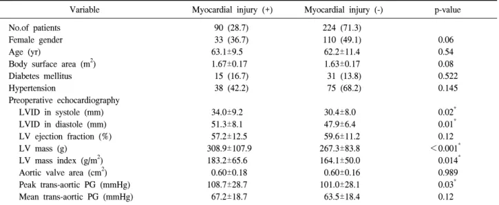
Table 1. Baseline characteristics of patients
Variable Myocardial injury (+) Myocardial injury (-) p-value
No.of patients Female gender Age (yr)
Body surface area (m
2) Diabetes mellitus Hypertension
Preoperative echocardiography LVID in systole (mm) LVID in diastole (mm) LV ejection fraction (%) LV mass (g)
LV mass index (g/m
2) Aortic valve area (cm
2) Peak trans-aortic PG (mmHg) Mean trans-aortic PG (mmHg)
90 (28.7) 33 (36.7) 63.1±9.5 1.67±0.17
15 (16.7) 38 (42.2)
34.0±9.2 51.3±8.1 57.2±12.5 308.9±107.9 183.2±65.6 0.60±0.18 108.7±28.7 67.2±18.7
224 (71.3) 110 (49.1) 62.2±11.4 1.63±0.17 31 (13.8) 75 (68.2)
30.4±8.0 47.9±6.4 59.6±11.2 267.3±83.8 164.1±50.0 0.60±0.16 101.0±28.1 63.5±18.4
0.06 0.54 0.08 0.522 0.145
0.02
*0.01
*0.12
<0.001
*0.014
*0.989 0.03
*0.12 Values are presented as number (%) or mean±standard deviation.
LVID, left ventricular internal dimension; LV, left ventricle; PG, pressure gradient.
*
p-value<0.05.
peak venous blood CK-MB or troponin-I levels of above 10 times the reference values; (2) postoperative low cardiac out- put syndrome (LCOS), which is defined as the documented low cardiac index (<2.0) despite adequate volume including the needs for mechanical circulatory support including in- tra-aortic balloon pump, extracorporeal membrane oxygenator (ECMO), and ventricular assisting device; (3) any episodes of ventricular tachycardia or fibrillation during the postoperative hospitalization period; and (4) LV dysfunction, which is de- fined as an LV ejection fraction (LVEF) of less than 55%
and a decrease in LVEF of more than 20% of the baseline value on early postoperative echocardiography. Clinical fol- low-up was performed via outpatient clinic visits. For the pa- tients who underwent follow-up at outside hospitals, clinical information was obtained by telephone contact. All deaths were regarded as cardiac origin unless a non-cardiac origin was diagnosed clinically or was determined at autopsy.
5) Statistical analysis
Categorical variables are expressed as frequencies and percentages. Chi-square test or Fisher’s exact test were used to compare. Continuous variables are presented as a mean with standard deviation or a median with ranges. Student’s unpaired t-test was used to compare. Kaplan- Meier curves
were used to delineate the overall survival. Univariable and multivariable risk factor analyses were performed by using the multiple logistic regression method. Variables with a p-value of 0.20 or less in the univariable analyses were can- didates for the multivariable models. Multivariable analyses involved a backward elimination technique, and variables with a p-value of less than 0.10 remained in the final model.
All reported p-values were two-sided, and a value of p<0.05 was considered statistically significant. For the statistical anal- ysis, PASW SPSS ver. 18.0 (SPSS Inc., Chicago, IL, USA) was used.
RESULTS 1) Baseline patient profiles
The baseline characteristics of the patients are listed in Tables 1, 2. Patients who were affected by PMI had larger LV dimension, greater LV mass/LV mass index, and greater peak trans-aortic pressure gradient than those without PMI.
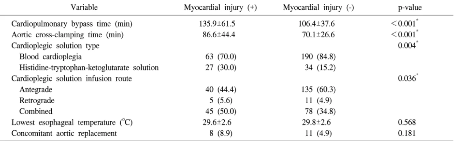
The HTK crystalloid cardioplegia was more frequently used
in patients with PMI, and the routes for cardioplegic delivery
were significantly different between the two groups of pa-
tients (Table 2).
Table 2. Baseline characteristics of the patients (operative factor)
Variable Myocardial injury (+) Myocardial injury (-) p-value
Cardiopulmonary bypass time (min) Aortic cross-clamping time (min) Cardioplegic solution type Blood cardioplegia
Histidine-tryptophan-ketoglutarate solution Cardioplegic solution infusion route Antegrade
Retrograde Combined
Lowest esophageal temperature (
oC) Concomitant aortic replacement
135.9±61.5 86.6±44.4
63 (70.0) 27 (30.0)
40 (44.4) 5 (5.6) 45 (50.0) 29.6±2.6
8 (8.9)
106.4±37.6 70.1±26.6
190 (84.8) 34 (15.2)
135 (60.3) 11 (4.9) 78 (34.8) 29.8±2.6
11 (4.9)
<0.001
*<0.001
*0.004
*0.036
*0.568 0.181 Values are presented as mean±standard deviation or number (%).
*