INTRODUCTION
The prevalence of drug hypersensitivity reactions is increas- ing. Detection of the culprit drug is a prerequisite for effective prevention by substituting safer alternatives. Aspirin and non- steroidal anti-inflammatory drugs (ASA/NSAIDs) are among the most common causes of hypersensitivity reactions, account- ing for 0.6%-2.5% of hypersensitivity reactions in the general population and 10%-20% in asthmatic patients.1,2 A previous study at a tertiary care hospital in Korea reported that ASA/
NSAIDs ranked first among the drugs provoking hypersensitiv- ity reactions.3 Nevertheless, a definitive diagnosis of ASA/NSAID- induced hypersensitivity is difficult because these are usually administered in combination with other drugs, making it nec- essary to test all drugs taken. Although hypersensitivity reactions to some drugs can be detected by topical provocation tests, re- actions to ASA/NSAIDs, except for aspirin-induced asthma, can be detected only by an oral provocation test. However, oral provocation tests are contraindicated in subjects who have ex- perienced severe reactions such as anaphylactic shock,4 and many subjects who have experienced drug hypersensitivity re- actions are not willing to risk an oral provocation test.
Flow Cytometry-Assisted Basophil Activation Test as a Safe Diagnostic Tool for Aspirin/NSAID Hypersenstivity
Myung Shin Kim,
1Young Joo Cho
2*
1Department of Internal Medicine, Soon Chun Hyang University Gumi Hospital, Gumi, Korea
2Department of Internal Medicine, Ewha Womans University School of Medicine, Seoul, Korea
Numerous methods have been proposed to diagnose ASA/
NSAIDs hypersensitivity. In vivo methods include the skin prick test (SPT),5 the autologous serum skin test,6,7 and measurement of urine leukotriene E4 before and after ingestion of the sus- pected drug.8 In vitro, analysis of genetic polymorphisms relat- ed to metabolism of arachidonic acid,9,10 measurement of 15- HETE in the reaction between ASA/NSAIDs and leukocytes,11 and the cellular allergen stimulation test (CAST) using baso- phils12,13 have been investigated.
Here, we used a flow cytometry-assisted basophil activation test (FAST) to analyze CD63 exposure upon basophil degranu- lation. CD63 is anchored in the basophilic granular membrane and can be detected on the outside of the cell upon the fusion of granules with plasma membranes.14 FAST has high sensitivi- Allergy Asthma Immunol Res. 2012 May;4(3):137-142.
http://dx.doi.org/10.4168/aair.2012.4.3.137 pISSN 2092-7355 • eISSN 2092-7363
Purpose: Aspirin and non-steroidal anti-inflammatory drugs (ASA/NSAIDs) are common causes of drug hypersensitivity. An oral provocation test is the only definitive diagnostic test. This study assessed the reliability of a flow cytometry-assisted basophil activation test (FAST) as a safe diagnostic method for ASA/NSAID-induced hypersensitivity, as its high sensitivity and specificity have been demonstrated for many other drugs. Methods: Eigh- teen patients and 11 controls were enrolled. Using a Flow-CAST kit® (Bühlmann Laboratories AG, Schönenbuch, Switzerland), 29 analyses with as- pirin, ibuprofen, and diclofenac were performed by flow cytometry to detect double-positive staining of anti-IgE and anti-CD63. The stimulation in- dex was defined as the activated basophil percentage after drug stimulation/basally active basophil percentage. A stimulation index≥2 and an ab- solute activated basophil percentage≥5 were considered positive. Results: Patients with hypersensitivity to ASA/NSAIDs were predominantly female, and the prevalence of atopy was higher in patients than in controls. A sensitivity of 61%, specificity of 91%, positive predictive value of 92%, and negative predictive value of 59% were achieved. Conclusions: FAST is a useful additional method for diagnosis of hypersensitivity re- actions to ASA/NSAIDs. Further development is required to increase the sensitivity of the test.
Key Words: Flow cytometry; basophil; aspirin; non-steroidal anti-inflammatory drugs; drug hypersensitivity
This is an Open Access article distributed under the terms of the Creative Commons Attribution Non-Commercial License (http://creativecommons.org/licenses/by-nc/3.0/) which permits unrestricted non-commercial use, distribution, and reproduction in any medium, provided the original work is properly cited.
Correspondence to: Young Joo Cho, MD, PhD, Department of Internal Medicine, Ewha Womans University Medical Center, 911-1 Mok-dong, Yangcheon-gu, Seoul 158-710, Korea.
Tel: +82-2-2650-5114; Fax: +82-2-2655-2076; E-mail: yjcho@ewha.ac.kr Received: December 25, 2010; Revised: September 28, 2011;
Accepted: October 12, 2011
•There are no financial or other issues that might lead to conflict of interest.
ty and specificity for the diagnosis of hypersensitivity reactions to inhalant allergens, bee venom, and latex.15 Some studies on hypersensitivity to ASA/NSAIDs have found high specificity of FAST,18-20 but reported sensitivities vary.
The present study was conducted to corroborate the applica- bility of FAST for detecting hypersensitivity to ASA/NSAIDs and to evaluate its efficacy in Koreans by determining its sensitivity, specificity, and positive/negative predictive values.
MATERIALS AND METHODS Study subjects
This study included 18 patients who showed hypersensitivity reactions to ASA/NSAIDs at our allergy clinic between August 2007 and August 2008. Skin reactions were provoked in 13 sub- jects; respiratory reactions, in two; and anaphylactic shock, in three. The control group comprised 11 subjects who were matched for age and gender (ratio, 2:1) with the study group.
The control subjects were patients admitted with other diseases (n=5) or were healthy subjects (n=3). Three subjects who showed hypersensitivity reactions to drugs other than ASA/
NSAIDs were also included as controls. We confirmed that the control subjects had experienced no hypersensitivity reactions to ASA/NSAIDs. All subjects were informed orally about the study and provided informed consent. The study was approved by the local institutional review board.
Clinical features, including the types of drugs producing hy- persensitivity reactions, time interval from drug ingestion to symptom onset, and types of hypersensitivity reactions, as well as past histories of bronchial asthma, allergic rhinitis, food al- lergy, and chronic urticaria, were collected for each subject. Se- rum total IgE was measured, and specific IgE was determined using the multiple allergen simultaneous test (MAST). SPTs were performed for common inhalant and food allergens, and atopy was defined as a positive SPT result.
Sample analysis
Blood samples were drawn from the subjects at least 2 weeks after hypersensitivity reactions and also at least 1 week after discontinuation of anti-allergic medications such as antihista- mines, antileukotrienes, and steroids. The cells were stimulated within 2 hours after blood sampling, or the samples were im- mediately refrigerated for up to 24 hours. Experiments were performed using a commercially available Flow-CAST kit® (Bühlmann Laboratories AG, Schönenbuch, Switzerland) fol- lowing the manufacturer’s instructions, as has been fully de- scribed and discussed elsewhere.14-23
For analysis, blood was collected in an EDTA tube and centri- fuged for 5 minutes at 1,100 rpm. The leukocyte-containing plasma fraction was then centrifuged for 10 minutes at 1,700 rpm to obtain a leukocyte pellet, which was carefully resuspend- ed with 100 μL of stimulation buffer per 1 mL of original blood
sample. Each patient’s cell suspension was divided among five test tubes (50 μL per tube). Stimulation buffer was added to one tube as a negative control; anti-IgE receptor antibody was add- ed to another tube as a positive control. To the remaining three tubes, 50 μL of aspirin (1.25 mg/mL), ibuprofen (50 μg/mL), or diclofenac (12.5 μg/mL) were added. The tubes were vortexed gently and incubated at 37°C for 40 minutes. The reaction was halted by adding 50 μL of pre-cooled blocking buffer to each tube. Leukocytes were labeled using FITC-conjugated anti-IgE and PE-conjugated anti-CD 63.
The samples were analyzed using CellQuest® containing 488- nm argon within 2 hours. The cutoff points for anti-IgE-FITC and anti-CD63-PE were determined for each subject based on the point where the initial fluorescence peak was observed in the positive control (Fig. 1). The percentage of basophils posi- tive for both IgE and CD 63 was obtained. The stimulation in- dex (SI) was calculated using the following formula:
SI=percentage of basophils activated by the drug/percentage of activated basophils in the negative control
The receiver operating characteristics (ROC) curve was used to determine a cutoff point for the SI. A positive result was de- fined as a SI≥2 (Fig. 2), which was similar to previous stud- ies.18-21,23 To account for nonspecific activation, only activated basophil percentages≥5% were considered positive.
Statistical analysis
Statistical analysis was performed using SPSS ver. 16.0 (SPSS Inc., Chicago, IL, USA). As the number of subjects was small, continuous variables were analyzed using the Mann-Whitey U test, and categorical variables were analyzed using Fisher’s ex- act test. A P value<0.05 was considered to indicate significance.
The FAST results are expressed as the percentage of basophils positive for both IgE and CD63.
RESULTS
Demographic and clinical characteristics of the study and control groups
Table 1 presents the demographic and clinical characteristics of the study and control groups. The ratio of males to females in the study group was 1:3.5, and the prevalence of atopy was sig- nificantly higher in the study group than in control group (61%
vs. 36%; P<0.05). Eight of the 18 study subjects (44%) had pre- viously experienced hypersensitivity reactions to ASA/NSAIDs.
Of these eight, one had also experienced hypersensitivity to a drug other than ASA/NSAIDs (an iron preparation). In the con- trol group, two subjects had a history of hypersensitivity to pen- icillin; and one, to cimetidine. Culprit drugs for the current re- actions in the study group were aspirin (n=6), propionic acid derivatives (ibuprofen, naproxen; n=8), acetic acid derivatives
(diclofenac, aceclofenac; n=2), piroxicam (n=1), and Joins® (Clematis mandshurica, Trichosanthes kirilowii, Prunella vul- garis; n=1).
Flow cytometry-assisted basophil activation test
The FAST results are shown in Tables 2 and 3. There was no significant difference in the mean FAST result for the negative test control (3.26%±1.68% vs. 2.25%±1.93%) or positive test
Table 1. Demographic features and clinical characteristics
Characteristic Study group Control group
Age, yr (mean±SD) 33±14 34±13
Gender, M:F 4:14 (22:78) 4:7 (36:64)
Atopy status* 11 (61) 4 (36)
History of drug allergy
None* 9 (50) 8 (73)
ASA/NSAIDs* 8 (44) 0
Other drugs 2 (11) 3 (27)
Culprit drugs
Salicylates (Aspirin, isopropylantipyrine) 6 (33) None Propionic acids (Ibuprofen, naproxen) 8 (44)
Acetic acids (Diclofenac) 2 (11)
Other NSAIDs† 2 (11)
Type of hypersensitivity
Respiratory 2 (11) None
Cutaneous 13 (72)
Anaphylaxis 3 (17)
Values are given as number (%), unless otherwise indicated.
NSAIDs, non-steroidal anti-inflammatory drugs.
*P<0.05.
†Other NSAIDs include piroxicam and Joins®.
100 101 102 103 104
15
10
5
0
Counts
anti-CD63-PE
anti-IgE-FITC
anti-CD63-PE
100 101 102 103 104 104
103 102 101 100
25.69%
100 101 102 103 104 104
103 102 101 100
2.11%
100 101 102 103 104 104
103 102 101 100
4.57%
A
B C D
anti-IgE-FITC anti-IgE-FITC
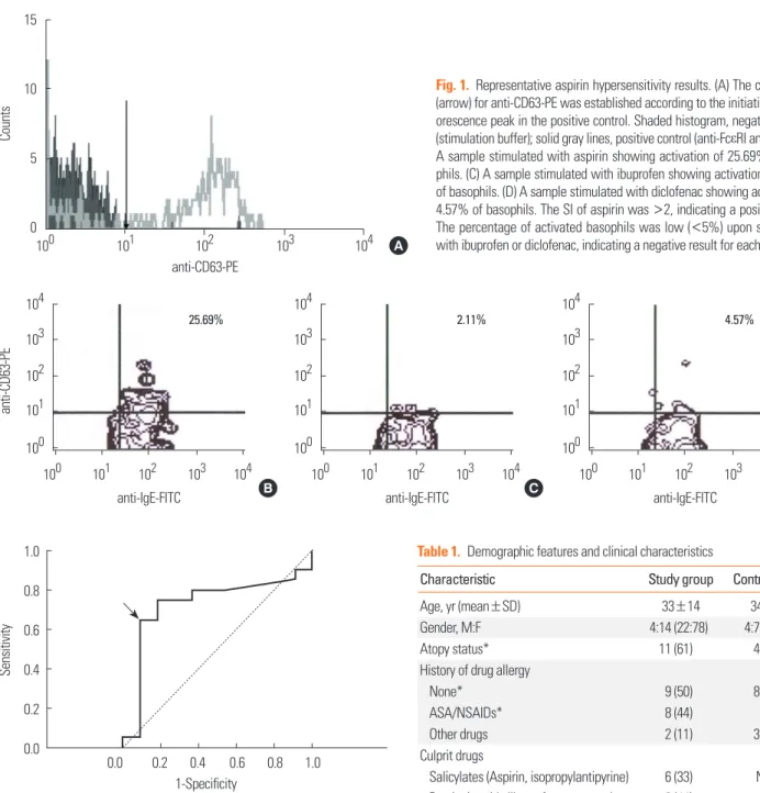
Fig. 1. Representative aspirin hypersensitivity results. (A) The cutoff point (arrow) for anti-CD63-PE was established according to the initiation of a flu- orescence peak in the positive control. Shaded histogram, negative control (stimulation buffer); solid gray lines, positive control (anti-FcεRI antibody). (B) A sample stimulated with aspirin showing activation of 25.69% of baso- phils. (C) A sample stimulated with ibuprofen showing activation of 2.11%
of basophils. (D) A sample stimulated with diclofenac showing activation of 4.57% of basophils. The SI of aspirin was >2, indicating a positive result.
The percentage of activated basophils was low (<5%) upon stimulation with ibuprofen or diclofenac, indicating a negative result for each.
control (73.73%±19.78% vs. 63.91%±26.03%) between the study and control groups. The FAST results showed a specificity 0.0 0.2 0.4 0.6 0.8 1.0
1.0 0.8 0.6 0.4 0.2 0.0
Sensitivity
1-Specificity
Fig. 2. The receiver operating characteristics (ROC) curve. According to the ROC curve, the optimal cutoff point for the stimulation index was 1.9 (arrow).
The observed area under the curve was 0.72 (95% Confidence Interval, 0.53- 0.93; P=0.039), which is considered fair.
of 91%, positive predictability of 92%, sensitivity of 61%, and negative predictability of 59%.
For patients with hypersensitivity reactions to salicylate, the FAST results for aspirin were positive in 50%. All patients who
showed hypersensitivity to propionic derivatives and acetic acid derivatives showed a negative response to ibuprofen and diclofenac, respectively.
Within the study group, atopy status did not affect the FAST results. The FAST results did not differ significantly between study subjects with cutaneous symptoms and those with respi- ratory symptoms, or between those with mild symptoms and those with severe symptoms (Table 4).
DISCUSSION
The aim of the present study was to evaluate the efficacy of FAST for the diagnosis of hypersensitivity to ASA/NSAIDs. There are many reports in the European literature regarding the use of Table 2. FAST results in study subjects with aspirin/ibuprofen/diclofenac hypersensitivity
Study
subject Reaction Gender/Age Culprit drug
% Activated basophils in FAST (SI)
Control value Test drug value
(-) (+) ASA IBU DIC
1 Asthma F/37 ASA 4.75 18.79 7.72 (1.63)
2 Urticaria F/43 ASA 4.68 74.38 4.19 (0.90)
3 Angioedema F/26 IBU 0.69 47.12 1.78 (2.58) 1.95 (2.83) 2.58 (3.74)
4 Angioedema F/22 NPX 1.13 46.65 5.28 (4.67)* 0.46 (0.41) 3.41 (3.02)
5 Anaphylactic shock F/24 ASA 4.49 41.27 1.72 (0.38) 1.05 (0.23) 0.73 (0.16)
6 Angioedema M/15 IBU 1.43 77.65 10.00 (6.99)* 2.94 (2.06) 0 (0)
7 Angioedema M/16 IBU 4.71 75.40 11.33 (2.41)* 4.92 (1.04) 9.29 (1.97)
8 Angioedema M/31 PRX 2.33 77.73 11.09 (4.76)* 0.29 (0.12)
9 Angioedema F/30 IBU 4.08 87.15 15.09 (3.70)*
10 Angioedema F/36 NPX 2.33 88.24 0.39 (0.17)
11 Angioedema F/18 NPX 0.83 82.76 25.69 (30.95)* 2.11 (2.54) 4.57 (5.51)
12 Angioedema F/26 IPA 2.44 84.30 7.49 (3.07)* 1.67 (0.68) 2.28 (0.93)
13 Asthma F/66 ASA 6.49 89.02 13.30 (2.05)* 4.56 (0.70) 18.77 (2.89)*
14 Anaphylactic shock F/46 Joins® 1.68 85.43 2.95 (1.76) 1.86 (1.11) 1.56 (0.93)
15 Anaphylactic shock F/43 DIC 2.41 92.25 8.11 (3.37)* 1.24 (0.51) 1.05 (0.44)
16 Angioedema M/49 IBU 4.59 90.45 8.35 (1.82) 1.58 (0.34) 5.43 (1.18)
17 Angioedema F/36 ASA 4.85 84.35 17.84 (3.68)* 4.13 (0.85) 2.85 (0.59)
18 Angioedema F/31 DIC 4.81 84.25 13.04 (2.71)*
SI, stimulation index; ASA, aspirin; IBU, ibuprofen; DIC, diclofenac; IPA, isopropylantipyrine; NPX, naproxen; PRX, piroxicam.
Positive results are indicated by* (% activated basophils≥5% and SI≥2).
Table 3. FAST results in the control group Control
subject Gender/
Age
% Activated basophils in FAST (SI) Control value Test drug value
(-) (+) ASA IBU DIC
1 F/29 5.07 87.50 6.41 (1.26)
2 M/16 4.37 25.94 2.79 (0.64) 1.47 (0.34) 1.59 (0.36)
3 M/22 0.32 72.37 1.57 (4.91)
4 M/43 2.19 66.92 3.71 (1.69) 1.84 (0.84) 3.06 (1.40) 5 F/18 4.61 49.62 8.45 (1.83) 3.49 (0.76) 3.82 (0.83)
6 F/57 4.17 91.43 5.56 (1.33)
7 F/29 1.32 64.85 4.53 (3.43)
8 F/32 1.02 93.47 10.92 (10.71)*
9 F/34 0.54 62.04 2.78 (5.15)
10 F/43 1.11 76.87 1.92 (1.73)
11 F/48 0.10 12.05 3.64 (36.40)
FAST, flow cytometry-assisted basophil activation test; SI, stimulation index;
ASA, aspirin; IBU, ibuprofen; DIC, diclofenac.
Positive results are indicated by* (% activated basophils≥5% and SI≥2).
Table 4. Comparison of FAST results according to the type of aspirin/ibupro- fen/diclofenac hypersensitivity
Type of NSAID hypersensitivity Positive results in FAST
Respiratory 1/2 (50)
Cutaneous 9/13 (69)
Anaphylactic shock 1/3 (33)
Values are given as No. (%).
FAST, flow cytometry-assisted basophil activation test; NSAID, non-steroidal anti-inflammatory drug.
FAST to test reactivity to ASA/NSAIDs,18-21 diclofenac,22 or met- amizol.23 These studies applied a SI≥2 as a cutoff on the basis of high specificity under the ROC curve, because the prevalence of hypersensitivity reactions to ASA/NSAIDs was relatively low.
Although the specificity and positive predictive value were re- ported to be 100% in all of these previous studies, the sensitivity varied between 40% and 70%.18-23
In the present study using a SI cutoff of 2, FAST showed a specificity of 91%, positive predictive value of 92%, sensitivity of 61%, and negative predictive value of 59%. The SI cutoff point, sensitivity, and specificity were similar to those in previous studies.18-23 Neither atopy status nor the type and severity of hy- persensitivity reactions affected the FAST results, which was also similar to previous results.18,21
Two earlier studies demonstrated that 27%-66% of subjects with hypersensitivity to one ASA/NSAID showed a positive FAST result with other ASA/NSAIDs. However, similar results were not obtained in the present study, probably owing to the small sample size.
In the present study, the specificity was relatively low, and all subjects showed positive FAST results only for aspirin, regard- less of the culprit drug. The control subject who showed a posi- tive FAST result had a negative oral aspirin provocation test re- sult. The discrepancy between the results of previous European studies and ours may be explained by differences in race and/
or drug concentration.
We employed commercially available concentrations of drugs based on the dose-reaction relationship. The concentrations of aspirin used in this study were similar to those used in previous studies, and the sensitivity and specificity were also similar. Pre- vious studies tested with 1-100 μg/mL of ibuprofen and 1-310 μg/mL of diclofenac, and one study reported a sensitivity of 29% for ibuprofen and 50% for diclofenac at concentrations lower than those used here.21 The maximum blood concentra- tions in vivo after ingestion of the medications were 15-55 and 0.5-2.5 μg/mL for ibuprofen and diclofenac, respectively.25 These values are lower than those obtained in our study. One report concluded that the use of higher drug concentrations appeared to increase the sensitivity of FAST because the high drug doses induced nonspecific basophil activation.26 We infer that in the present study, the drug concentrations did not affect the FAST results.
The low sensitivity of 65% for ASA/NSAID-induced hypersen- sitivity may be a drawback of FAST. Two studies from the U.S.
and Turkey showed negative FAST results.26,27 The U.S. study re- ported sensitivities of 30% for aspirin at 0.3-1.25 mg/mL and 80% at 5 mg/mL; however, high aspirin concentrations also stimulated basophils in the control group, indicating poor spec- ificity.26 The Turkish study found very low sensitivity (16.7%- 33.3%).27 The low sensitivities may be attributable to the fact that mast cells and eosinophils are also involved in hypersensi- tivity reactions to ASA/NSAIDs. There is no direct evidence for
the involvement of basophils in ASA/NSAIDs hypersensitivity, although many studies have demonstrated increased levels of leukotriene C4 (LTC4) in serum and bronchial and nasal secre- tions, as well as LTE4 and 15-HETE in urine, after aspirin chal- lenge in hypersensitive patients. LTC4, which is a mainstay of ASA/NSAID-induced hypersensitivity, is produced by LTC4 synthase, which is distributed mainly in mast cells and baso- phils. These cells contain many glycogen granules filled with various chemical mediators, including histamine, and are de- granulated by direct or indirect stimulation, with subsequent induction of immediate-type hypersensitivity reactions.24
There are several confounding factors derived from the het- erogeneous study and control subjects in the present study.
Further investigations with a larger sample size, particularly in- cluding subjects with hypersensitivity reactions of the respira- tory system, and studies aiming to establish the relationship be- tween FAST results and ethnic groups are needed to confirm our results.
The sensitivity of FAST must be improved before it can be ap- plied for the clinical evaluation of drug sensitivity reactions. For example, additional studies have been explored the use of a combination of anaphylatoxin12 and simultaneous measure- ment of CD63 and CD20326-29 or CRTH2/DP2.30 With improved sensitivity, FAST may provide a safe, convenient, and rapid method for the diagnosis of hypersensitivity to ASA/NSAIDs.
ACKNOWLEDGMENTS
We thank Yoo Jin Lee for technical support.
REFERENCES
1. Szczeklik A, Stevenson DD. Aspirin-induced asthma: advances in pathogenesis, diagnosis, and management. J Allergy Clin Immunol 2003;111:913-21.
2. Kasper L, Sladek K, Duplaga M, Bochenek G, Liebhart J, Gladysz U, Malolepszy J, Szczeklik A. Prevalence of asthma with aspirin hyper- sensitivity in the adult population of Poland. Allergy 2003;58:1064-6.
3. Choi JH, Shin YS, Suh CH, Nahm DH, Park HS. The frequency of adverse drug reactions in a tertiary care hospital in Korea. Korean J Med 2004;67:290-6.
4. Nizankowska-Mogilnicka E, Bochenek G, Mastalerz L, Swierczyn@
ska M, Picado C, Scadding G, Kowalski ML, Setkowicz M, Ring J, Brockow K, Bachert C, Wöhrl S, Dahlén B, Szczeklik A. EAACI/GA- 2LEN guideline: aspirin provocation tests for diagnosis of aspirin hypersensitivity. Allergy 2007;62:1111-8.
5. Palma-Carlos AG, Medina M, Palma-Carlos ML. Skin tests in NSAIDS hypersensitivity. Eur Ann Allergy Clin Immunol 2006;38:
182-5.
6. Asero R. Predictive value of autologous plasma skin test for multi- ple nonsteroidal anti-inflammatory drug intolerance. Int Arch Al- lergy Immunol 2007;144:226-30.
7. Asero R, Tedeschi A, Lorini M. Autoreactivity is highly prevalent in patients with multiple intolerances to NSAIDs. Ann Allergy Asthma Immunol 2002;88:468-72.
8. Mastalerz L, Setkowicz M, Sanak M, Szczeklik A. Hypersensitivity to aspirin: common eicosanoid alterations in urticaria and asthma.
J Allergy Clin Immunol 2004;113:771-5.
9. Kim SH, Jeong HH, Cho BY, Kim M, Lee HY, Lee J, Wee K, Park HS.
Association of four-locus gene interaction with aspirin-intolerant asthma in Korean asthmatics. J Clin Immunol 2008;28:336-42.
10. Choi JH, Lee KW, Oh HB, Lee KJ, Suh YJ, Park CS, Park HS. HLA as- sociation in aspirin-intolerant asthma: DPB1*0301 as a strong marker in a Korean population. J Allergy Clin Immunol 2004;113:
562-4.
11. Kowalski ML, Ptasinska A, Jedrzejczak M, Bienkiewicz B, Cieslak M, Grzegorczyk J, Pawliczak R, Dubuske L. Aspirin-triggered 15-HETE generation in peripheral blood leukocytes is a specific and sensi- tive Aspirin-Sensitive Patients Identification Test (ASPITest). Aller- gy 2005;60:1139-45.
12. May A, Weber A, Gall H, Kaufmann R, Zollner TM. Means of in- creasing sensitivity of an in vitro diagnostic test for aspirin intoler- ance. Clin Exp Allergy 1999;29:1402-11.
13. Pierzchalska M, Mastalerz L, Sanak M, Zazula M, Szczeklik A. A moderate and unspecific release of cysteinyl leukotrienes by aspi- rin from peripheral blood leucocytes precludes its value for aspirin sensitivity testing in asthma. Clin Exp Allergy 2000;30:1785-91.
14. Boumiza R, Debard AL, Monneret G. The basophil activation test by flow cytometry: recent developments in clinical studies, stan- dardization and emerging perspectives. Clin Mol Allergy 2005;3:9.
15. Ebo DG, Bridts CH, Hagendorens MM, Aerts NE, De Clerck LS, Ste- vens WJ. Basophil activation test by flow cytometry: present and future applications in allergology. Cytometry B Clin Cytom 2008;74:
201-10.
16. Torres MJ, Padial A, Mayorga C, Fernández T, Sanchez-Sabate E, Cornejo-García JA, Antúnez C, Blanca M. The diagnostic interpre- tation of basophil activation test in immediate allergic reactions to betalactams. Clin Exp Allergy 2004;34:1768-75.
17. Monneret G, Benoit Y, Debard AL, Gutowski MC, Topenot I, Bien- venu J. Monitoring of basophil activation using CD63 and CCR3 in allergy to muscle relaxant drugs. Clin Immunol 2002;102:192-9.
18. Gamboa P, Sanz ML, Caballero MR, Urrutia I, Antépara I, Esparza R, de Weck AL. The flow-cytometric determination of basophil acti- vation induced by aspirin and other non-steroidal anti-inflamma- tory drugs (NSAIDs) is useful for in vitro diagnosis of the NSAID hypersensitivity syndrome. Clin Exp Allergy 2004;34:1448-57.
19. Sanz ML, Gamboa P, de Weck AL. A new combined test with flow- cytometric basophil activation and determination of sulfidoleukot-
rienes is useful for in vitro diagnosis of hypersensitivity to aspirin and other nonsteroidal anti-inflammatory drugs. Int Arch Allergy Immunol 2005;136:58-72.
20. Sainte-Laudy J, Touraine F, Boumediene A, Bonnaud F, Cogné M.
Clinico-biological characteristics of flow cytometry applied to hy- persensitivity to NSAIDs. Inflamm Res 2007;56 Suppl 1:S63-4.
21. Rodríguez-Trabado A, Cámara-Hijón C, Ramos-Cantariño A, Por- cel-Carreño SL, Jiménez-Timón S, Pereira-Navarro G, Hernández- Arbeiza FJ, Fernández-Pereira L. Basophil activation test for the in vitro diagnosis of nonsteroidal anti-inflammatory drug hypersen- sitivity. Allergy Asthma Proc 2008;29:241-9.
22. Malbrán A, Yeyati E, Rey GL, Galassi N. Diclofenac induces baso- phil degranulation without increasing CD63 expression in sensi- tive patients. Clin Exp Immunol 2007;147:99-105.
23. Gamboa PM, Sanz ML, Caballero MR, Antépara I, Urrutia I, Jáu- regui I, González G, Diéguez I, De Weck AL. Use of CD63 expres- sion as a marker of in vitro basophil activation and leukotriene de- termination in metamizol allergic patients. Allergy 2003;58:312-7.
24. Adkinson NF, Yunginger JW, Busse WW, Bochner BS, Holgate ST, Simons FER. Middleton’s allergy: principles & practice. 6th ed.
Philadelphia: Mosby; 2003. 1695-710.
25. McEvoy GK. AHFS drug information. Wisconsin: American Society of Health-System Pharmacists; 2006. 2010, 2021, 2038.
26. Celik GE, Schroeder JT, Hamilton RG, Saini SS, Adkinson NF. Effect of in vitro aspirin stimulation on basophils in patients with aspirin- exacerbated respiratory disease. Clin Exp Allergy 2009;39:1522-31.
27. Bavbek S, Ikinciog(ullari A, Dursun AB, Gulog(lu D, Arikan M, Elhan AH, Misirligil Z. Upregulation of CD63 or CD203c alone or in com- bination is not sensitive in the diagnosis of nonsteroidal anti-in- flammatory drug intolerance. Int Arch Allergy Immunol 2009;150:
261-70.
28. Boumiza R, Monneret G, Forissier MF, Savoye J, Gutowski MC, Powell WS, Bienvenu J. Marked improvement of the basophil acti- vation test by detecting CD203c instead of CD63. Clin Exp Allergy 2003;33:259-65.
29. Sainte-Laudy J, Belon P. Improvement of flow cytometric analysis of basophil activation inhibition by high histamine dilutions. A novel basophil specific marker: CD 203c. Homeopathy 2006;95:3-8.
30. Ebo DG, Sainte-Laudy J, Bridts CH, Mertens CH, Hagendorens MM, Schuerwegh AJ, De Clerck LS, Stevens WJ. Flow-assisted aller- gy diagnosis: current applications and future perspectives. Allergy 2006;61:1028-39.