Copyright © 2019. Anatomy & Cell Biology
Introduction
Cyclosporins are a group of closely related cyclic under- capeptides produced as secondary metabolites in strains of fungi including Cylindrocarpon lucidum and Trichoderma polysporum isolated from soil samples [1]. Among various cy-
closporins, cyclosporin A (CsA) is one of the most commonly used immunosuppressive drugs in the treatment of patients with organ transplantation and autoimmune diseases includ- ing acquired immune deficiency syndrome owing to its supe- rior T-cell specificity and low myelotoxicity [2]. After entering into recipient cells, CsA can bind to cyclophilins known to peptidylpropyl isomerase activity through catalyzing isomeri- zation of peptide bonds from trans form to cis form at proline residues in protein folding pathway [3]. Such binding of CsA to cyclophilins can block their peptidylpropyl isomerase ac- tivity. Thus, CsA has shown immunosuppressive effects in adipocytes [4], myocytes [5], and lymphocytes [6]. Although CsA is an extremely valuable immunosuppressive agent for
Corresponding author:
Jinu Kim
Department of Anatomy, Jeju National University School of Medicine, 102 Jejudaehak-ro, Jeju 63243, Korea
Tel: +82-64-754-8181, Fax: +82-64-702-2687, E-mail: jinu.kim@jejunu.ac.kr
Cyclosporin A aggravates hydrogen peroxide–
induced cell death in kidney proximal tubule epithelial cells
Daeun Moon
1, Jinu Kim
1,21Interdisciplinary Graduate Program in Advanced Convergence Technology & Science, Jeju National University, Jeju, 2Department of Anatomy, Jeju National University School of Medicine, Jeju, Korea
Abstract: Cyclosporin A (CsA) does not only exert a toxic effect on kidney parenchymal cells, but also protects them against necrotic cell death by inhibiting opening of mitochondrial permeability transition pore. However, whether CsA plays a role in hydrogen peroxide–induced kidney proximal tubular cell death is currently unclear. In the present study, treatment with CsA further increased apoptosis and necrosis in HK-2 human kidney proximal tubule epithelial cells during exposure to hydrogen peroxide. In addition, hydrogen peroxide–induced p53 activation and BH3 interacting-domain death agonist (BID) expression were higher in CsA-treated cells than those in non-treated cells, whereas hydrogen peroxide–induced activation of mitogen- activated protein kinases including p38, c-Jun N-terminal kinase, and extracellular signal-regulated kinase and activation of protein kinase B were not significantly altered by treatment with CsA. In oxidant-antioxidant system, reactive oxygen species (ROS) production induced by hydrogen peroxide was further enhanced by treatment with CsA. However, expression levels of antioxidant enzymes including manganese superoxide dismutase, copper/zinc superoxide dismutase, and catalase were not altered by treatment with hydrogen peroxide or CsA. Treatment with CsA further enhanced mitochondrial membrane potential induced by exposure to hydrogen peroxide, although it did not alter endoplasmic reticulum stress based on expression of glucose-regulated protein 78 and 94. Taken together, these data suggest that CsA can aggravate hydrogen peroxide-induced cell death through p53 activation, BID expression, and ROS production.
Key words: Cyclosporin, Cell death, Hydrogen peroxide, p53, Reactive oxygen species Received November 6, 2018; Revised March 7, 2019; Accepted March 12, 2019
organ transplant recipients, unfortunately CsA has a number of serious side effects in various tissues, including kidney damage which is the most frequent and severe side effect of CsA [7]. Moderate to severe kidney dysfunction occurs in approximately 30% of patients treated with CsA, significantly limiting its clinical application [7]. Nephrotoxicity induced by CsA is characterized by reduced glomerular filtration rates and pathological changes including kidney proximal tubular damage, macrophage infiltration, and interstitial fibrosis [8, 9]. On the other hand, cyclophilin D located within the mi- tochondrial matrix can bind to the complex between adenine nucleotide translocator and voltage-dependent anion channel in the outer membrane of mitochondria, and form a mito- chondrial permeability transition pore [10]. Mitochondrial permeability transition can induce mitochondrial swelling, rupture of mitochondrial outer membrane, and release of apoptotic stimulators, leading to apoptotic and necrotic cell death [10]. Because CsA can bind to cyclophilin D and subse- quently blocks the mitochondrial permeability transition pore formation, it can inhibit mitochondria-mediated cell death [10]. These findings indicate that CsA has opposite functions as a double-edged sword. However, intracellular actions of CsA in kidneys, especially kidney parenchymal cells in vitro, remain still unclear.
Incomplete reduction of oxygen during various biologi- cal processes can generate reactive oxygen species (ROS) [11]. ROS are produced rapidly in response to extracellular stimuli followed by subsequent degradation. They mediate diverse cellular functions in the early phase of injury [11].
Compared with other members of ROS, hydrogen peroxide (H2O2) formed by either enzyme-catalyzed or spontaneous dismutation of superoxide anion is more stable and mem- brane-permeable. Thus, H2O2 has been used to understand the pathogenesis of various tissue damages and the induction of cell death [12]. Furthermore, exogenous and endogenous H2O2 is considered to be an important mediator of kidney tubular injury in a variety of situations including kidney isch- emia reperfusion injury [13-15], cisplatin nephrotoxicity [16- 18], glomerulonephritis [19], and ureteral obstruction [20-22].
Among kidney tubular segments, kidney proximal tubules are more susceptible to cell injury and death than kidney distal tubules during these situations [23]. Based on these findings, exogenous H2O2 has been used to induce ROS-mediated oxidant injury in kidney proximal tubule epithelial cells.
However, little is known about the mechanism of actions of CsA in kidney proximal tubule epithelial cells during H2O2
injury. Therefore, the objective of this study was to determine whether exogenous CsA could affect H2O2-induced cell death in kidney proximal tubule epithelial cells and identify proteins implicated in alteration of cell death following treatment with CsA.
Materials and Methods
Cell culture and treatmentHK-2 human kidney proximal tubule epithelial cell line as purchased from the American Type Culture Collection (Rockville, MD, USA) was cultured in RPMI 1640 medium (Welgene, Daegu, Korea), supplemented with 10% fetal bo- vine serum (Welgene) at 37°C with 5% CO2, as described previously [24-26]. After reaching 80% confluence on culture dishes, the culture medium was changed to a serum-free medium. After that, cells were treated with either CsA (1, 10, or 100 nM; Sigma, St. Louis, MO, USA) or vehicle (1%
dimethyl sulfoxide [DMSO], Sigma) for 60 minutes and sub- sequently exposed to 1 mM H2O2 (Sigma) or distilled water (control) for 0, 30, 60, or 120 minutes. To obtain intracellular proteins, cultured cells were washed with phosphate buffered saline (PBS) and harvested in M-PER mammalian protein extraction reagent (Thermo Fisher Scientific, Waltham, MA, USA) in the presence of 1% protease inhibitor cocktail set III (Merck Millipore, Billerica, MA, USA), 0.5% phosphatase inhibitor cocktail 2 (Sigma), and 0.5% phosphatase inhibitor cocktail 3 (Sigma). Apoptotic and necrotic cells were analyzed by CytoFLEX flow cytometer (Beckman Coulter, Brea, CA, USA) with an EzWay annexin V-FITC apoptosis detection kit (Komabiotech, Seoul, Korea) [14].
Cell viability
A yellow water soluble tetrazolium dye thiazolyl blue tet- razolium bromide (MTT; Biosesang, Seongnam, Korea) was used to measure cell viability as described previously [27, 28].
After removing the culture medium, cells were incubated at 37°C with 5 mg/ml MTT in PBS for 30 minutes. After remov- ing the MTT solution, 300 μl of DMSO was added to each well and incubated for 5 minutes. To quantifying the purple- colored formazan product, absorbance of 100 μl solution from wells incubated with DMSO was measured on a 96-well plate (SPL, Pocheon, Korea) at wavelength of 595 nm and ref- erence wavelength of 620 nm using a VERSA max plate leader (Molecular Devices, Sunnyvale, CA, USA).
Western blot
Electrophoresis of 20 μg protein in cell lysate was per- formed using Any kD or 7.5% Mini-PROTEAN TGX precast gels (Bio-Rad, Hercules, CA, USA) and tris-glycine buf- fer systems. These proteins on gels were then blotted onto polyvinylidene fluoride membranes as described previously [29-32]. These membranes were incubated with antibodies against phosphorylated p53 (p-p53; 1:2,500 dilution, catalog No. GTX70218, GeneTex, Irvine, TX, USA), total p53 (t-p53;
1:2,500 dilution, catalog No. sc-135630, Santa Cruz Biotech- nology, Santa Cruz, CA, USA), BH3 interacting-domain death agonist (BID; 1:2,500 dilution, catalog No. sc-11423, Santa Cruz Biotechnology), phosphorylated-p38 (p-p38;
1:2,500 dilution, catalog No. 9211, Cell Signaling Technology, Beverly, MA, USA), total p38 (t-p38; 1:2,500 dilution, cata- log No. 9212, Cell Signaling Technology), phosphorylated c- Jun N-terminal kinase (p-JNK; 1:2,500 dilution, catalog No.
9251, Cell Signaling Technology), total JNK (t-JNK; 1:5,000 dilution, catalog No. 9252, Cell Signaling Technology), phos- phorylated extracellular signal-regulated kinase (p-ERK;
1:5,000 dilution, catalog No. 4370, Cell Signaling Technol- ogy), total ERK (t-ERK; 1:5,000 dilution, catalog No. sc-93, Santa Cruz Biotechnology), phosphorylated protein kinase B (p-AKT; 1:2,500 dilution, catalog No. 3787, Cell Signaling Technology), total AKT (t-AKT; 1:2,500 dilution, catalog No.
sc-8312, Santa Cruz Biotechnology), manganese superoxide dismutase (MnSOD; 1:10,000 dilution, catalog No. sc-30080, Santa Cruz Biotechnology), copper/zinc superoxide dis- mutase (CuZnSOD; 1:10,000 dilution, catalog No. sc-11407, Santa Cruz Biotechnology), catalase (1:5,000 dilution, catalog No. sc-271803, Santa Cruz Biotechnology), 94 kDa glucose- regulated protein (GRP94; 1:5,000 dilution, catalog No. CSB- PA00109A0Rb, CUSABIO, Wuhan, China), 78 kDa glucose- regulated protein (GRP78; 1:10,000 dilution, catalog No.
CSB-PA010827YA01MO, CUSABIO), and β-actin (1:5,000 dilution, catalog No. sc-47778, Santa Cruz Biotechnology) overnight at 4°C, respectively. After washing, membranes were incubated with peroxidase anti-rabbit IgG antibodies (1:5,000 dilution, catalog No. WB-1000, Vector Laboratories, Burlingame, CA, USA) against p-p53, p-p38, t-p38, p-JNK, t-JNK, p-ERK, t-ERK, p-AKT, t-AKT, MnSOD, CuZnSOD, GRP94, and GRP78 antibodies or peroxidase anti-mouse IgG antibodies (1:5,000 dilution, catalog no. WB-2000, Vector Laboratories) against t-p53, catalase, and β-actin antibod- ies at room temperature for 60 minutes. After that, Western Lighting chemiluminescence reagent (NEL101, PerkinElmer,
Boston, MA, USA) was used to detect proteins with an Azure c300 imaging system (Azure Biosystems, Dublin, CA, USA).
Anti–β-actin antibody was used as a loading control for stripped membranes. Intensities of bands were quantified us- ing AzureSpot analysis software (Azure Biosystems).
ROS production
An oxidative sensitive dye 2ʹ,7ʹ-dichlorodihydrofluorescein diacetate (DCFDA) was used to measure ROS production as described previously [33]. Briefly, HK-2 cells were seeded onto a 24-well plate at a density of 105 cells/well and treated with or without H2O2 plus/minus CsA as indicated. After that, they were incubated with 20 μM DCFDA for 45 minutes at 37°C. After washing twice with PBS, 1% Triton X-100 was added to each well. Then 2ʹ,7ʹ-dichlorofluorescein intensity of 200 μl of cell lysate on a Nunc 96-well black plate (Thermo Fisher Scientific) was quantified with a SpectraMax i3 plate reader (Molecular Devices) using 485 nm for excitation and 535 nm for emission.
Mitochondrial membrane potential
After cells were seeds onto a 24-well plate at a density of 105 cells/well, mitochondrial membrane potential was mea- sured as described previously [34, 35]. Briefly, at the indicated time points, cells were incubated with 200 nM tetramethyl- rhodamine ethyl ester perchlorate (TMRE) in 0.1% DMSO for 20 minutes at 37°C. After washing six times with PBS, 1%
Triton X-100 was added to each well. TMRE intensity for 200 μl cell lysate on a Nunc 96-well black plate (Thermo Fisher Scientific) was then quantified with the SpectraMax i3 plate reader (Molecular Devices) using 549 nm for excitation and 575 nm for emission.
Statistical analysis
Results are expressed as mean±standard deviation. Analy- sis of variance was used to compare data among groups using Systat SigmaPlot (Systat Software Inc., San Jose, CA, USA).
Differences between two groups were assessed by two-tailed unpaired Student’s t tests. P-values <0.05 were considered sta- tistically significant.
Results
CsA enhances cell death induced by H2O2 injury in kidney proximal tubule epithelial cells
To determine whether CsA affects H2O2-induced cell death
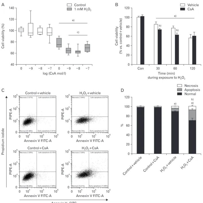
Fig. 1. Cyclosporin A (CsA) enhances hydrogen peroxide (H2O2) injury in human kidney proximal tubule epithelial cells. Human kidney proximal tubule epithelial HK2 cells were cultured in RPMI 1640 until reaching 80% confluence. (A) HK2 cells were treated with either CsA (1, 10, or 100 nM) or 1% dimethyl sulfoxide (vehicle) for 1 hour and then exposed to 1 mM H2O2 or distilled water (control) for 60 minutes. Cell viability was measured using MTT assay (n=9 wells from 3 experiments per condition). In box plots, whiskers represent the minimum and maximum;
bases represent the interquartile range between the first and third quartiles; and midlines represent the median. (B) HK2 cells were treated with either 10 nM CsA or vehicle for 60 minutes and then exposed to 1 mM H2O2 or control for 30, 60, or 120 minutes. Cell viability was measured using MTT assay (n=9 wells from 3 experiments per condition). (C, D) HK2 cells were treated with either 10 nM CsA or vehicle for 60 minutes and then exposed to 1 mM H2O2 or control for 60 minutes. Cell death was analyzed by flow cytometry with an annexin VFITC detection kit after treatment with FITCconjugated annexin V and propidium iodide. The flow cytometry assay distinguishes among normal (lower left), early apoptosis (lower right), late apoptosis (upper right), and necrosis (upper left). Three experiments were performed to evaluate the cell death. In each experiment, three samples per experimental condition were included. a)P<0.05 vs. control. b)P<0.05 vs. vehicle. c)P<0.01 vs. 0 mol/l.
in kidney proximal tubule epithelial cells, viabilities of HK-2 cells undergoing pretreatment with CsA and subsequent ex- posure to H2O2 were determined. Consistent with previous results [36], 60-minute exposure to 1 mM H2O2 markedly decreased cell viability based on MTT assay results (Fig. 1A).
Treatment with CsA at final concentrations of 1 nM to 100 nM did not significantly alter viabilities of control cells, but exogenous CsA further decreased viabilities of H2O2-exposed cells (Fig. 1A). The decline in viability after 30-minute expo- sure to H2O2 in CsA-treated cell was more severe than that in control cells (Fig. 1B). However, there was no significant difference in cell viability between CsA- and vehicle-treated groups after 120-minute exposure to H2O2 (Fig. 1B). To dis- tinguish between apoptosis and necrosis in dead cells, flow cytometry was performed on HK-2 cells stained with FITC- conjugated annexin V and propidium iodide. Exposure to 1 mM H2O2 significantly induced apoptosis and necrosis (Fig.
1C, D). Upon H2O2 injury, treatment with 10 nM CsA further
increased apoptosis and necrosis rather than vehicle-treated cells (Fig. 1C, D). However, exogenous CsA did not induce apoptosis and necrosis in control cells (Fig. 1C, D). These data suggest that CsA enhances apoptotic and necrotic cell deaths during early phase of H2O2 injury in kidney proximal tubule epithelial cells.
CsA increases p53 activation and BID expression after H2O2 injury in kidney proximal tubule epithelial cells
Tumor suppressor p53 is known to be upregulated and activated by phosphorylation in response to a number of cellular stresses including H2O2 injury [37]. The upregula- tion and activation of p53 subsequently induce cell death through apoptosis or necrosis [37]. It has been reported that treatment with CsA upregulates and activates p53 in rat C6 glioma cells and mouse embryo fibroblasts [38], while it con- versely decreases the number of p53-positive cells induced by ultraviolet-B irradiation in mouse dermis [39]. To exam-
p53phosphorylation (foldvs.control+vehicle)
Control 0
6 5 4 3 2 1
p53activation (foldvs.control+vehicle)
Control 0
7
5 4 3 2 1
a) 6
p53expression (foldvs.control+vehicle)
Control 0.0
1.4 1.2 1.0 0.8 0.6 0.4 0.2 p-p53
t-p53
-Actin
Veh CsA Veh CsA Control
p-p53 t-p53 p-p53/t-p53
b)
b) a)
b)
a) a) b)
b) Vehicle CsA
Vehicle CsA
Vehicle CsA
BID
-Actin
Veh CsA Veh CsA Control
BIDexpression (foldvs.control+vehicle)
Control 0.0
2.5
2.0
1.5
1.0
0.5
a) a) b)
# Vehicle CsA
A B C D
E F
Fig. 2. Cyclosporin A (CsA) increases p53 activation and BH3 interactingdomain death agonist (BID) expression after H2O2 injury in human kidney proximal tubule epithelial cells. HK2 cells were treated with either 10 nM CsA or vehicle for 60 minutes and then exposed to 1 mM H2O2
or control for 60 minutes (n=6 experiments per condition). (A) Phosphorylation level of p53 (pp53, 53 kDa) and total expression level of p53 (tp53, 53 kDa) were measured by western blot analysis. Antibody of βactin (43 kDa) as a loading control was used for normalization. (B–D) Intensities of pp53 and tp53 protein bands were quantified using the AzureSpot software. (E) BID (22 kDa) expression was measured by western blot analysis. Antibody of βactin (43 kDa) as a loading control was used for normalization. (F) Intensity of BID protein expression was quantified using the AzureSpot software. a)P<0.05 vs. control. b)P<0.05 vs. vehicle.
ine whether treatment with CsA might alter the activation and expression of p53 in kidney proximal tubule epithelial cells, we measured phosphorylated and total forms of p53 protein in CsA-treated HK-2 cells. Phosphorylation level of p53 was significantly increased in control cells at 60 minutes after treatment with CsA compared to that in vehicle-treated control cells (Fig. 2A, B). In contrast, total expression level of p53 was significantly decreased by CsA under the same con- dition (Fig. 2A, C). Although CsA decreased p53 expression in control cells, the ratio of p53 phosphorylation to its total expression was increased by treatment with CsA in control cells, indicating CsA-induced p53 activation (Fig. 2A, D).
Next, we assessed CsA-induced p53 activation upon H2O2
injury. Consistent with previous data in mouse kidney me- sangial cells [40], our results revealed that H2O2 significantly induced p53 phosphorylation, but not its total expression, in vehicle-treated cells (Fig. 2A–C), resulting in an increment of p53 activation based on the ratio of p53 phosphorylation to its total expression (Fig. 2A, D). Upon H2O2 injury, treatment with CsA markedly augmented the phosphorylation level of p53, but not its total expression (Fig. 2A–C). Because of this, CsA-treated cells showed striking increment in p53 activation after H2O2 injury (Fig. 2A, D), indicating that CsA increases H2O2-induced p53 activation in kidney proximal tubule epi- thelial cells. It is known that p53 activation results in apopto- sis and that p53 transcriptionally transactivates a number of proapoptotic proteins [41]. Thus, we tested whether BID of p53 downstream genes was further upregulated by CsA after H2O2 injury in HK-2 cells. As shown in Fig. 2E and F, CsA caused further increase in BID upregulation induced by H2O2
in these cells. However, CsA did not alter BID expression in control cells (Fig. 2E, F). Taken together, these data suggest that CsA enhances H2O2-induced p53 signaling in kidney proximal tubule epithelial cells.
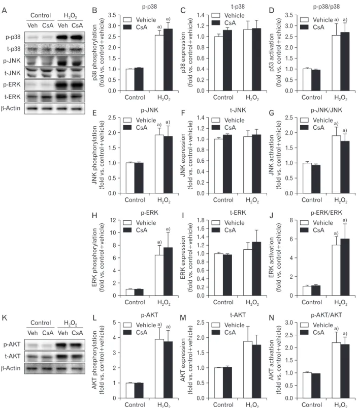
CsA does not alter activation of mitogen-activated protein kinases or AKT in kidney proximal tubule epithelial cells
One well-documented result of H2O2 injury is mitogen- activated protein kinase (MAPK) activation in various cell types [42]. Our results showed that H2O2 significantly en- hanced phosphorylation levels of p38, JNK and ERK, but did not their expression levels, resulting in significant activations of MAPKs based on the high ratio of phosphorylation level to total expression level in MAPKs (Fig. 3A–J). However, CsA did not alter their phosphorylation, total expression, or
consequential activation in H2O2- or vehicle-treated cells (Fig.
3A–J). On the other hand, CsA can also activate AKT in hu- man keratinocyte cells [43]. Thus, we determined whether CsA induced the activation of AKT in kidney proximal tubule epithelial cells. Although H2O2-induced AKT phosphoryla- tion, expression, and consequential activation in HK-2 cells were confirmed in a previous report [44], treatment with CsA did not alter its phosphorylation, expression, or conse- quential activation in kidney proximal tubule epithelial cells with or without H2O2 injury (Fig. 3K–N). These data indicate that CsA does not affect the activation of MAPKs or AKT in kidney proximal tubule epithelial cells, suggesting that CsA- enhanced cell death in H2O2-exposed cells is not implicated in their activations.
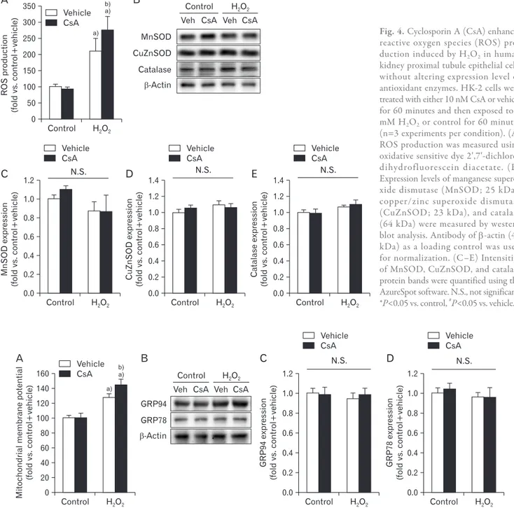
CsA increases ROS production after H2O2 injury in kidney proximal tubule epithelial cells
It is well-known that exogenous CsA induces oxidative stress in kidneys in vivo, leading to rapid loss of kidney func- tion [45]. To examine whether CsA could also induce oxida- tive stress in kidney proximal tubule epithelial cells in vitro, we measured ROS levels in HK-2 cells with or without H2O2
injury using the oxidative sensitive DCFDA dye. The produc- tion of ROS was significantly increased by exposure to H2O2
in vehicle-treated cells (Fig. 4A). Upon H2O2 injury, CsA- treated cells had greater ROS production than vehicle-treated cells (Fig. 4A). However, treatment with CsA did not alter the level of ROS in control cells (Fig. 4A). We further determined expression levels of antioxidant enzymes using western blot analysis. Our results showed that expression levels of anti- oxidant enzymes were not altered by short exposure to H2O2
and/or treatment with CsA (Fig. 4B–E). Taken together, these data indicate that CsA treatment after H2O2 injury causes greater ROS production than H2O2 injury alone.
CsA increases mitochondrial depolarization induced by H2O2 injury in kidney proximal tubule epithelial cells
CsA can induce mitochondrial dysfunction by decreas- ing mitochondrial respiration in rat skeletal muscle cells [46]
while it can conversely improve mitochondrial respiratory function by attenuating mitochondrial permeability transition in cardiomyocyte cells derived from dogs with heart failure [47]. To investigate the impact of CsA on mitochondrial func- tion in kidney proximal tubule epithelial cells with or without H2O2 injury, we monitored mitochondrial membrane poten- tials using TMRE dye. It is known that exposure to H2O2 can
Fig. 3. Cyclosporin A (CsA) does not alter activations of mitogenactivated protein kinases and protein kinase B (AKT) after H2O2 injury in human kidney proximal tubule epithelial cells. HK2 cells were treated with either 10 nM CsA or vehicle for 60 minutes and then exposed to 1 mM H2O2 or control for 60 minutes (n=6 experiments per condition). (A) Phosphorylation levels of p38 (pp38, 43 kDa), cJun Nterminal kinase (pJNK, 46 and 55 kDa), and extracellular signalregulated kinase (pERK, 42 and 44 kDa), and their total expression levels (tp38, pJNK, and pERK) were measured by western blot analysis. Antibody of βactin (43 kDa) as a loading control was used for normalization. (B, C, E, F, H, I) Intensities of pp38, tp38, pJNK, total JNK (tJNK), pERK, and total ERK (tERK) protein bands were quantified using the AzureSpot software. (D, G, J) Activations of p38, JNK, and ERK indicated by their respective ratio of phosphorylation level to total expression level. (K) AKT phosphorylation (pAKT) and total expression (tAKT) with a molecular mass of 56 to 62 kDa were measured by western blot analysis. Antibody of βactin (43 kDa) as a loading control was used for normalization. (L, M) Intensities of pAKT and tAKT protein bands were quantified using the AzureSpot software. (N) AKT activation based on the ratio of phosphorylation to total expression. a)P<0.05 vs. control.
p-p38 t-p38 p-JNK t-JNK p-ERK t-ERK
-Actin
Veh CsA Veh CsA Control
A
p38expression (foldvs.control+vehicle)
Control 0.0
1.4 1.2 1.0 0.8 0.6 0.4 0.2
t-p38
#
Vehicle CsA
C
p38phosphorylation (foldvs.control+vehicle)
Control 0.0
3.5 3.0 2.5 2.0 1.5 1.0 0.5
p-p38 Vehicle CsA
B
a) a)
p53activation (foldvs.control+vehicle)
Control 0.0
3.5 3.0 2.5 2.0 1.5 1.0 0.5
p-p38/p38 Vehicle CsA
D
a) a)
JNKexpression (foldvs.control+vehicle)
Control 0.0
1.4 1.2 1.0 0.8 0.6 0.4 0.2
t-JNK Vehicle CsA
F
JNKphosphorylation (foldvs.control+vehicle)
Control 0.0
2.5 2.0 1.5 1.0 0.5
p-JNK Vehicle CsA
E
a) a)
JNKactivation (foldvs.control+vehicle)
Control 0.0
2.5 2.0 1.5 1.0 0.5
p-JNK/JNK Vehicle CsA
G
a) a)
ERKexpression (foldvs.control+vehicle)
Control 0.0
1.8 1.6 1.4 1.2 1.0 0.8 0.6 0.4 0.2
t-ERK Vehicle CsA
I
ERKphosphorylation (foldvs.control+vehicle)
Control 0
12 10 8 6 4 2
p-ERK Vehicle CsA
H
a) a)
ERKactivation (foldvs.control+vehicle)
Control 0
8
6
4
2
p-ERK/ERK Vehicle CsA
J
a) a)
p-AKT t-AKT
-Actin
Veh CsA Veh CsA Control
K
AKTexpression (foldvs.control+vehicle)
Control 0.0
2.5 2.0 1.5 1.0 0.5
t-AKT Vehicle CsA
M
AKTphosphorylation (foldvs.control+vehicle)
Control 0
5 4 3 2 1
p-AKT Vehicle CsA
L
a) a)
AKTactivation (foldvs.control+vehicle)
Control 0.0
3.0 2.5 2.0 1.5 1.0 0.5
p-AKT/AKT Vehicle CsA
N
a) a)
increase the membrane potential of mitochondria during ear- ly phase in rat pulmonary microvascular endothelial cells, and then rapidly decreases the mitochondrial membrane potential after that [48]. In the present study, 60 minutes of exposure to
1 mM H2O2 significantly increased the mitochondrial mem- brane potential in vehicle-treated cells (Fig. 5A). The mito- chondrial membrane potential was significantly increased by CsA in H2O2-exposed cells, compared to that in vehicle-treat-
CuZnSODexpression (foldvs.control+vehicle)
Control 0.0
1.4 1.2 1.0 0.8 0.6 0.4 0.2
# Vehicle CsA
D
MnSODexpression (foldvs.control+vehicle)
Control 0.0
1.2 1.0 0.8 0.6 0.4 0.2
Vehicle CsA
C
Catalaseexpression (foldvs.control+vehicle)
Control 0.0
1.4 1.2 1.0 0.8 0.6 0.4 0.2
Vehicle CsA
E
ROSproduction (foldvs.control+vehicle)
Control 0
350 300 250 200 150 100 50
Vehicle CsA
A
a) a)
B
MnSOD CuZnSOD Catalase
-Actin
Veh CsA Veh CsA Control
b)
N.S. N.S. N.S.
Fig. 4. Cyclosporin A (CsA) enhances reac tive oxygen species (ROS) pro
duc tion induced by H2O2 in human kidney proximal tubule epithelial cells without altering expression level of antioxidant en zymes. HK2 cells were treated with either 10 nM CsA or vehicle for 60 minutes and then exposed to 1 mM H2O2 or control for 60 minutes (n=3 experiments per condition). (A) ROS production was measured using oxidative sensitive dye 2ʹ,7ʹdichloro
dihydrofluorescein diacetate. (B) Expression levels of manganese supero
xide dismutase (MnSOD; 25 kDa), copper/zinc superoxide dismutase (CuZnSOD; 23 kDa), and catalase (64 kDa) were measured by western blot analysis. Antibody of βactin (43 kDa) as a loading control was used for normalization. (C–E) Intensities of MnSOD, CuZnSOD, and catalase protein bands were quantified using the AzureSpot software. N.S., not significant.
*P<0.05 vs. control, #P<0.05 vs. vehicle.
GRP94expression (foldvs.control+vehicle)
Control 0.0
1.2 1.0 0.8 0.6 0.4 0.2
# Vehicle CsA
C
GRP78expression (foldvs.control+vehicle)
Control 0.0
1.2 1.0 0.8 0.6 0.4 0.2
# Vehicle CsA
D
Mitochondrialmembranepotential (foldvs.control+vehicle)
Control 0
160 140 120 100 80 60 40 20
a) a) b)
# Vehicle CsA
A
GRP94 GRP78
-Actin
Veh CsA Veh CsA Control
B N.S. N.S.
Fig. 5. Cyclosporin A (CsA) increases mitochondrial depolarization induced by H2O2 in human kidney proximal tubule epithelial cells. HK2 cells were treated with either 10 nM CsA or vehicle for 60 minutes and then exposed to 1 mM H2O2 or control for 60 minutes. (A) Percentage of mitochondrial membrane potential was measured using tetramethylrhodamine ethyl ester perchlorate dye (n=9 wells per 3 experiments per condition). The mitochondrial membrane potential in the group with control plus vehicle was taken as 100%. a)P<0.05 vs. control. b)P<0.05 vs.
vehicle. (B) Expression levels of 94 kDa glucoseregulated protein (GRP94) and 78 kDa glucoseregulated protein (GRP78) were measured by western blot analysis. Antibody of βactin (43 kDa) as a loading control was used for normalization. (C, D) Intensities of GRP94 and GRP78 protein bands were quantified using the AzureSpot software (n=6 experiments per condition). N.S., not significant.
ed H2O2-exposed cells (Fig. 5A). However, the mitochondrial membrane potential was not significantly different between vehicle-treated and CsA-treated cells in the control group (Fig.
5A). These data indicate that CsA worsen the mitochondrial depolarization induced by H2O2 injury in kidney proximal tu- bule epithelial cells. In addition to mitochondrial dysfunction, we assessed whether CsA was implicated in endoplasmic re- ticulum (ER) stress in kidney proximal tubule epithelial cells after H2O2 injury, as identified by alterations in GRP94 and GRP78 expressions. Expression levels of GRP94 or GRP78 were not altered by either exposure to H2O2 or treatment with CsA in HK-2 cells (Fig. 5B–D), suggesting that H2O2- or CsA- induced cell death did not involve ER stress in kidney proxi- mal tubule epithelial cells.
Discussion
ROS has an important role in a variety of kidney disease models. H2O2, a diffusible reactive oxygen metabolite formed by either enzyme-catalyzed or spontaneous dismutation of superoxide anion, has been implicated in the pathogenesis of tissue damage in acute kidney injury including ischemia reperfusion injury [49-51] and cisplatin nephrotoxicity [16- 18]. H2O2 inflicts oxidative stress at multiple cellular sites either directly or indirectly through generation of more reac- tive intermediates [12], acting as an indirect activator of p53, MAPK, and AKT [52, 53], and modulating mitochondrial function [54]. Thus, H2O2 is toxic when it is administered to intact kidney or cultured kidney tubular cells [55], leading to cell death. Our present data consistently revealed H2O2- induced p53 activation, MAPK activation, AKT activation, oxidative stress, mitochondrial dysfunction, and subsequent cell death in kidney proximal tubule epithelial cells.
CsA can inhibit mitochondria-mediated cell death, since it binds to cyclophilin D and subsequently blocks the for- mation of mitochondrial permeability transition pore [10], but can conversely kill kidney parenchymal cells, since it induces nephrotoxicity through glomerular filtration rate reduction and immune cell infiltration [8, 9]. If so, how does CsA contribute to the fate of kidney tubular cells upon H2O2 injury developed in acute kidney injury? In a previous study, exogenous H2O2 induced the permeability transition pore formation in mitochondria of subcutaneous connective tissue-derived cells while pretreatment with CsA significantly attenuated the formation of mitochondrial permeability tran- sition pore and subsequently apoptotic cell death [56]. Con-
trary to our expectations, the present study revealed that pre- treatment with CsA worsened mitochondrial dysfunction and cell mortality induced by exposure to H2O2 in human kidney proximal tubule epithelial cells. These data suggest that, even if CsA can efficiently bind to cyclophilin D, CsA-induced other signaling pathways may cause more severe damage and higher mortality in kidney proximal tubule epithelial cells.
The activation of p53 or the expression of its transcrip- tional targets including Bax and BID occurs in the response to stress-inducing agents including H2O2 injury [37, 57, 58]. Al- though one previous study has reported that CsA can protect mouse dermis against ultraviolet-B irradiation through p53 downregulation [39], several investigators have shown that CsA activates p53 in various cell types including rat C6 glio- ma cells [38], mouse embryo fibroblasts [38], human kerati- nocytes [59], and human skin fibroblasts [59]. In the present study, we found that CsA activated p53 reflected by increases in its phosphorylation and its target gene BID expression, thus triggering cell death of kidney proximal tubule epithelial cells after exposure to H2O2. The p53 protein is required for cellular apoptotic response to oxidative stress by hydrogen peroxide, supporting the idea that p53 function is critical for cell death induced by oxidative stress [60]. Oxidative stress is also an important mediator of CsA-induced cell injury. It is increased in failing transplanted kidneys from patients treated with CsA [45, 61, 62]. In support of these reports, the present study showed that CsA-induced p53 activation was impli- cated in ROS production increased by treatment with CsA during H2O2 injury. In response to oxidative stress, activated p53 does not only binds DNA to trigger apoptotic cell death by transactivating several genes [63], but also accumulates in the mitochondrial matrix to trigger necrotic cell death by opening the mitochondrial permeability transition pore [37]. CsA is a well-known cyclophilin D inhibitor which is a downstream player in cell death induced by the formation of mitochondrial permeability transition pore [10]. However, it has been reported that even in cyclophilin D–deficient cells, the mitochondrial permeability transition pore can still open in response to a strong stimulus such as oxidative stress [10, 64], indicating that cyclophilin D is a regulator but not neces- sarily a component of the mitochondrial permeability transi- tion pore. Based on previous reports, our data suggest that the fate of kidney proximal tubule epithelial cells is determined by CsA-induced cell death through the ROS-p53 axis rather than CsA-induced cell survival by inhibiting cyclophilin D- dependent mitochondrial permeability transition.
Antioxidants including vitamin E, melatonin, and other in- dolic compounds inhibit lipid peroxidation in animal models of CsA-induced nephrotoxicity [65-67], suggesting a role of oxidative stress in CsA-induced damage. Furthermore, it has been reported that CsA induces a dose-dependent increase in oxidants and lipid peroxidation [68]. Consistent with previous studies, the present study showed that ROS production was more enhanced by treatment with CsA than that by vehicle in H2O2-exposed kidney proximal tubule epithelial cells. Con- sidering the well-known relationship between oxidative stress and kidney tubular cell death [69-71], our data suggest that CsA-induced ROS production worsen kidney proximal tubu- lar cell damage after exposure to H2O2 through cell death.
Conclusively, the present study shows that CsA aggravates cell death induced by H2O2 injury. This phenomenon is im- plicated in p53 activation and ROS production stimulated by CsA. These findings may be of clinical importance for pharmacological intervention of CsA-treated organ transplant patients.
ORCID
Daeun Moon: https://orcid.org/0000-0002-0181-1513 Jinu Kim: https://orcid.org/0000-0002-1313-4791
Author Contributions
Conceptualization: JK. Data acquisition: DM. Data analysis or interpretation: DM, JK. Critical revision of the manuscript:
JK. Approval of the final version of the manuscript: all au- thors.
Conflicts of Interest
No potential conflict of interest relevant to this article was reported.
Acknowledgements
This work was supported by a research grant from the Jeju National University Hospital Research Fund of Jeju National University in 2017.
References
1. Borel JF, Feurer C, Gubler HU, Stähelin H. Biological effects of cyclosporin A: a new antilymphocytic agent. Agents Actions
1976;6:468-75.
2. White DJ, Calne RY. The use of cyclosporin A immunosuppres- sion in organ grafting. Immunol Rev 1982;65:115-31.
3. Takahashi N, Hayano T, Suzuki M. Peptidyl-prolyl cis-trans isomerase is the cyclosporin A-binding protein cyclophilin. Na- ture 1989;337:473-5.
4. Pereira MJ, Palming J, Rizell M, Aureliano M, Carvalho E, Svens- son MK, Eriksson JW. The immunosuppressive agents rapamy- cin, cyclosporin A and tacrolimus increase lipolysis, inhibit lipid storage and alter expression of genes involved in lipid metabo- lism in human adipose tissue. Mol Cell Endocrinol 2013;365:
260-9.
5. Nazareth W, Yafei N, Crompton M. Inhibition of anoxia-induced injury in heart myocytes by cyclosporin A. J Mol Cell Cardiol 1991;23:1351-4.
6. Bunjes D, Hardt C, Rollinghoff M, Wagner H. Cyclosporin A mediates immunosuppression of primary cytotoxic T cell re- sponses by impairing the release of interleukin 1 and interleukin 2. Eur J Immunol 1981;11:657-61.
7. Myers BD, Ross J, Newton L, Luetscher J, Perlroth M. Cyclospo- rine-associated chronic nephropathy. N Engl J Med 1984;311:
699-705.
8. Thomas SE, Andoh TF, Pichler RH, Shankland SJ, Couser WG, Bennett WM, Johnson RJ. Accelerated apoptosis characterizes cyclosporine-associated interstitial fibrosis. Kidney Int 1998;53:
897-908.
9. Young BA, Burdmann EA, Johnson RJ, Alpers CE, Giachelli CM, Eng E, Andoh T, Bennett WM, Couser WG. Cellular prolifera- tion and macrophage influx precede interstitial fibrosis in cyclo- sporine nephrotoxicity. Kidney Int 1995;48:439-48.
10. Baines CP, Kaiser RA, Purcell NH, Blair NS, Osinska H, Hamble- ton MA, Brunskill EW, Sayen MR, Gottlieb RA, Dorn GW, Rob- bins J, Molkentin JD. Loss of cyclophilin D reveals a critical role for mitochondrial permeability transition in cell death. Nature 2005;434:658-62.
11. Circu ML, Aw TY. Reactive oxygen species, cellular redox sys- tems, and apoptosis. Free Radic Biol Med 2010;48:749-62.
12. Salahudeen AK, Clark EC, Nath KA. Hydrogen peroxide- induced renal injury: a protective role for pyruvate in vitro and in vivo. J Clin Invest 1991;88:1886-93.
13. Kim J, Kil IS, Seok YM, Yang ES, Kim DK, Lim DG, Park JW, Bonventre JV, Park KM. Orchiectomy attenuates post-ischemic oxidative stress and ischemia/reperfusion injury in mice: a role for manganese superoxide dismutase. J Biol Chem 2006;281:
20349-56.
14. Kim J, Kim KY, Jang HS, Yoshida T, Tsuchiya K, Nitta K, Park JW, Bonventre JV, Park KM. Role of cytosolic NADP+-depen- dent isocitrate dehydrogenase in ischemia-reperfusion injury in mouse kidney. Am J Physiol Renal Physiol 2009;296:F622-33.
15. Kim J, Seok YM, Jung KJ, Park KM. Reactive oxygen species/
oxidative stress contributes to progression of kidney fibrosis fol- lowing transient ischemic injury in mice. Am J Physiol Renal Physiol 2009;297:F461-70.
16. Kim J, Long KE, Tang K, Padanilam BJ. Poly(ADP-ribose) poly-
merase 1 activation is required for cisplatin nephrotoxicity. Kid- ney Int 2012;82:193-203.
17. Park S, Yoon SP, Kim J. Cisplatin induces primary necrosis through poly(ADP-ribose) polymerase 1 activation in kidney proximal tubular cells. Anat Cell Biol 2015;48:66-74.
18. Yoon SP, Kim J. Exogenous spermidine ameliorates tubular ne- crosis during cisplatin nephrotoxicity. Anat Cell Biol 2018;51:
189-99.
19. Kim J, Imig JD, Yang J, Hammock BD, Padanilam BJ. Inhibition of soluble epoxide hydrolase prevents renal interstitial fibrosis and inflammation. Am J Physiol Renal Physiol 2014;307:F971- 80.
20. Kim J, Padanilam BJ. Loss of poly(ADP-ribose) polymerase 1 attenuates renal fibrosis and inflammation during unilateral ure- teral obstruction. Am J Physiol Renal Physiol 2011;301:F450-9.
21. Kim J, Padanilam BJ. Renal nerves drive interstitial fibrogenesis in obstructive nephropathy. J Am Soc Nephrol 2013;24:229-42.
22. Kim J, Yoon SP, Toews ML, Imig JD, Hwang SH, Hammock BD, Padanilam BJ. Pharmacological inhibition of soluble epoxide hydrolase prevents renal interstitial fibrogenesis in obstructive nephropathy. Am J Physiol Renal Physiol 2015;308:F131-9.
23. Pabla N, Dong Z. Cisplatin nephrotoxicity: mechanisms and renoprotective strategies. Kidney Int 2008;73:994-1007.
24. Yoon SP, Kim J. Exogenous CGRP upregulates profibrogenic growth factors through PKC/JNK signaling pathway in kidney proximal tubular cells. Cell Biol Toxicol 2018;34:251-62.
25. Kim J. Spermidine rescues proximal tubular cells from oxida- tive stress and necrosis after ischemic acute kidney injury. Arch Pharm Res 2017;40:1197-208.
26. Kim J, Padanilam BJ. Renal denervation prevents long-term se- quelae of ischemic renal injury. Kidney Int 2015;87:350-8.
27. Ammar NM, El-Hawary SS, Mohamed DA, Afifi MS, Ghanem DM, Awad G. Phytochemical and biological studies of Tribulus terrestris L. growing in Egypt. Int J Pharmacol 2018;14:248-59.
28. Ali A, Kim MJ, Kim MY, Lee HJ, Roh GS, Kim HJ, Cho GJ, Choi WS. Quercetin induces cell death in cervical cancer by reducing O-GlcNAcylation of adenosine monophosphate-activated pro- tein kinase. Anat Cell Biol 2018;51:274-83.
29. Kim J. Spermidine is protective against kidney ischemia and re- perfusion injury through inhibiting DNA nitration and PARP1 activation. Anat Cell Biol 2017;50:200-6.
30. Kim J. Poly(ADP-ribose) polymerase activation induces high mobility group box 1 release from proximal tubular cells during cisplatin nephrotoxicity. Physiol Res 2016;65:333-40.
31. Lee JS, Lim JY, Kim J. Mechanical stretch induces angiotensino- gen expression through PARP1 activation in kidney proximal tubular cells. In Vitro Cell Dev Biol Anim 2015;51:72-8.
32. Park Y, Tae HJ, Cho JH, Kim IS, Ohk TG, Park CW, Moon JB, Shin MC, Lee TK, Lee JC, Park JH, Ahn JH, Kang SH, Won MH, Cho JH. The relationship between low survival and acute in- crease of tumor necrosis factor alpha expression in the lung in a rat model of asphyxial cardiac arrest. Anat Cell Biol 2018;51:128- 35.
33. Wang H, Joseph JA. Quantifying cellular oxidative stress by di-
chlorofluorescein assay using microplate reader. Free Radic Biol Med 1999;27:612-6.
34. Song H, Yoon SP, Kim J. Poly(ADP-ribose) polymerase regulates glycolytic activity in kidney proximal tubule epithelial cells. Anat Cell Biol 2016;49:79-87.
35. Yoon SP, Kim J. Poly(ADP-ribose) polymerase 1 contributes to oxidative stress through downregulation of sirtuin 3 during cis- platin nephrotoxicity. Anat Cell Biol 2016;49:165-76.
36. Ye J, Li J, Yu Y, Wei Q, Deng W, Yu L. L-carnitine attenuates oxi- dant injury in HK-2 cells via ROS-mitochondria pathway. Regul Pept 2010;161:58-66.
37. Vaseva AV, Marchenko ND, Ji K, Tsirka SE, Holzmann S, Moll UM. p53 opens the mitochondrial permeability transition pore to trigger necrosis. Cell 2012;149:1536-48.
38. Pyrzynska B, Serrano M, Martínez-A C, Kaminska B. Tumor suppressor p53 mediates apoptotic cell death triggered by cyclo- sporin A. J Biol Chem 2002;277:14102-8.
39. Sugie N, Fujii N, Danno K. Cyclosporin-A suppresses p53- dependent repair DNA synthesis and apoptosis following ultra- violet-B irradiation. Photodermatol Photoimmunol Photomed 2002;18:163-8.
40. Kume S, Haneda M, Kanasaki K, Sugimoto T, Araki S, Isono M, Isshiki K, Uzu T, Kashiwagi A, Koya D. Silent information regu- lator 2 (SIRT1) attenuates oxidative stress-induced mesangial cell apoptosis via p53 deacetylation. Free Radic Biol Med 2006;40:
2175-82.
41. Yu J, Zhang L, Hwang PM, Rago C, Kinzler KW, Vogelstein B.
Identification and classification of p53-regulated genes. Proc Natl Acad Sci U S A 1999;96:14517-22.
42. Guyton KZ, Liu Y, Gorospe M, Xu Q, Holbrook NJ. Activation of mitogen-activated protein kinase by H2O2. Role in cell survival following oxidant injury. J Biol Chem 1996;271:4138-42.
43. Han W, Ming M, He TC, He YY. Immunosuppressive cyclospo- rin A activates AKT in keratinocytes through PTEN suppres- sion: implications in skin carcinogenesis. J Biol Chem 2010;285:
11369-77.
44. Andreucci M, Fuiano G, Presta P, Lucisano G, Leone F, Fuiano L, Bisesti V, Esposito P, Russo D, Memoli B, Faga T, Michael A.
Downregulation of cell survival signalling pathways and in- creased cell damage in hydrogen peroxide-treated human renal proximal tubular cells by alpha-erythropoietin. Cell Prolif 2009;
42:554-61.
45. Wolf A, Clemann N, Frieauff W, Ryffel B, Cordier A. Role of re- active oxygen formation in the cyclosporin-A-mediated impair- ment of renal functions. Transplant Proc 1994;26:2902-7.
46. Hokanson JF, Mercier JG, Brooks GA. Cyclosporine A decreases rat skeletal muscle mitochondrial respiration in vitro. Am J Respir Crit Care Med 1995;151:1848-51.
47. Sharov VG, Todor A, Khanal S, Imai M, Sabbah HN. Cyclospo- rine A attenuates mitochondrial permeability transition and improves mitochondrial respiratory function in cardiomyocytes isolated from dogs with heart failure. J Mol Cell Cardiol 2007;42:
150-8.
48. Madesh M, Hawkins BJ, Milovanova T, Bhanumathy CD, Joseph
SK, Ramachandrarao SP, Sharma K, Kurosaki T, Fisher AB. Se- lective role for superoxide in InsP3 receptor-mediated mitochon- drial dysfunction and endothelial apoptosis. J Cell Biol 2005;170:
1079-90.
49. Yoon SP, Kim J. Poly(ADP-ribose) polymerase 1 activation links ischemic acute kidney injury to interstitial fibrosis. J Physiol Sci 2015;65:105-11.
50. Kim J, Kim JI, Na YK, Park KM. Intra-renal slow cell-cycle cells contribute to the restoration of kidney tubules injured by isch- emia/reperfusion. Anat Cell Biol 2011;44:186-93.
51. Kim J, Kim JI, Jang HS, Park JW, Park KM. Protective role of cy- tosolic NADP(+)-dependent isocitrate dehydrogenase, IDH1, in ischemic pre-conditioned kidney in mice. Free Radic Res 2011;
45:759-66.
52. Niwa K, Inanami O, Yamamori T, Ohta T, Hamasu T, Kuwabara M. Redox regulation of PI3K/Akt and p53 in bovine aortic en- dothelial cells exposed to hydrogen peroxide. Antioxid Redox Signal 2003;5:713-22.
53. Wu GS. The functional interactions between the p53 and MAPK signaling pathways. Cancer Biol Ther 2004;3:156-61.
54. Nulton-Persson AC, Szweda LI. Modulation of mitochondrial function by hydrogen peroxide. J Biol Chem 2001;276:23357-61.
55. Yoshioka T, Bills T, Moore-Jarrett T, Greene HL, Burr IM, Ichikawa I. Role of intrinsic antioxidant enzymes in renal oxi- dant injury. Kidney Int 1990;38:282-8.
56. Takeyama N, Miki S, Hirakawa A, Tanaka T. Role of the mito- chondrial permeability transition and cytochrome C release in hydrogen peroxide-induced apoptosis. Exp Cell Res 2002;274:16- 24.
57. Chen QM, Liu J, Merrett JB. Apoptosis or senescence-like growth arrest: influence of cell-cycle position, p53, p21 and bax in H2O2 response of normal human fibroblasts. Biochem J 2000;
347(Pt 2):543-51.
58. Levine AJ. p53, the cellular gatekeeper for growth and division.
Cell 1997;88:323-31.
59. Voskamp P, Bodmann CA, Koehl GE, Rebel HG, Van Olderen MG, Gaumann A, El Ghalbzouri A, Tensen CP, Bavinck JN, Willemze R, Geissler EK, De Gruijl FR. Dietary immunosup- pressants do not enhance UV-induced skin carcinogenesis, and reveal discordance between p53-mutant early clones and carci- nomas. Cancer Prev Res (Phila) 2013;6:129-38.
60. Yin Y, Terauchi Y, Solomon GG, Aizawa S, Rangarajan PN, Yaza-
ki Y, Kadowaki T, Barrett JC. Involvement of p85 in p53-depen- dent apoptotic response to oxidative stress. Nature 1998;391:707- 10.
61. Mohamadin AM, El-Beshbishy HA, El-Mahdy MA. Green tea extract attenuates cyclosporine A-induced oxidative stress in rats. Pharmacol Res 2005;51:51-7.
62. Satyanarayana PS, Singh D, Chopra K. Quercetin, a bioflavonoid, protects against oxidative stress-related renal dysfunction by cyclosporine in rats. Methods Find Exp Clin Pharmacol 2001;23:
175-81.
63. Gu W, Roeder RG. Activation of p53 sequence-specific DNA binding by acetylation of the p53 C-terminal domain. Cell 1997;
90:595-606.
64. Basso E, Fante L, Fowlkes J, Petronilli V, Forte MA, Bernardi P.
Properties of the permeability transition pore in mitochondria devoid of cyclophilin D. J Biol Chem 2005;280:18558-61.
65. Wang C, Salahudeen AK. Lipid peroxidation accompanies cyclo- sporine nephrotoxicity: effects of vitamin E. Kidney Int 1995;47:
927-34.
66. Longoni B, Migliori M, Ferretti A, Origlia N, Panichi V, Boggi U, Filippi C, Cuttano MG, Giovannini L, Mosca F. Melatonin prevents cyclosporine-induced nephrotoxicity in isolated and perfused rat kidney. Free Radic Res 2002;36:357-63.
67. Wang C, Salahudeen AK. Cyclosporine nephrotoxicity: attenua- tion by an antioxidant-inhibitor of lipid peroxidation in vitro and in vivo. Transplantation 1994;58:940-6.
68. Longoni B, Boschi E, Demontis GC, Marchiafava PL, Mosca F.
Regulation of Bcl-2 protein expression during oxidative stress in neuronal and in endothelial cells. Biochem Biophys Res Com- mun 1999;260:522-6.
69. Kim J, Jung KJ, Park KM. Reactive oxygen species differently regulate renal tubular epithelial and interstitial cell prolifera- tion after ischemia and reperfusion injury. Am J Physiol Renal Physiol 2010;298:F1118-29.
70. Kim J, Jang HS, Park KM. Reactive oxygen species generated by renal ischemia and reperfusion trigger protection against subsequent renal ischemia and reperfusion injury in mice. Am J Physiol Renal Physiol 2010;298:F158-66.
71. Kim J, Park JW, Park KM. Increased superoxide formation in- duced by irradiation preconditioning triggers kidney resistance to ischemia-reperfusion injury in mice. Am J Physiol Renal Physiol 2009;296:F1202-11.