J Korean Soc Radiol 2018;78(2):130-134 https://doi.org/10.3348/jksr.2018.78.2.130
INTRODUCTION
Amyloidosis is a heterogeneous group of diseases, character- ized by misfolding of extracellular proteins, which generates in- soluble fibrils that result in the disruption of tissue structure and function (1). Numerous studies have reported that various malignancies are associated with amyloidosis, including renal cell carcinoma (RCC), which is associated with systemic amy- loidosis relatively frequently. However, there has been few ra- diologic reports of amyloidosis in a patient with RCC (2).
Herein, we report a case of secondary systemic amyloidosis in a 21-year-old female patient with RCC.
CASE REPORT
A 21-year-old woman presented to our hospital, with gross
hematuria, diarrhea, severe dizziness, anemia, and loss of 10 kg over a 1-month period. She had no previous significant medical or family history. She underwent contrast-enhanced abdominal computed tomography (CT) scan, which revealed a 6-cm-sized well-defined heterogeneous enhanced mass in the right kidney, suggestive of non-conventional RCC (Fig. 1A). In addition, multiple metastases were identified in the retrocaval and aorto- caval lymph nodes (LNs) (Fig. 1A), and a small amount of asci- tes and mild peritoneal thickening were noted.
Subsequently, she underwent right radical nephrectomy with regional lymph node dissection. The pathology results revealed papillary RCC, and metastatic lesions in the retrocaval and aorto- caval LNs. Sarcomatoid differentiation of RCC and amyloidosis in the adjacent renal parenchyma were also noted (Fig. 1B, C).
Following surgery, the patient’s symptoms of diarrhea and vomiting persisted for 5 days, and postoperative CT scans dem-
Radiologic Manifestation of Intra-Abdominal Involvement of Systemic Amyloidosis Secondary to Renal Cell Carcionma
신세포암에 의한 이차적 아밀로이드증의 복강내 침범의 영상학적 소견You Jin Lee, MD
1, Ga Jin Han, MD
1*, Suk Kim, MD
1, Nam Kyung Lee, MD
1, Ho Jin Shin, MD
2, Hong Koo Ha, MD
3, Tae Un Kim, MD
4Departments of 1Radiology, 2Internal Medicine, 3Urology, Pusan National University Hospital, Pusan National University School of Medicine, Busan, Korea
4Department of Radiology, Pusan National University Yangsan Hospital, Pusan National University School of Medicine, Yangsan, Korea
Amyloidosis has a wide spectrum of manifestations in numerous abdominal organs.
It can be categorized into primary or secondary disease according to the presence of accompanying conditions, such as rheumatoid arthritis, tuberculosis, or neoplasms.
Herein, we present a rare case of amyloidosis associated with renal cell carcinoma that showed nonspecific radiologic features such as diffuse bowel wall thickening, ascites and peritoneal thickening.
Index terms Amyloidosis Abdomen
Renal Cell Carcinoma Radiology
Received July 20, 2017 Revised August 22, 2017 Accepted October 16, 2017
*Corresponding author: Ga Jin Han, MD Department of Radiology, Pusan National University Hospital, Pusan National University School of Medicine, 179 Gudeok-ro, Seo-gu, Busan 49241, Korea.
Tel. 82-51-240-7354 Fax. 82-51-244-7534 E-mail: bbr322@naver.com
This is an Open Access article distributed under the terms of the Creative Commons Attribution Non-Commercial License (http://creativecommons.org/licenses/by-nc/4.0) which permits unrestricted non-commercial use, distri- bution, and reproduction in any medium, provided the original work is properly cited.
A
C
E
B
D
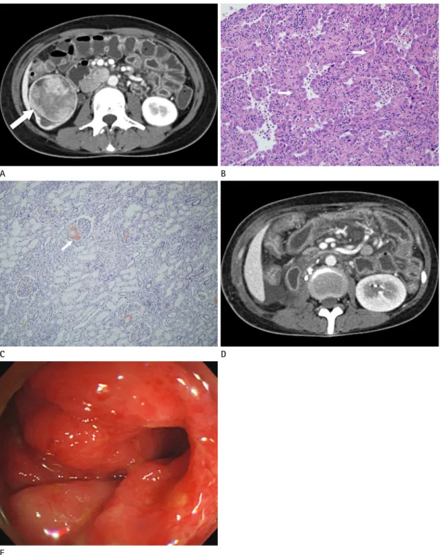
Fig. 1. A 21-year-old woman of systemic amyloidosis secondary to RCC.
A. Axial CT scan shows a 6 cm–sized enhancing mass (arrow) at the right kidney, suggestive of RCC and multiple metastatic lymph nodes at the aortocaval and retrocaval areas.
B. Photomicrograph (× 100, hematoxylin-eosin stain) shows eosinophilic cytoplasm and sparse capillaries (arrows), Histologic diagnosis is papil- lary RCC.
C. Photomicrograph (× 100, Congo red stain) shows strong affinity for the stain (arrow), suggestive of amyloidosis.
D. Postoperative CT scan reveals diffuse wall thickening of small bowel, colon and ascites, peritoneal thickening. These findings are worse than the findings of preoperative CT scan.
E. Colonoscopy image reveals scarring, redness and diffuse mucosal thickening of ascending colon and the histologic diagnosis is amyloidosis.
CT = computed tomography, RCC = renal cell carcinoma
onstrated exacerbation of the diffuse wall thickening of the small bowel, colon, and stomach (Fig. 1D). In addition, CT demon- strated increased ascites with peritoneal thickening, suggestive of peritoneal carcinomatosis.
For further evaluation, esophagogastroduodenoscopy (EGD) and colonoscopy were performed. The EGD identified diffuse mucosal thickening and redness, along with fine mucosal gran- ules in the gastroesophageal junction, gastric antrum, and duo- denal bulb. Colonoscopy revealed diffuse thickening and red- ness of the mucosal folds, friability of the colon and scarring (Fig. 1E). The endoscopic biopsy results demonstrated eosino- philic amorphous material deposition in the lamina propria of the stomach and colon, consistent with amyloidosis. Protein electrophoresis, immunoelectrophoresis, immunofixation, and bone marrow biopsy did not identify the presence of parapro- teins or plasma cell dyscrasia. Immunohistochemical analysis of the biopsy specimen revealed diffuse expressions of amyloid A and amyloid P. Thus, a diagnosis of amyloid A protein (AA) amyloidosis involving the kidney and gastrointestinal tract as- sociated with RCC was confirmed.
Transesophageal echocardiography identified diastolic dys- function and impaired relaxation of the left ventricle, whilst the left ventricular systolic pressure and function were preserved.
This is representative of early stage restrictive cardiomyopathy.
To determine whether the patient had cardiac amyloidosis, car- diac magnetic resonance imaging was performed, which re- vealed no evidence of amyloidosis.
The patient received 4 cycles of first-line chemotherapy with thalidomide, cyclophosphamide and dexamethasone as treat- ment of amyloidosis for 3 months. Follow-up CT at 5 months demonstrated decreased diffuse bowel wall thickening, nodular peritoneal thickening, and amount of ascites, indicating that the extent of gastrointestinal amyloidosis was reduced. In addi- tion, EGD and colonoscopy demonstrated improvements in the diffuse mucosal thickening and redness.
DISCUSSION
Herein, we discussed a case of amyloidosis associated with RCC involving the gastrointestinal tract and kidney. Amyloido- sis is a rare disease, characterized by the extracellular deposition of amorphous and fibrillar proteins known as amyloids. Pro-
gressive deposition of amyloids affects the function of normal tissues, leading to organ dysfunction and other clinical mani- festations, many of which are severe (1). The clinical and radio- logic manifestations of amyloidosis are wide-ranging and fre- quently nonspecific, providing a diagnostic challenge for clinicians and radiologists.
Amyloidosis is typically primary or secondary systemic amy- loidosis, with localized amyloidosis accounting for only 10–20%
of cases (3). Amyloid light-chain (AL) amyloidosis (primary systemic amyloidosis) is associated with monoclonal plasma cell dyscrasia and more than 30% of the patients with primary amyloidosis consequently progress to multiple myeloma (4).
The precipitating protein in secondary amyloidosis is always the amino acid terminus of the acute phase protein serum amy- loid A. Several diseases including chronic infection and inflam- mation, such as rheumatoid arthritis, tuberculosis, osteomyeli- tis, and pyelonephritis, are associated with AA amyloidosis (5).
As in the present case, systemic amyloidosis can occur simul- taneously with malignant disease, particularly RCC. The most common malignant tumor types associated with systemic amy- loidosis are multiple myeloma, Hodgkin’s lymphoma, and RCC (6). A previous study reported that systemic amyloidosis occurs in 3% of all RCC cases. Conversely, RCC occurs in 8% of system- ic amyloidosis cases (6).
There are three proposed mechanisms for how RCC triggers the development of systemic amyloidosis; however, these have not yet been proven. In the first mechanism, chronic inflamma- tion from tumor necrosis and chronic infection provides the stimuli for secondary amyloidosis (7). In the second mecha- nism, amyloids are produced by RCC cells, which either secrete an amyloidogenic substance or modify a serum precursor pro- tein into an amyloidogenic fragment (8). Finally, in the third proposed mechanism, renal tubular epithelial lysosomal en- zymes partially break circulating light-chain proteins into in- soluble polymers and amyloid-like fibrils (9).
Amyloids may infiltrate all organs and present as diverse ra- diographic findings. To the best of our knowledge, there are no reports about the specific imaging features of different types of amyloidosis. In patients with RCC or other malignancies, the gastrointestinal tract is the most commonly involved organ of amyloidosis. Although gastric involvement in systemic amyloi- dosis is common, symptomatic gastric amyloidosis is rare. The
endoscopic appearance of gastric amyloidosis can closely re- semble that of gastric malignancy, and biopsy is thus essential to establish a diagnosis of gastric amyloidosis. The radiologic features of the upper gastrointestinal series include fine muco- sal granules, mucosal or intramural filling defects, mucosal fold thickening, and single or multiple polypoid masses (1).
In the gastrointestinal tract, the small intestine is the most common site of histological and radiographic abnormalities caused by amyloidosis. CT demonstrates diffuse or nodular wall thickening, intussusception, and bowel dilatation depend- ing on the degree of hypomotility, gastrointestinal bleeding, and luminal narrowing due to amyloid infiltration or secondary to ischemia. In AL amyloidosis, polypoid protrusions and muco- sal fold thickening have been identified to be positively corre- lated with the deposition of amyloids in the muscularis mucosa and submucosa (1). In contrast, in secondary amyloidosis, the small bowel series typically exhibits a coarse mucosal pattern with innumerable fine granular densities due to AA deposition at the lamina propria (1). Although the small bowel series was not investigated in the current case, amyloid deposits were identified at the lamina propria of the bowel through histology and diffuse bowel wall thickening was demonstrated on CT.
Amyloid deposit in the colon, although common in biopsy specimens, are rarely observed radiologically. The CT findings of colon involvement of amyloidosis include thickening of the colonic wall, luminal narrowing, distension and perforated gi- ant diverticulum, secondary to ischemia or amyloid infiltration (1). The differential diagnoses for gastrointestinal amyloidosis include infectious enterocolitis, bowel ischemia, other causes of gastrointestinal bleeding such as gastric ulcer, gastric cancer, gastric varix; and other infiltrating diseases, such as small bowel lymphoma.
As in the present case, renal disease is a common manifesta- tion of systemic amyloidosis, and the imaging findings for this disease are wide-ranging, including renal atrophy with cortical thinning, renal enlargement, amorphous renal calcifications and focal renal mass lesions resulting from the amyloid deposition (3). This case exhibited normal kidney configuration and atten- uation, excluding RCC, on CT. This may be due to age of pa- tient, disease duration, and renal function.
Approximately 70% of patients with amyloidosis reportedly have liver involvement, however, their presenting symptoms
and radiologic findings are non-specific (10). Amyloidosis rarely involves intra-abdominal organs, including the gallbladder, bili- ary tree, pancreas, spleen, genitourinary tract, retroperitoneum, and LNs. The treatment for AL amyloidosis is chemotherapy, while the treatment of AA amyloidosis is controlling the pri- mary disease.
In conclusion, we presented a case of systemic amyloidosis associated with RCC. Although the underlying mechanisms how this the neoplasm triggers amyloidosis are uncertain, RCC is a relatively common cause of secondary amyloidosis. Second- ary systemic amyloidosis associated with RCC presents a wide- range of clinical and radiologic features. Therefore, when there are suspicious imaging findings, such as bowel wall thickening, ischemic changes, edema of the gastrointestinal tract, or hepa- tomegaly in patients with RCC, secondary amyloidosis should be considered as a differential diagnosis and a tissue biopsy is recommended.
Acknowledgments
This research was supported by clinical research grant from Pusan National University Hospital in 2017.
REFERENCES
1. Kim SH, Han JK, Lee KH, Won HJ, Kim KW, Kim JS, et al. Ab- dominal amyloidosis: spectrum of radiological findings.
Clin Radiol 2003;58:610-620
2. Babu A, Lachmann H, Pickett T, Boddana P, Ludeman L. Re- nal cell carcinoma presenting as AA amyloidosis: a case re- port and review of the literature. CEN Case Rep 2014;3:68- 74
3. Kawashima A, Alleman WG, Takahashi N, Kim B, King BF Jr, LeRoy AJ. Imaging evaluation of amyloidosis of the urinary tract and retroperitoneum. Radiographics 2011;31:1569- 1582
4. Rajkumar SV, Dispenzieri A, Kyle RA. Monoclonal gammop- athy of undetermined significance, Waldenström macro- globulinemia, AL amyloidosis, and related plasma cell disor- ders: diagnosis and treatment. Mayo Clin Proc 2006;81:693- 703
5. Kyle RA, Bayrd ED. Amyloidosis: review of 236 cases. Med- icine (Baltimore) 1975;54:271-299
6. Pras M, Franklin EC, Shibolet S, Frangione B. Amyloidosis associated with renal cell carcinoma of the AA type. Am J Med 1982;73:426-428
7. Malle E, Sodin-Semrl S, Kovacevic A. Serum amyloid A: an acute-phase protein involved in tumour pathogenesis. Cell Mol Life Sci 2009;66:9-26
8. Altaffer LF 3rd, Chenault OW Jr. Paraneoplastic endocri- nopathies associated with renal tumors. J Urol 1979;122:
573-577
9. Tan M, Epstein W. Polymer formation during the degrada- tion of human light chain and Bence-Jones proteins by an extrct of the lysosomal fraction of normal human kidney.
Immunochemistry 1972;9:9-16
10. Georgiades CS, Neyman EG, Barish MA, Fishman EK. Amy- loidosis: review and CT manifestations. Radiographics 2004;
24:405-416
신세포암에 의한 이차적 아밀로이드증의 복강내 침범의 영상학적 소견
이유진
1· 한가진
1* · 김 석
1· 이남경
1· 신호진
2· 하홍구
3· 김태언
4아밀로이드증은 다양한 복강내 기관에서 다양하게 나타난다. 아밀로이드증은 일차적인 것과 류마티스 관절염, 결핵, 종양 과 연관되어 발생하는 이차적인 것으로 분류된다. 이 보고는 미만성 장벽비후와 복수, 복막비후와 같은 비특이적인 영상의 학적 소견을 보였던 신세포암과 연관된 아밀로이드증의 증례이다.
부산대학교 의학전문대학원 부산대학교병원 1영상의학과, 2내과, 3비뇨기과,
4부산대학교 의학전문대학원 양산부산대학교병원 영상의학과