- 41 - 대한두경부종양학회지, 제34권 제1호, 2018. pp.41-43
Korean Journal of Head & Neck Oncology, Vol.34, No.1
http://doi.org/10.21593/kjhno/2018.34.1.41 ISSN 1229-5183(Print) / ISSN 2586-2553(Online)
측경부에 발생한 방추세포 / 다형성 지방종1예
김영윤⋅윤성호⋅박종민⋅이동훈+
전남대학교 의과대학 화순전남대학교병원 이비인후-두경부외과학교실
A case of a Spindle cell/pleomorphic lipoma in the lateral neck
Young Yoon Kim, MD, Sung Ho Yoon, MD, Jong Min Park, MD, Dong Hoon Lee, MD, PhD+
Department of Otorhinolaryngology-Head and Neck Surgery, Chonnam National University Medical School &
Chonnam National University Hwasun Hospital, Hwasun, South Korea
= Abstract =
The Spindle cell lipoma is a slow-growing benign tumor seen generally in the shoulders, upper back, and back of the neck of male. The Pleomorphic (giant-cell) lipoma is a benign tumor of adipose tissue with atypical histo- logical features. It is mainly seen in the same lesion as the Spindle cell lipoma. The Pleomorphic lipoma is cytoge- netically similar to spindle cell lipoma with a consistent loss of chromosome 16q material. For this reason, these two entities are regarded as a similar spectrum in the adipose tumors. Herein, we present a 53-year old man with Spindle cell/pleomorphic lipoma in the lateral neck. Physical and radiologic examinations of the Spindle cell/pleomorphic lipoma in the lateral neck are not specific and preoperative diagnosis is usually difficult. Therefore, clinicians should consider the possibility that Spindle cell/pleomorphic lipoma may occur in the lateral neck mim- icking the other more frequently observed lesions.
Key W ords : Spindle cell lipoma; pleomorphic lipoma; adipose tumors
R eceived R e v i s e d A ccepted
: November 28, 2017 : March 8, 2018 : March 12, 2018 +Corresponding author: 이동훈
전남 화순군 화순읍일심리 160번지 전남대학교 의과대학 화순전남대학교병원, 이비인후-두경부외과학교실 Tel: 061-379-8190 Fax: 061-379-8199
E-mail: [email protected]
서 론
방추 세포 / 다형성 지방종은 대부분 어깨, 등, 뒷목에 발생하는 지방성 양성종양으로, 주로 남성에게서 발견 되고 여성에서의 발생은 드물다.1-3)방추 세포 지방종과 다형성 지방종은 세포유전학적으로 염색체 16q 물질의 지속적인 손실 소견으로 인해 유사한 특성을 보이며 또 한 조직학, 면역 조직학 특징에 있어서도 중첩되는 특성 을 갖는다.4,5) 이러한 이유로 두 종양은 지방종의 같은 개체로 간주되고 있다.6)국내에서는 최근에 두피에 발생
한 방추 세포 지방종 1예가 보고되었다.7)최근 저자들은 좌측 측경부에서 발생한 방추 세포 / 다형성 지방종을 발견하여, 치험 하였기에 문헌고찰과 함께 보고하는 바 이다.
증 례
53세 남자가 2년전부터 발생한 좌측 측경부 종물을 주소로 타병원에서 경부 초음파를 시행 후 본원으로 전 원 되었다. 과거력상 4년전 하지의 지방종으로 타병원에 서 제거술 받았으며, 당뇨로 10개월전부터 약물 치료 중 이었다. 이학적 검사상 좌측 측경부, 악하선 부위에 2cm 가량의 무통성의 단단한 종괴가 만져졌고 그 외에 다른 신체부위에 저명한 이상 소견은 보이지 않았다.
본원에서 시행한 세침흡인검사 결과 무정형 물질을 포 함한 약간의 염증 세포로 진단되었고 경부 초음파 시행 결과 좌측 경부 천근막 부위에 약 2.5 x 1.2 x 2.1 cm 크기의
- 42 -
Fig. 1. Neck US shows a 2.5 x 1.2 x 2.1 cm lobulated heteroge- neously hyperechoic mass with partially obscured margin, perilesional infiltration and increased vascularity in the left su- perficial cervical fascia.
Fig. 2. Neck CT axial scan show A 2.3 x 1.3 cm lobulated subtle enhancing mass in subcutaneous fat layer of left chin.
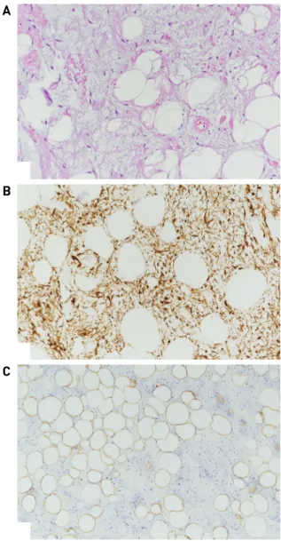
A
B
C
Fig. 3. (A) There are composed of a variable mixture of spindle cells and adipose tissue. The spindle cells are usually small and elongated and arranged haphazardly. The pleomorphic lip- oma is a circumscribed nodule within an otherwise typical lipoma. Areas resembling spindle cell lipoma may sometimes be present within pleomorphic lipomas (H & E, x200). (B) Spindle cells stain for CD34 (CD 34+, x200). (C) Immunohistochemical staining for S-100 (+) (S100, x200).
분엽된 비균질성 고음영의 종괴가 관찰되었다. (Fig. 1).
전산화 단층 촬영 결과 좌측 턱의 피하 지방층에 약 2.3 x 1.3 cm 크기의 분엽성의 조영 증강 되는 종양이 관찰되 었다 (Fig. 2). 이에 양성 연부 조직 종양으로 의심하고 국소 마취 하에 수술적인 치료를 계획하였다.
국소마취하에 좌측 악하선과 흉쇄 유돌근 사이를 경계 로 피부 절개 후에 피하층에 존재하는 종양을 확인하였 다. 주변 조직과 비교적 잘 분리되었으며, 종양의 경계를 확인후 주변 조직의 손상을 최소화하면서 종양을 제거하 였다. 조직 병리 소견상 작고 긴 방추 세포와 핵이 중첩되 는 다양한 수의 거대 세포가 존재하는 방추 세포 / 다형성 지방종으로 진단되었다 (Fig. 3). 환자는 술 후 특별한 합병증 없이 퇴원 후 21개월째 재발 없이 외래 통원 치료 중이다.
고 찰
방추 세포 지방종은 1975년 Enzinger와 Harvey에 의해 처음 기술 되었다.1)방추 세포 지방종과 다형성 지방종 은 유사한 임상학 및 조직학적, 면역조직화학 및 세포유 전학적 특성을 갖고 있어 동일한 개체로 간주되고 있 다.4,8)방추 세포 / 다형성 지방종은 보통 성장속도가 느 린 양성 종양으로 일반적으로 어깨, 등, 뒷목에서 주로 발견된다.1,2) 보통은 남성에서 흔하고 여성에서 발견되 는 경우는 10% 미만이다.3) 방추 세포 / 다형성 지방종 크기는 보통 3~5cm 정도이며 일부에서 14cm이상의 종 양이 보고되기도 하였다. 주변피하조직과 경계를 잘 이
- 43 - 루고 쉽게 구분되어 수술 시 제거가 잘되는 편이다.1,2)
현미경학적으로, 방추 세포 / 다형성 지방종은 방추 세포, 다형성 세포, 성숙한 지방 조직의 비율에서 다양한 조직 학적 특징을 나타낼 수 있다.9)고전적인 방추 세포 지방종은 성숙한 지방과 방추 세포가 비슷한 비율로 구 성된다. 방추 세포는 하나의 길쭉한 핵과 좁은 세포질로 이루어진 동일 형태이며, 핵의 유사분열에 있어 증가된 양상을 보이진 않는다.10)고전적인 다형성 지방종은 호 산구가 있는 세포질에 여러 가지 과대염색체성 핵이 작 은 꽃과 같은 동심 배열(floret-like)을 가지고 있는 산란된 기괴한 거대 세포의 존재를 특징으로 한다.11)
전형적인 방추 세포 지방종은 말초신경초종양, 결절 근막염, 고립성 섬유종 및 근섬유모세포종과 구별하기 어렵다. 이에 CD34 염색법은 S-100 평활근의 액틴, CD99, desmin과 함께 진단에 도움을 줄 수 있는 도구이다. CD34 염색을 통해 방추 세포 / 다형성 지방종이 실제 지방 형성 종양 보다는 수지상 간질성 신생물임을 확인할 수 있다.
S-100단백질은 성숙한 지방 세포의 핵에 염색되는 특징 을 보이며, 방추 세포나 비정형 또는 거대세포에서는 염 색되지 않는다. BCL-2과 CD10 또한 방추 세포 / 다형성 지방종에서 양성 소견을 보이는 경우가 많으나 염색이 특이적이지 않아 감별에 있어 활용적이지 못하다.12) 종 종 방추 세포 지방종은 경화성의 변칙적 지방 종성 종양 과 고분화 지방 육종과 혼동되기도 한다.13)
방추 세포 / 다형성 지방종의 최선의 치료법은 완전한 절제술을 통한 제거이며, 양성 종양으로 전 절제술 후 재발은 드문 편으로 알려져 있다.14) 방추 세포 / 다형성 지방종은 종양의 발생 위치가 다양하고 전형적으로 나타 나는 위치가 아닌 경우에 진단이 어려울 수도 있고 세침 흡인 검사에서 다양한 양상으로 보여져 종양의 완전 절 제술이 확진에 추천 되어진다.15)
앞서 언급한 종양들과의 감별을 위해서도 종양의 제거 를 통한 조직검사가 필요하겠으며, 이전에도 두경부에 서 발생한 방추 세포 / 다형성 지방종 사례는 몇 차례 보고되었으나 국내에서 측경부에서 발생한 경우는 보고 되지 않아 문헌고찰과 함께 보고하는 바이다. 측경부에 발생한 종양의 경우에 방추 세포 / 다형성 지방종 가능성 을 염두에 두어야 할 것이다.
References
1) Enzinger FM, Harvey DA. Spindle cell lipoma. Cancer. 1975;
36:1852-1859.
2) Duve S, Müller-Höcker J, Worret WI. Spindle-cell lipoma of the skin. Am J Dermatopathol. 1995;17:529-533.
3) Billings SD, Henley JD, Summerlin D-J, Valkiki S, Tomich CE.
Spindle cell lipoma of the oral cavity. Am J Dermatopathol.
2006;28:28-31.
4) Shmookler BM, Enzinger FM. Pleomorphic lipoma – A benign tumor simulating liposarcoma. A clinicopathologic analysis of 48 cases. Cancer. 1981;47:126-133.
5) Rubin BP, Fletcher CD. The cytogenetics of lipomatous tumours.
Histopathology. 1997;30:507-511.
6) Reis-Filho JS, Milanezi F, Soares MF, Fillus-Neto J, Schmitt FC.
Intradermal spindle cell/pleomorphic lipoma of the vulva: Case report and review of the literature. J Cutan Pathol. 2002;29:
59-62.
7) Jeon JH, Kim JH, Yu DS, Song HJ, Oh CH. A Case of Spindle Cell Lipoma of the Scalp. Korean J Dermatol. 2014;52:430-432.
8) Dal Cin P, Sciot R, Polito P, Stas M, de Wever I, Comelis A, et al.
Lesions of 13q may occur independently of deletion of 16q in spindle cell/pleomorphic lipomas. Histopathology. 1997;31:
222-225.
9) Reis-Filho JS, Milanezi F, Soares MF, Fillus-Neto J, Schmitt FC.
Intradermal spindle cell/pleomorphic lipoma of the vulva: case report and review of the literature. J Cutan Pathol. 2002;29:
59-62.
10) Fletcher CD, Martin-Bates E. Spindle cell lipoma: a clin- icopathological study with some original observations.
Histopathology. 1987;11:803-817.
11) Diaz-Cascajo C, Borghi S, Weyers W. Pleomorphic lipoma with pseudopapillary structures: a pleomorphic counterpart of pseu- doangiomatous spindle cell lipoma. Histopathology. 2000;36:
75-476.
12) Suster S, Fisher C. Immunoreactivity for the human hema- topoietic progenitor cell antigen (CD34) in lipomatous tumors.
Am J Surg Pathol. 1997;21:195-201.
13) Rubin BP, Dal Cin P. The genetics of lipomatous tumors. Semin Diagn Pathol. 2001;18:286-293.
14) French CA, Mentzel T, Kutzner H, Fletcher CD. Intradermal spindle cell/pleomorphic lipoma. Am J Dermatopathol. 2000;
22:496-502.
15) Domanski HA, Carlen B, Jonsson K, Mertens F, Akerman M.
Distinct cytologic features of spindle cell lipoma. A cytologic-his- tologic study with clinical, radiologic, electron microscopic, and cytogenetic correlations. Cancer. 2001;93:381-389.