• 검색 결과가 없습니다.

Morusin induces cell death through inactivating STAT3 signaling in prostate cancer cells

N/A
N/A
Protected

Academic year: 2021

Share "Morusin induces cell death through inactivating STAT3 signaling in prostate cancer cells"

Copied!
12
0
0

로드 중.... (전체 텍스트 보기)

전체 글

(1)

Original Article

Morusin induces cell death through inactivating STAT3 signaling in prostate cancer cells

Sung-Lyul Lim

1

, Sang-Yoon Park

1

, Sukmin Kang

1

, Dain Park

1

, Sung-Hoon Kim

1,2

, Jae-Young Um

1,2

, Hyeung-Jin Jang

2

, Jun-Hee Lee

3,4

, Chul-Ho Jeong

5

, Jung-Hee Jang

6

, Kwang Seok Ahn

1,2

, Seok-Geun Lee

1,2,4

1

Cancer Preventive Material Development Research Center,

2

Department of Science in Korean Medicine,

3

De- partment of Sasang Constitutional Medicine, College of Korean Medicine, Kyung Hee University, Seoul 130-701, Republic of Korea;

4

Korean Medicine Clinical Trial Center, Kyung Hee University Korean Medicine Hospital, Seoul 130-872, Republic of Korea;

5

Institute for New Drug Development, College of Pharmacy,

6

Department of Pharma- cology, School of Medicine, Keimyung University, Daegu 704-701, Republic of Korea

Received October 30, 2014; Accepted November 29, 2014; Epub December 15, 2014; Published January 1, 2015 Abstract: STAT3 has been recognized as an efficacious drug target for prostate cancer because of its constitutive activation in this fatal disease. We recently identified the root bark of Morus alba Linn. as a potential STAT3 inhibitor among 33 phytomedicines traditionally used in Korea. Morusin, an active compound isolated from the root bark of Morus alba, has shown anti-oxidant and anti-inflammatory effects. In the present study, we examined whether morusin has a potential as an anti-cancer agent in prostate cancer. We found that morusin suppressed viability of prostate cancer cells, but little effect in normal human prostate epithelial cells. Morusin also reduced STAT3 activity by inhibiting its phosphorylation, nuclear accumulation, and DNA binding activity. In addition, morusin down-regu- lated expression of STAT3 target genes encoding Bcl-xL, Bcl-2, Survivin, c-Myc and Cyclin D1, which are involved in regulation of apoptosis and cell cycle. Furthermore, morusin induced apoptosis in human prostate cancer cells by reducing STAT3 activity. Taken together, these results suggest that morusin could be a potentially therapeutic agent for prostate cancer by reducing STAT3 activity and inducing apoptosis.

Keywords: Morusin, prostate cancer, apoptosis, STAT3, SHP1, traditional phytomedicine

Introduction

The root bark of Morus alba Linn., also known and read as Sang Bek Pi in Korea, has been traditionally used in oriental medicine including Korean medicine and Chinese medicine as a diuretic, anti-tussive, anti-diabetic, or anti- inflammatory drug [1, 2]. The root bark of Morus alba Linn. has several active compounds such as prenylated flavonoids, stilbenes, benzofu- rans, alkaloids, and other phenolic compounds [3]. Morusin, a prenylated flavonoid isolated from the root bark of Morus alba Linn. [4], dis- played anti-microbial activity, scavenging activ- ity against superoxide anion radical, and anti- inflammatory activity [5-7]. Two recent studies showed that morusin induced apoptosis in can- cer cells [8, 9]. One showed that morusin can inhibit cervical cancer stem cell growth and migration through reducing NF-κB and inducing apoptosis [9]. Another noticed that morusin

may induce apoptosis via activating caspases and inhibiting NF-κB in human colorectal can- cer HT-29 cells [8].

Prostate cancer is the most common cancer

among men in the United States and is the sec-

ond leading cause of cancer death, affecting

28% of all male cancer cases and 10% of all

male cancer deaths [10]. In addition, the inci-

dence of prostate cancer in South Korea is the

fifth and mortality of prostate cancer is the sev-

enth among men [11]. However, the incidence

of prostate cancer in South Korea is rapidly

increasing [12]. Although different types of

treatment including surgery, radiotherapy, che-

motherapy and hormonal therapy are available

for patients with primary prostate cancer, the

complications frequently associated with these

conventional treatments diminish positive clini-

cal outcomes [13]. In addition, few options are

still available for treating patients with advanced

(2)

or metastatic stages of the disease [14].

Therefore, more efficient and advanced treat- ment strategies are required for cure of patients from the fatal disease.

STAT3, a member of the STAT family, is a tran- scription factor [15]. Various signals to stimu- late STAT3 induce phosphorylation and dimer- ization (homodimers or heterodimers) of STAT3, resulting in its translocation from the cytoplasm to the nucleus to bind each promoter of target genes that are involved in apoptosis, prolifera- tion, differentiation, angiogenesis, and survival [15]. Constitutive activation of STAT3 induces deregulation of cell proliferation and survival that mediates inflammation and tumorigenesis in many cancers [15, 16]. Interestingly, some studies have suggested a correlation between STAT3 and prostate cancer [17, 18]. Recent studies also indicated that inhibition of STAT3 can induce apoptosis in prostate cancer cells and that STAT3 promotes metastasis in pros- tate cancer [19, 20]. Therefore, the STAT3- targeted therapy has shown a great promise of a therapeutic strategy for prostate cancer [15, 21]. Based on these results, we developed the system to screen potential STAT3 inhibitors and found that the root bark of Morus alba Linn.

among 33 phytomedicines traditionally used in Korean medicine decreased STAT3 activity (unpublished data). Thus we have tried to find which active compound in the root bark of Morus alba Linn. plays a role as a STAT3 inhibi- tor, and could be used as an anti-prostate can- cer agent. In the present study, we suggest that morusin, one of active compounds in the root bark of Morus alba Linn. could be a potential anti-cancer agent for prostate cancer by inhibit- ing STAT3 activity.

Materials and methods Cell lines

DU145, PC3 and LNCaP and RWPE-1 were pre- viously described [22, 23], and M2182 was kindly provided by Dr. Paul B. Fisher (Virginia Commonwealth University School of Medicine, Richmond, VA, USA). RWPE-1 was cultured in keratinocyte growth medium-gold bullet kit (Lonza, Inc., Allendale, NJ, USA). DU145, PC3, M2182 and LNCaP were cultured in RPMI-1640 (Lonza) supplemented with 10% fetal bovine serum (Lonza) and 100 U/ml of the antibiotics

and antimycotics (Lonza). All cells were cultured in humidified incubator with 5% CO

2

at 37°C.

Reagents

Morusin purchased from Biopurify Phytoche- micals Ltd. (Chengdu, Sichuan, China) was dis- solved in DMSO (Sigma-Aldrich, St. Louis, Mo, USA) to make 50 mM stock solutions. 3-(4,5- dimethylthiazol-2-yl)-2,5-diphenyltetrazolium bromide (MTT) and sodium orthovanadate were purchased from Sigma-Aldrich, and H

2

O

2

was from Junsei Chemical (Tokyo, Japan).

Cell viability assays

Cell viability was measured by MTT assays as described previously [24]. Cells (3-10 × 10

3

cells/well) were seeded in 96-well plates, and treated with different concentrations of mor- usin for 24 hours. Cell viability was presented as percentage compared to the control.

Western blot analysis

Whole cell extracts were prepared, and Western blotting was carried out as described [25].

Primary antibodies STAT3 (1:1000), p (phos- pho)-STAT3 (1:1000), JAK2 (1:1000), SRC (1:

1000), p-SRC (1:1000), C (cleaved)-CASP3 (1:

1000), C-CASP8 (1:1000), C-CASP9 (1:1000), Bcl-xL (1:1000), c-Myc (1:1000) and Cyclin D1 (1:1000) from Cell Signaling Technology (Danvers, MA, USA), p-JAK2 (1:1000), Bcl-2 (1:1000), Survivin (1:1000), PARP (1:1000), SHP1 (1:1000) and SHP2 (1:1000) from Santa Cruz Biotechnology (Santa Cruz, CA, USA), β-actin (1:10000) from Sigma-Aldrich, and sec- ondary antibodies HRP–conjugated anti-mouse IgG or anti-rabbit IgG (1:5000; Cell Signaling Biotechnology) were used for immunoblotting.

Then the membranes were detected using a chemiluminescence system (Corebio, Seoul, Republic of Korea).

Immunofluorescence staining

Cells were seeded in 8-well chamber slides and

treated with morusin for 6 hours. After the cells

were fixed and permeabilized, the cells were

stained with rabbit polyclonal anti-p-STAT3 anti-

body (1:100), Alexa Flour 488 goat anti-rabbit

antibody (1:1000) (Invitrogen, Carlsbad, CA,

USA) and 4’,6-diamidino-2-phenylindole (DAPI)

(Sigma-Aldrich) as described [26]. The images

(3)

were observed by the FluoView FV1000 confo- cal microscope (Olympus, Tokyo, Japan).

Transient transfection and luciferase (Luc) as- says

Cells were transfected with pSTAT3-Luc report- er containing STAT3 binding sites to measure STAT3 activity (Clontech, Palo Alto, CA, USA) in a 60-mm plate using Lipofectamine 2000 (Invitrogen). One day after transfection, cells were re-plated into a 48-well plate. Then the cells were treated with morusin for 24 hours.

Luc assays were performed using a Luc assay system (Promega, Madison, WI, USA) according to the manufacturer’s instructions. Luc activity was normalized by protein concentration.

Reverse transcription-polymerase chain reaction (RT-PCR)

Total RNA was isolated using PureHelix™ RNA Extraction Solution (Nanohelix, Seoul, Republic of Korea). 1 μg of total RNA was used for RT-PCR using Maxime™ RT PreMix Kit (iNtRON Biotech- nology, Kyounggi, Republic of Korea) and HiPi Figure 1. Effects of morusin on cell viability in human prostate cancer cells. (A) DU145, (B) M2182, (C) PC3, (D) LNCaP and (E) RWPE-1 cells were treated with morusin for 24 hours as indicated. Cell viability was determined by MTT assays. Data in the graphs are pre- sented as the mean ± SEM (*, P < 0.05; and ***, P <

0.001 versus mock-treated control).

(4)

Plus 5X PCR Master Mix (ELPIS Biotech, Daejeon, Republic of Korea) according to the manufacturer’s protocol. Sequences of the primers used for RT-PCR are as following: BCL2 sense, 5’-ATGGCGCACGCTGGGAGAAC-3’; BCL2 antisense, 5’-GGTGTGCAGGTGCCGGTTGA-3’;

BCL-XL sense, 5’-TACCAGCCTGACCAATATGGC- 3’; BCL-XL antisense, 5’-TGGGTTCAAGTGATTC- TCCTG-3’; Survivin sense, 5’-TTGGCCCAGTGTT- TCTTCTGCTTC-3’; Survivin antisense, 5’-GCAC- TTTCTCCGCAGTTTCCTCAA-3’; Cyclin D1 sense, 5’-AGAAGCTGTGCATCTACACCGACA-3’; Cyclin D1 antisense, 5’-TGATCTGTTTGTTCTCCTCCGC- CT-3’; GAPDH sense, 5’-ATGGGGAAGGTGAAGG- TCGGAGTC-3’; and GAPDH antisense, 5’-GC- TGATGATCTTGAGGCTGTTGTC-3’.

Transcription factor activity assays

Nuclear extracts were prepared and 2 µg of nuclear extracts in each sample was analyzed for the STAT3 DNA binding activity using the TransAM STAT3 kit (Active Motif, Carlsbad, CA, USA) according to the manufacturer’s protocol.

Apoptosis analysis

Cell cycle and apoptosis were observed respec- tively by Propidium Iodide (PI) (Invitrogen) and Annexin V (ApoScan from BioBud, Seoul, Republic of Korea) staining followed by flow cytometry analysis using FACS Calibur (BD Biosciences, San Jose, CA, USA) as described [27]. TUNEL assays were also performed using a DeadEnd™ Fluorometric TUNEL assay kit (Promega) according to the manufacturer’s protocol.

Statistical analysis

Data were presented as mean ± standard error of the mean (SEM) from at least three indepen- dent experiments in triplicate or more and ana-

lyzed for statistical significance using the unpaired Student’s t-test. P < 0.05 was consid- ered significant.

Results

Morusin suppresses cell viability in human prostate cancer cells

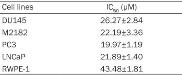
In order to investigate the possible therapeutic effect of morusin in prostate cancer, we first examined whether morusin affects viability of human prostate cancer cells. Human prostate cancer cell lines DU145, M2182, PC3 and LNCaP, and a normal immortalized prostate epithelial cell line RWPE-1 were treated with morusin for 24 hours, and cell viability was measured by MTT assays. Intriguingly, morusin suppressed cell viability in a dose-dependent manner in all the prostate cancer cell lines but less effect on RWPE-1 cells (Figure 1A-E). IC

50

values also indicated that morusin is a more potent cytotoxic reagent in prostate cancer cells compared to that in normal cells (Table 1).

These results suggested that morusin is a potentially favorable therapeutic phytochemi- cal for prostate cancer.

Morusin induces apoptosis in human prostate cancer cells

Since apoptosis is a crucial mechanism by which phytochemicals regulate cancer cell via- bility, we then investigated whether morusin can affect cell cycle and apoptosis in human prostate cancer cells DU145 and M2182. As shown in Figure 2A and Supplementary Figure 1A, morusin increased the cell population of the sub-G

1

phase in both DU145 and M2182 which is indicative of apoptosis. To further con- firm whether morusin induces apoptosis, we performed Annexin-V staining assays. As shown in Figure 2B and Supplementary Figure 1B, morusin increased Annexin-V positive cell pop- ulation in both cell lines, corroborating that morusin induced apoptosis in prostate cancer cells. These results were also confirmed by TUNEL assays (Figure 2C). In addition, we found that morusin increased cleavage of both initia- tors (CASP8 and CASP9) and executioner (CASP3) caspases and also PARP cleavage, indicating their activation (Figure 2D). Together these results indicated that morusin induces apoptosis in prostate cancer cells.

Table 1. IC

50

value of each prostate cancer cells

Cell lines IC

50

(µM)

DU145 26.27±2.84

M2182 22.19±3.36

PC3 19.97±1.19

LNCaP 21.89±1.40

RWPE-1 43.48±1.81

IC

50

was determined using the data morusin treatment

for 24 hours. Data are presented as the mean ± SEM.

(5)

Morusin inhibits the STAT3 signaling pathway in human prostate cancer cells

Since morusin is an active compound of the root bark of Morus alba Linn. [4], which we found as a potential inhibitor of STAT3 (data not shown), we expected that morusin could inhibit the STAT3 signaling pathway that might be the mechanism by which morusin induced apopto- sis and reduced viability of prostate cancer cells. In order to investigate the effect of

morusin on the STAT3 pathway, we analyzed its effect on the STAT3 phosphorylation in pros- tate cancer cells. As shown in Figure 3A, morusin reduced the phosphorylation levels of STAT3 in DU145 and M2182 cells. In addition, morusin decreased the phosphorylation levels of upstream signaling molecules of STAT3, SRC and JAK2 (Figure 3A). After phosphorylation, STAT3 dimerizes and translocates to the nucle- us where STAT3 act as a transcriptional activa- tor [16]. Therefore, we next analyzed the effect Figure 2. Effect of morusin on apoptosis in human prostate cancer cells. DU145 and M2182 cells were treated with the indicated concentration of morusin for 24 hours. A. The cells were stained with PI, and analyzed by flow cytom- etry. B. The cells were stained with Annexin V, and analyzed by flow cytometry. C. DU145 and M2182 cells were treated with morusin (30 μM) for 24 hours. The TUNEL and DAPI staining were analyzed by confocal microscopy.

Scale bar, 70 μm. D. DU145 and M2182 cells were treated with morusin for 24 hours. Whole cell lysates were sub-

jected to Western blotting with the indicated antibodies. β-actin was used as an internal control. Data in the graphs

are presented as the mean ± SEM (*, P < 0.05; **, P < 0.01; and ***, P < 0.001 versus mock-treated control).

(6)

of morusin on nuclear accumulation and DNA binding activity of STAT3 in prostate cancer cells. As shown in Figure 3B and 3C, morusin inhibited the nuclear accumulation of STAT3, and also reduced its DNA binding activity in DU145 and M2182 cells. These results noticed that morusin is a potent STAT3 inhibitor, and suggested that STAT3 might be a target of morusin to decrease cell viability in prostate cancer.

Morusin represses transcriptional activity of STAT3 in human prostate cancer cells

Since STAT3 is a well-known transcription fac- tor which regulates target genes involved in

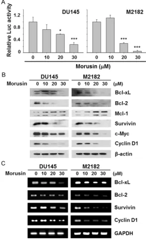
apoptosis, cell proliferation, and other cellular functions [28, 29], we next examined whether morusin regulates the STAT3-mediated tran- scriptional activation in prostate cancer cells using the pSTAT3-Luc reporter system. As shown in Figure 4A, morusin repressed the transcriptional activity of STAT3 in both DU145 and M2182 cells. Furthermore, morusin reduced protein levels of Bcl-xL, Bcl-2, Survivin, c-Myc, and Cyclin D1 (Figure 4B), which are STAT3 target genes involved in regulation of apoptosis and cell proliferation [30, 31].

Accordingly, morusin down-regulated mRNA

levels of BCL-XL, BCL-2, Survivin, and Cyclin D1

(Figure 4C). These results suggested that

morusin might regulate apoptosis and cell via-

Figure 3. Effect of morusin on the STAT3 signaling pathway in human prostate cancer cells. A. DU145 and M2182

cells were treated with morusin for 6 hours. Cell lysates were subjected to Western blotting with the indicated an-

tibodies. β-actin was used as an internal control. B. DU145 and M2182 cells were treated with morusin (30 μM)

for 6 hours. The p-STAT3 and DAPI staining were analyzed by confocal microscopy. Scale bar, 10 μm. C. DU145 and

M2182 cells were treated with morusin for 24 hours. Nuclear extracts were subjected for measuring the STAT3 DNA

binding activity. Data in the graphs are presented as the mean ± SEM (**, P < 0.01; and ***, P < 0.001 versus

mock-treated control).

(7)

sion of these proteins in morusin treated pros- tate cancer cells. As shown in Figure 5A, morusin dramatically upregulated SHP1 in a dose-dependent manner, but not SHP2 in DU145 and M2182 prostate cancer cells. Next, we confirmed whether SHP1 dominantly partici- pates in morusin-mediated inhibition of STAT3 using by treatment of PTPs inhibitor, pervana- date. Treatment of pervanadate reversed morusin-mediated inhibition of STAT3 phos- phorylation and also reduced morusin-mediat- ed apoptosis in DU145 and M2182 cells (Figure 5B and 5C), indicating the critical role of SHP1 in morusin-induced STAT3 inhibition.

Discussion

Prostate cancer is the second most frequently diagnosed cancer and the sixth leading cause of cancer death worldwide in males, but the most frequent cancer among males in the developed countries because of the wide utili- zation of prostate-specific antigen (PSA) testing [34]. The increase of PSA testing led to earlier diagnosis, which may increase the survival time of prostate cancer patients but does not neces- sarily reduce mortality [34]. During the earlier stages, most prostate cancers are dependent on androgen hormone, and tumor treatment can be obtained by surgery, radiotherapy, and androgen deprivation [35, 36]. However, these treatments are not effective for advanced and metastatic prostate cancer. Therefore, recent studies have noticed effective phytochemicals for advanced stage of prostate cancer [37-39].

In the cytotoxicity assay to verify whether morusin could be a novel therapeutic agent for prostate cancer, we found that morusin sup- pressed cell viability in human prostate cancer cells, but interestingly had less toxic effect in normal prostate cells (Figure 1). Although we found that morusin induced apoptosis in pros- tate cancer cells (Figure 2 and Supplementary Figure 1), its rate (15~20% at 30 μM of morusin) was lower than cytotoxicity (~80% at the same concentration) (Figures 1 and 2). These results indicate that morusin may induce other cell death such as necrosis, autophagy and etc. in prostate cancer cells. A recent report stated that another type of prenylated flavonoid xan- thohumol has participated in autophagosome maturation via direct interaction with autopha- gosome protein [40] supports this hypothesis.

Figure 4. Effect of morusin on transcriptional activity of STAT3 in human prostate cancer cells. A. DU145 and M2182 cells were transfected with pSTAT3-Luc plasmid and the transfected cells were treated with morusin for 24 hours. Data in the graphs are pre- sented as the mean of fold-normalized activities ± SEM to that of the untreated cells taken as 1 (*, P <

0.05; and ***, P < 0.001 versus mock-treated con- trol). B and C. DU145 and M2182 cells were treated with morusin for 24 hours. B. The treated cells were analyzed for protein levels of the STAT3 target genes.

β-actin was used as an internal control. C. The treat- ed cells were analyzed for mRNA levels of the STAT3 target genes. GAPDH was used as an internal control.

bility of prostate cancer cells by inhibiting tran- scriptional activity of STAT3.

Morusin induces apoptosis by inhibiting STAT3 in human prostate cancer cells

Non-transmembrane protein tyrosine phospha-

tases (PTPs) have critical roles in the inhibitory

regulation of the JAK/STAT signaling pathway in

various type of cancer cells [32, 33]. To verify

whether inactivation of JAK2/STAT3 signaling

by morusin was mediated by induction of PTPs,

such as SHP1 and SHP2, we quantified expres-

(8)

Since STAT3 has been found constitutively acti- vated in many cancers including prostate can- cer, STAT3 has been recognized as a valuable drug target [28, 29]. For this reason, we devel- oped the system to screen potential STAT3 inhibitors in traditionally used Korean medi- cines, and morusin is one of candidates screened as a STAT3 inhibitor (unpublished data). Therefore we verified whether morusin can affect STAT3 activity in prostate cancer

cells at first. Our results clearly showed that

morusin inhibited the SRC/JAK2/STAT3 signal-

ing pathway by blocking their phosphorylation,

and consequential loss of nuclear accumula-

tion and transcriptional activity of STAT3

(Figures 3 and 4A). These STAT3 inhibition

resulted in suppression of the downstream tar-

get genes encoding anti-apoptotic proteins

such as Bcl-xL, Bcl-2 and Survivin and cell cycle

regulator proteins such as c-Myc and Cyclin D1

Figure 5. Effect of pervanadate on morusin-mediated STAT3 inhibition and apoptosis in human prostate cancer

cells. A. DU145 and M2182 cells were treated with morusin for 6 hours. B. DU145 and M2182 cells were treated

with pervanadate (0.5 μM) and morusin (30 μM) for 6 hours. A and B. Cell lysates were subjected to Western blotting

with the indicated antibodies. β-actin was used as an internal control. C. DU145 and M2182 cells were treated with

pervanadate (0.5 μM) and morusin (30 μM) for 24 hours. The TUNEL and DAPI staining were analyzed by confocal

microscopy (C, control; M, morusin; and P, pervanadate). Scale bar, 70 μm. Quantitative analysis of TUNEL positive

cells are expressed as relative amount of the treated cells compared to that of the mock-treated control. Data in the

graphs are presented as the mean ± SEM (*, P < 0.05; and **, P < 0.01 versus morusin-treated control).

(9)

(Figure 4B and 4C). Moreover, morusin signifi- cantly induced SHP1 expression and inhibition of SHP1 by PTPs inhibitor pervanadate partially restored p-STAT3 level (Figure 5). These results suggest that morusin may inhibit STAT3 signal- ing via direct participation in regulation of tyro- sine kinases, such as SRC, JAK2 and EGFR, as well as SHP1. Taken together these results indi- cate that morusin induces apoptosis through inhibiting STAT3 by induction of SHP1 in pros- tate cancer cells, and suggest that morusin is a potentially useful anti-cancer agent for pros- tate cancer. Further studies including in vivo animal studies and other types of human tumors with increased active STAT3 are war- ranted and some are currently in progress to test its anti-cancer effect and to develop improved therapies for prostate cancer and methods for ameliorating its pathogenesis.

Acknowledgements

We thank Dr. Paul B. Fisher (Virginia Common- wealth University School of Medicine, Rich- mond, VA, USA) for the cell line M2182. This work was supported by a research grant from the National Research Foundation of Korea (2007-0054931).

Disclosure of conflict of interest

The authors declare no conflict of interests.

Address correspondence to: Seok-Geun Lee, Cancer Preventive Material Development Research Center, Department of Science in Korean Medicine, College of Korean Medicine, Kyung Hee University, Seoul 130-701, Republic of Korea. Tel: +82-2-961-2355;

Fax: +82-2-961-2355; E-mail: seokgeun@khu.ac.kr References

[1] Asano N, Yamashita T, Yasuda K, Ikeda K, Kizu H, Kameda Y, Kato A, Nash RJ, Lee HS and Ryu KS. Polyhydroxylated alkaloids isolated from mulberry trees (Morusalba L.) and silkworms (Bombyx mori L.). J Agric Food Chem 2001; 49:

4208-4213.

[2] Singab AN, El-Beshbishy HA, Yonekawa M, No- mura T and Fukai T. Hypoglycemic effect of Egyptian Morus alba root bark extract: effect on diabetes and lipid peroxidation of strepto- zotocin-induced diabetic rats. J Ethnopharma- col 2005; 100: 333-338.

[3] Lee HJ, Lyu da H, Koo U, Nam KW, Hong SS, Kim KO, Kim KH, Lee D and Mar W. Protection of prenylated flavonoids from Mori Cortex Radi- cis (Moraceae) against nitric oxide-induced

cell death in neuroblastoma SH-SY5Y cells.

Arch Pharm Res 2012; 35: 163-170.

[4] Zong YY, Ip SP, Dong TX and Che CT. [Determi- nation of morusin in cortex mori]. Zhongguo Zhong Yao Za Zhi 2007; 32: 1038-1040.

[5] Bellik Y, Boukraa L, Alzahrani HA, Bakhotmah BA, Abdellah F, Hammoudi SM and Iguer-Oua- da M. Molecular mechanism underlying anti- inflammatory and anti-allergic activities of phy- tochemicals: an update. Molecules 2012; 18:

322-353.

[6] Fukai T, Satoh K, Nomura T and Sakagami H.

Antinephritis and radical scavenging activity of prenylflavonoids. Fitoterapia 2003; 74: 720- [7] Sohn HY, Son KH, Kwon CS, Kwon GS and 724.

Kang SS. Antimicrobial and cytotoxic activity of 18 prenylated flavonoids isolated from medici- nal plants: Morus alba L., Morus mongolica Schneider, Broussnetia papyrifera (L.) Vent, Sophora flavescens Ait and Echinosophora ko- reensis Nakai. Phytomedicine 2004; 11: 666- [8] Lee JC, Won SJ, Chao CL, Wu FL, Liu HS, Ling P, 672.

Lin CN and Su CL. Morusin induces apoptosis and suppresses NF-kappaB activity in human colorectal cancer HT-29 cells. Biochem Bio- phys Res Commun 2008; 372: 236-242.

[9] Wang L, Guo H, Yang L, Dong L, Lin C, Zhang J, Lin P and Wang X. Morusin inhibits human cer- vical cancer stem cell growth and migration through attenuation of NF-kappaB activity and apoptosis induction. Mol Cell Biochem 2013;

379: 7-18.

[10] Siegel R, Naishadham D and Jemal A. Cancer statistics, 2013. CA Cancer J Clin 2013; 63:

11-30.

[11] Jung KW, Won YJ, Kong HJ, Oh CM, Seo HG and Lee JS. Cancer statistics in Korea: incidence, mortality, survival and prevalence in 2010.

Cancer Res Treat 2013; 45: 1-14.

[12] Park SK, Sakoda LC, Kang D, Chokkalingam AP, Lee E, Shin HR, Ahn YO, Shin MH, Lee CW, Lee DH, Blair A, Devesa SS and Hsing AW. Ris- ing prostate cancer rates in South Korea. Pros- tate 2006; 66: 1285-1291.

[13] Michaelson MD, Cotter SE, Gargollo PC, Ziet- man AL, Dahl DM and Smith MR. Management of complications of prostate cancer treatment.

CA Cancer J Clin 2008; 58: 196-213.

[14] Bhatnagar V and Kaplan RM. Treatment op- tions for prostate cancer: evaluating the evi- dence. Am Fam Physician 2005; 71: 1915- 1922.

[15] Yu H, Pardoll D and Jove R. STATs in cancer in- flammation and immunity: a leading role for STAT3. Nat Rev Cancer 2009; 9: 798-809.

[16] Timofeeva OA, Tarasova NI, Zhang X, Chaso-

vskikh S, Cheema AK, Wang H, Brown ML and

Dritschilo A. STAT3 suppresses transcription of

(10)

proapoptotic genes in cancer cells with the in- volvement of its N-terminal domain. Proc Natl Acad Sci U S A 2013; 110: 1267-1272.

[17] DeMiguel F, Lee SO, Lou W, Xiao X, Pflug BR, Nelson JB and Gao AC. Stat3 enhances the growth of LNCaP human prostate cancer cells in intact and castrated male nude mice. Pros- tate 2002; 52: 123-129.

[18] Ni Z, Lou W, Leman ES and Gao AC. Inhibition of constitutively activated Stat3 signaling path- way suppresses growth of prostate cancer cells. Cancer Res 2000; 60: 1225-1228.

[19] Abdulghani J, Gu L, Dagvadorj A, Lutz J, Leiby B, Bonuccelli G, Lisanti MP, Zellweger T, Alanen K, Mirtti T, Visakorpi T, Bubendorf L and Neva- lainen MT. Stat3 promotes metastatic progres- sion of prostate cancer. Am J Pathol 2008;

172: 1717-1728.

[20] Barton BE, Karras JG, Murphy TF, Barton A and Huang HF. Signal transducer and activator of transcription 3 (STAT3) activation in prostate cancer: Direct STAT3 inhibition induces apop- tosis in prostate cancer lines. Mol Cancer Ther 2004; 3: 11-20.

[21] Bowman T, Garcia R, Turkson J and Jove R.

STATs in oncogenesis. Oncogene 2000; 19:

2474-2488.

[22] Bhutia SK, Dash R, Das SK, Azab B, Su ZZ, Lee SG, Grant S, Yacoub A, Dent P, Curiel DT, Sarkar D and Fisher PB. Mechanism of autophagy to apoptosis switch triggered in prostate cancer cells by antitumor cytokine melanoma differ- entiation-associated gene 7/interleukin-24.

Cancer Res 2010; 70: 3667-3676.

[23] Chiang SY, Kim SM, Kim C, Um JY, Park KR, Kim SW, Lee SG, Jang HJ, Nam D, Ahn KS, Kim SH, Choi SH, Shim BS, Na YC, Jeong EK, Cho SK and Ahn KS. Antiproliferative effects of Dangyuja (Citrus grandis Osbeck) leaves through suppression of constitutive signal transducer and activator of transcription 3 ac- tivation in human prostate carcinoma DU145 cells. J Med Food 2012; 15: 152-160.

[24] Lee SG, Jeon HY, Su ZZ, Richards JE, Vozhilla N, Sarkar D, Van Maerken T and Fisher PB. As- trocyte elevated gene-1 contributes to the pathogenesis of neuroblastoma. Oncogene 2009; 28: 2476-2484.

[25] Lee SG, Kim K, Kegelman TP, Dash R, Das SK, Choi JK, Emdad L, Howlett EL, Jeon HY, Su ZZ, Yoo BK, Sarkar D, Kim SH, Kang DC and Fisher PB. Oncogene AEG-1 promotes glioma-induced neurodegeneration by increasing glutamate excitotoxicity. Cancer Res 2011; 71: 6514- 6523.

[26] Park SY, Lim SL, Jang HJ, Lee JH, Um JY, Kim SH, Ahn KS and Lee SG. Embelin induces apoptosis in human glioma cells through inac-

tivating NF-kappaB. J Pharmacol Sci 2013;

121: 192-199.

[27] Lee SG, Su ZZ, Emdad L, Sarkar D, Franke TF and Fisher PB. Astrocyte elevated gene-1 acti- vates cell survival pathways through PI3K-Akt signaling. Oncogene 2008; 27: 1114-1121.

[28] Coffer PJ, Koenderman L and de Groot RP. The role of STATs in myeloid differentiation and leu- kemia. Oncogene 2000; 19: 2511-2522.

[29] Garcia R, Bowman TL, Niu G, Yu H, Minton S, Muro-Cacho CA, Cox CE, Falcone R, Fairclough R, Parsons S, Laudano A, Gazit A, Levitzki A, Kraker A and Jove R. Constitutive activation of Stat3 by the Src and JAK tyrosine kinases par- ticipates in growth regulation of human breast carcinoma cells. Oncogene 2001; 20: 2499- 2513.

[30] Aggarwal BB, Sethi G, Ahn KS, Sandur SK, Pan- dey MK, Kunnumakkara AB, Sung B and Ichikawa H. Targeting signal-transducer-and- activator-of-transcription-3 for prevention and therapy of cancer: modern target but ancient solution. Ann N Y Acad Sci 2006; 1091: 151- [31] Zhang X, Zhang J, Wang L, Wei H and Tian Z. 169.

Therapeutic effects of STAT3 decoy oligodeoxy- nucleotide on human lung cancer in xenograft mice. BMC Cancer 2007; 7: 149.

[32] Han Y, Amin HM, Franko B, Frantz C, Shi X and Lai R. Loss of SHP1 enhances JAK3/STAT3 sig- naling and decreases proteosome degradation of JAK3 and NPM-ALK in ALK+ anaplastic large-cell lymphoma. Blood 2006; 108: 2796- 2803.

[33] Tang TL, Freeman RM Jr, O’Reilly AM, Neel BG and Sokol SY. The SH2-containing protein-tyro- sine phosphatase SH-PTP2 is required up- stream of MAP kinase for early Xenopus devel- opment. Cell 1995; 80: 473-483.

[34] Jemal A, Bray F, Center MM, Ferlay J, Ward E and Forman D. Global cancer statistics. CA Cancer J Clin 2011; 61: 69-90.

[35] Arnold JT and Isaacs JT. Mechanisms involved in the progression of androgen-independent prostate cancers: it is not only the cancer cell’s fault. Endocr Relat Cancer 2002; 9: 61-73.

[36] Denis LJ and Griffiths K. Endocrine treatment in prostate cancer. Semin Surg Oncol 2000;

18: 52-74.

[37] Diallo JS, Betton B, Parent N, Peant B, Lessard L, Le Page C, Bertrand R, Mes-Masson AM and Saad F. Enhanced killing of androgen-indepen- dent prostate cancer cells using inositol hexakisphosphate in combination with protea- some inhibitors. Br J Cancer 2008; 99: 1613- 1622.

[38] Tang L, Jin T, Zeng X and Wang JS. Lycopene

inhibits the growth of human androgen-inde-

pendent prostate cancer cells in vitro and in

(11)

BALB/c nude mice. J Nutr 2005; 135: 287- 290.

[39] Teiten MH, Gaascht F, Eifes S, Dicato M and Diederich M. Chemopreventive potential of curcumin in prostate cancer. Genes Nutr 2010;

5: 61-74.

[40] Sasazawa Y, Kanagaki S, Tashiro E, Nogawa T, Muroi M, Kondoh Y, Osada H and Imoto M.

Xanthohumol impairs autophagosome matura-

tion through direct inhibition of valosin-con-

taining protein. ACS Chem Biol 2012; 7: 892-

900.

(12)

Supplementary Figure 1. Effect of morusin on apoptosis in human prostate cancer cells. DU145 and M2182 cells

were treated with the indicated concentration of morusin for 24 hours. A. The cells were stained with PI, and ana-

lyzed by flow cytometry. B. The cells were stained with Annexin V, and analyzed by flow cytometry.

수치

Table 1. IC 50  value of each prostate cancer  cells Cell lines IC 50  (µM) DU145 26.27±2.84 M2182 22.19±3.36 PC3 19.97±1.19 LNCaP 21.89±1.40 RWPE-1 43.48±1.81
Figure 4. Effect of morusin on transcriptional activity  of STAT3 in human prostate cancer cells

참조

관련 문서

These results suggest that the bilobalide inhibits the cell proliferation and induces the apoptotic cell death in FaDu human pharyngeal squamous cell carcinoma via both

Concentration-Dependent inhibition of cell viability by adenosine in human oral fibroblast and FaDu human head and neck squamous cell carcinoma · · ·

Differential Expression of Desmoglein1, Desmoglein3, Epithelial Membrane Antigen, Ber-EP4 and CD10 in Basal Cell Carcinoma and Squamous Cell Carcinoma

1) Effects of methanol extracts of Capsella bursa-pastoris on cyclooxygenase-2 (COX-2) and Inducible Nitric oxide synthase (iNOS) expression in human prostate cancer cell

Inhibitory effects of classified methanol extracts of Smilax china L on the COX-2 and iNOS expression of human colorectal cancer cell lines... Inhibitory

In vitro cell migration in APE or JAG1 siRNA-treated M059K cell line Fig,11 Down-regulation of JAG1 induces S phase arrest in

Green (University of New South Wales), and Reuben Collins (Colorado School of Mines) MRS Bulletin (2008)... World

 Often found connected to other molecules on the outsides of cells --- cellular recognition, cell signaling, cell