• 검색 결과가 없습니다.

Analysis of ROS and Apoptosis of Porcine Skin-derived Stem-like Cells after Differentiation Induction into Mesodermal Cell Types

N/A
N/A
Protected

Academic year: 2021

Share "Analysis of ROS and Apoptosis of Porcine Skin-derived Stem-like Cells after Differentiation Induction into Mesodermal Cell Types"

Copied!
7
0
0

로드 중.... (전체 텍스트 보기)

전체 글

(1)

Analysis of ROS and Apoptosis of Porcine Skin-derived Stem-like Cells after Differentiation Induction into Mesodermal Cell Types

Hyo-Kyung Bae

1,*

, Hwa-Yeon Lee

1,*

, Yeo-Reum Park

1

, Choon-Keun Park

2

, Boo-KeunYang

2

and Hee-Tae Cheong

1,†

1

College of Veterinary Medicine, Kangwon National University, Chuncheon 24375, Korea

2

College of Animal Life Sciences, Kangwon National University, Chuncheon24375, Korea

ABSTRACT

The present study was conduct to examine the H

2

O

2

expression level and apoptosis-related gene expression levels inporcineskin-derived stem cell-like cells (pSSCs) after adipogenic, chondrogenic, and osteogenic differentiation induction. The pSSCs were obtained by digestion of porcine ear skin biopsy and cultured in each induction medium for 21 to 26 days to induce adipogenic, chondrogenic, and osteogenic differentiation, respectively. The H

2

O

2

levels of pSSCs after induction culture were evaluated by staining with 2’7’-dichlorodihydrofluorescein diacetate (H

2

DCFDA).

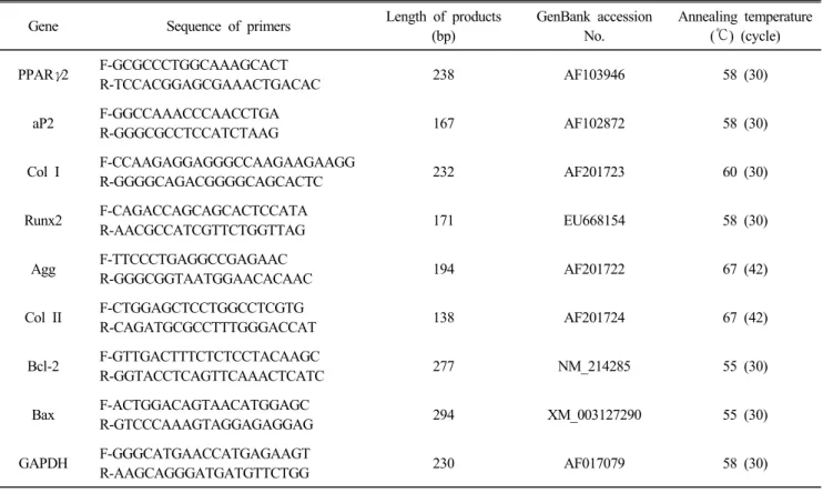
The apoptotic gene expression of pSSCs after induction culture was also estimated by RT-PCR. The pSSCs have a potential to differentiate into three mesodermal cell types (adipocytes, chondrocytes, and osteoblasts). Non-induced control and chondrogenic-induced cells were showed higher H

2

DCFDA intensity (P<0.05) than adipogenic- and osteogenic-induced cells. The relative expression of Bax/Bcl-2 level was significantly low (P<0.05) in adipogenic- and osteogenic-induced cells compared to non-induced control. However, there was no difference in the relative expression of Bax/Bcl-2 level among differentiation induction groups. The result of the present study shows that the apoptosis of pSSCs is not detrimentally increased by differentiation induction culture, although chondrogenic-induced pSSCs showed high ROS generation level and apoptotic index similarly to those of non-induced cells.

(Key words : porcine skin-derived stem-like cells (pSSCs), differentiation induction, ROS, apoptosis)

This study was supported by 2014 Research Grant from Kangwon National University (No. C1009754-01-01)

These authors contributed equally to this work.

†Correspondence : [email protected]

INTRODUCTION

Mesenchymal stem cells (MSCs) have emerged as promising tools for regenerative medicine and tissue engineering (Badylak and Nerem, 2010). MSCs are characterized as undifferentiated cells, able to self-renewal with a high proliferation capacity and supporting cellular therapy (Kern et al., 2006). MSCs have several advantages including their excellent safety and the potential as autologous cells for transplantation into humans (Trounson, 2009). In particular, the quantity and accessibility of subcutaneous adipose tissue in humans makes it an attrac- tive alternative to other tissues as a source of adult stem cells (Fraser et al., 2006; Zuk, 2010). The successful extraction and cultivation of MSCs was achieved from porcine skin (Dick and Scott, 1992; Dyce et al., 2004), adipose tissues (Simon and Maibach, 2000), bone marrow (Kern et al., 2006). The multipotent stem cell component of MSCs isolates is able to differentiate of mesodermal lineage including adipocyte, chon-

drocyte and osteoblasts (Atashi et al., 2015). Recently, we also reported that successful extraction and differentiation induction into three mesenchymal cell types and further in vivo cartilage formation from porcine skin-derived stem cell-like cells (pSSCs) as well as porcine adipose-derived stem cells (Hwang et al., 2015).

It was reported that MSC cells in chondrogenic differentia- tion induction culture generate hypoxia (Zhu et al., 2006), and undergo apoptosis with increasing cell density (Wang et al., 2009). ROS-derived apoptosis might restrict the differentiation of MSC cells to different cell types as well as chondrogenic differentiation. Exogenous reactive oxygen species (ROS) are one of the important factors that induce apoptosis of MSCs.

The high levels of ROS cause cell damage by oxidation and

nitration of macromolecules including RNA, DNA and protein

(Schwartz et al., 2004). According to the previous study it was

clarified that ROS acts as an important signaling molecular

pathway in the regulation of cell growth and differentiation

(2)

(Lambeth, 2004). ROS such as hydrogen peroxide (H

2

O

2

), superoxide anion (O

2

), and hydroxyl radical (OH —) are gene- rally considered to be damage to cells and tissues. Hypoxia can activate p38 expression and caspase-8 and induce the apoptosis of MSCs via the mitochondrial death pathway and endoplasmic reticulum stress (Wei et al., 2010). Apoptosis is controlled by regulators, Bcl-2 family of apoptosis regulator proteins, which have either an inhibitory effect on programmed cell death or block the protective effect of inhibitors (Tsujimoto et al., 1984; Cleary et al., 1986). Bcl-2, encoded by the BCL2 gene, is the founding member of the Bcl-2 family and speci- fically considered an important anti-apoptotic protein. Bax (bcl-2-like protein 4), encoded by the BAX gene, is one of the Bcl-2 family member, and functions as an apoptotic activator (pro-apoptotic protein).

In the present study, we examined the H

2

O

2

expression level and apoptosis-related gene (Bax/Bcl-2) expression levels in pSSCs after adipogenic, chondrogenic and osteogenic differen- tiation induction.

MATERIALS AND METHODS

1. Isolation of pSSCs

The pSSCs were isolated according to the method of previous study (Hwang et al., 2015). Briefly, porcine skin samples were obtained from ear tissues of pigs (6 month-old females). Car- tilage tissue was completely removed from the ear skin sample, and the epidermis and dermis of the skin tissue were finely chopped and digested by treatment with 10 ml digestion solu- tion containing 0.2% trypsin-EDTA (Sigma-Aldrich, St. Louis, MO, USA) and 0.2% collagenase type I in Dulbecco’s phosphate buffered saline (DPBS; Gibco, Grand Island, NY, USA) for 30 min at 37 ℃ with agitation. Cells were recovered and plated in an 100-mm culture dish (BD, San Diego, CA, USA) in DMEM supplemented with 10% fetal bovine serum (FBS; Gibco) and 1% P/S, in humidified air with 5% (v/v) CO

2

at 37 ℃, and cultured for more than 10 days. The cells were passaged one or two times with changing the culture medium every 2 days.

The MSC characteristics of the pSSCs established in this study were confirmed by fluorescence activated cell sorting (FACS) analysis in our previous study (Hwang et al., 2015).

2. In Vitro Differentiation Induction into Three Mesodermal Cell Types The pSSCs were seeded at a density of 2×10

4

cells/cm

2

for

adipogenic and osteogenic differentiation, or 1×10

4

cells/cm

2

for chondrogenic differentiation in 24-well plates (BD) and were grown for 2 ∼3 days in standard DMEM with 10% FBS.

At 70 ∼80% confluence of cells, the medium was replaced in test wells by each induction medium. To induce adipogenic differentiation, the cells were cultured in DMEM containing 10

% FBS, 500 μM isobutylmethlxanthine (Sigma-Aldrich), 100 μM indomethacin (Sigma-Aldrich), 1% antibiotics, 1 μM dexame- thasone (Sigma-Aldrich), and 10 μg/ml insulin (Sigma-Aldrich) for 21 days. To induce osteogenic differentiation, the cells were cultured in DMEM containing 10% FBS, 50 μM ascorbic acid (Sigma-Aldrich), 10 mM glycerol 2-phosphate (Sigma-Aldrich), 1% antibiotics (Sigma-Aldrich), and 0.1 μM dexamethasone for 26 days. To induce chondrogenic differentiation, the cells were cultured for 26 days in DMEM containing 10% FBS, 1% anti- biotics, 1% Insulin-Transferrin-Selenium-A Supplement (Gibco), 50 μM ascorbic acid, 0.1 μM dexamethasone and 10 ng/ml transforming growth factor-beta1(TGFβ 1, ProSpec-Tany Techno- Gene, Rehovot, Israel). The induction medium was changed every 2 ∼3 days. Cells cultured in DMEM supplemented with 10% FBS and 1% P/S for 26 days were considered as non- induced controls.

At the end of induction culture, differentiation induced cells were fixed with 4% paraformaldehyde (Sigma-Aldrich) for 15 min at room temperature (RT) prior to staining. For adipogenic differentiation, induced cells were stained with 0.6% (w/v) Oil Red O solution (Sigma-Aldrich) for 20 min at RT. For osteo- genic differentiation, the cells were stained by von Kossa. The cells were treated with 5% silver nitrate (Sigma-Aldrich) and exposed to strong light for 1 h. The reaction was stopped by treatment with 5% sodium thiosulfate nitrate (Sigma-Aldrich) for 2 ∼3 min. For chondrogenic differentiation, the cells were stained with 0.5% Alcian blue (Sigma-Aldrich) in 0.1 N HCl (pH 1.0) for 30 min at RT. Stained cells were then examined under a light microscope (TE300, Nikon, Tokyo, Japan).

3. Quantification of Intracellular ROS Levels

The ROS levels of induced and control cells were measured using a modified method previously described (Bae et al., 2015).

Briefly, the induction media of cells was completely removed

and washed with DPBS. Cells were stained in 10 µM 2’7’-

dichlorodihydrofluorescein diacetate (H

2

DCFDA, Invitrogen,

Karlsruhe, Germany) dye for 30 min in the dark at 39 ℃ to

measure the H

2

O

2

levels. After incubation, the cells were wash-

(3)

Table 1. Nucleotide sequences of primers used in an RT-PCR assay

Gene Sequence of primers Length of products

(bp)

GenBank accession No.

Annealing temperature (℃) (cycle)

PPAR γ 2 F-GCGCCCTGGCAAAGCACT

R-TCCACGGAGCGAAACTGACAC 238 AF103946 58 (30)

aP2 F-GGCCAAACCCAACCTGA

R-GGGCGCCTCCATCTAAG 167 AF102872 58 (30)

Col I F-CCAAGAGGAGGGCCAAGAAGAAGG

R-GGGGCAGACGGGGCAGCACTC 232 AF201723 60 (30)

Runx2 F-CAGACCAGCAGCACTCCATA

R-AACGCCATCGTTCTGGTTAG 171 EU668154 58 (30)

Agg F-TTCCCTGAGGCCGAGAAC

R-GGGCGGTAATGGAACACAAC 194 AF201722 67 (42)

Col II F-CTGGAGCTCCTGGCCTCGTG

R-CAGATGCGCCTTTGGGACCAT 138 AF201724 67 (42)

Bcl-2 F-GTTGACTTTCTCTCCTACAAGC

R-GGTACCTCAGTTCAAACTCATC 277 NM_214285 55 (30)

Bax F-ACTGGACAGTAACATGGAGC

R-GTCCCAAAGTAGGAGAGGAG 294 XM_003127290 55 (30)

GAPDH F-GGGCATGAACCATGAGAAGT

R-AAGCAGGGATGATGTTCTGG 230 AF017079 58 (30)

ed twice with DPBS and immediately examined under a fluorescence microscope (IX71, Olympus, Tokyo, Japan) with filters at 450 ∼480 nm for excitation and at 515 nm for emi- ssion. The fluorescent images were obtained at random from at least three clear areas of individual cells per well and recorded as JPEG files using a digital camera (Cellsens entry software 1.6, Olympus, Japan) and analyzed the intensity of fluorescence in each cells using ImageJ software 1.37 (NIH, Bethesda, MD, USA).

4. RNA Extraction and Reverse Transcription-Polymerase Chain Reaction (RT-PCR)

Total RNA was extracted from control and induced cells using Trizol (Invitrogen) and 1 μg total RNA was used for cDNA synthesis with RT-PreMix (Bioneer, Daejeon, Korea).

Polymerase chain reaction (PCR) was performed with PCR- PreMix (Bioneer) under standard PCR conditions. The primers used, peroxisome proliferator-activated receptor gamma 2 (PPARγ2), adipocyte fatty acid binding protein 2 (aP2), colla- gen type I (Col I), runt-related transcription factor 2 (Runx2), collagen type II (Col II), aggrecan (Agg), BCL2-like 1 (BCL2),

BCL2-associated X protein (BAX) and glyceraldehyde 3- pho- sphate dehydrogenase (GAPDH) are shown in Table 1. PCR cycles consisted of an initial denaturation step at 95 ℃ for 5 min, followed by 34 amplification cycles consisting of 30 s of denaturation at 95 ℃, 30 s of annealing at 62℃, and 1 min of extension at 72 ℃. A final extension was performed at 72℃ for 10 min. The PCR products were analyzed by UV irradiation on a 2% agarose gel (Amresco, Solon, OH, USA) stained with ethidium bromide (Sigma-Aldrich). Gels were read using a UV transilluminator (CN-08, Vilber Lourmat, Torcy, France). For apoptotic genes, the relative amount of each mRNA species was calculated by dividing the intensity of target primers by the intensity of the corresponding GAPDH.

5. Statistical Analysis

At least three independent sets of experiments for each con- dition were performed in triplicate. Statistical analyses were performed using ANOVA and Duncan’s multiple range test with the SAS software (SAS Institute, Cary, NC, USA).

RESULTS

(4)

1. Multipotency of pSSCs

Following differentiation induction, pSSCs were stained po- sitively by Oil red O with the accumulation of neutral lipid vacuoles for adipocyte induction, von Kossa with the extent of calcium accumulation for osteoblast induction, or Alcian blue with forming cell nodules for chondrocyte induction (Fig. 1).

In RT-PCR, the expressions of PPARγ2 and aP2 in adipogenic- induced cells, Col I and Runx2 mRNA level in osteogenic- induced cells, and Col II and Agg in chondrogenic-induced cells were highly expressed from pSSCs (Fig. 1).

2. H

2

O

2

Levels of pSSCs after Induction Culture

Each differentiation induced and non-induced control of pSSCs were observed a marked induction of H

2

O

2

generation by fluorescence microscopy (Fig. 2). Non-induced control and chondrogenic-induced cells were showed higher H

2

DCFDA intensity than adipogenic-andosteogenic-induced cells, mean- while lowest in osteogenic-induced cells (P<0.05).

Fig. 1. Morphologies and marker gene expressions of three meso- dermal cell types. (A) Adipogenic-induced cells stained by Oil red O, (B) Chondrogenic-induced cells stained by von Kossa, (C) Osteogenic-induced cells stained by Alcian blue.

Scale bar: 100 µm (×200).

3. Apoptotic Gene Expression in pSSCs after Induction Culture The expressions of anti-apoptotic gene Bcl-2 and the pro- apoptotic gene Bax were detected in all groups (Fig. 3). The relative expression of Bax/Bcl-2 level was significantly low (P<0.05) in adipogenic- and osteogenic-inducted cells com- pared to non-induced control. However, there was no difference in the relative expression of Bax/Bcl-2 level among differen- tiation induction groups.

DISCUSSION

Fig. 2. Fluorescence images and H

2

O

2

levels of pSSCs stained with

H

2

DCFDA after differentiation induction culture. (A) Un-indu-

ced control, (B) Adipogenic-induced cells, (C) Chondrogenic-

induced cells, (D) Osteogenic-induced cells. Scale bar =

50 μm. (E) H

2

O

2

levels of pSSCs were evaluated by the

fluorescence intensity in each cells (mean±SEM).

a∼c

Values

with different letters differ significantly (P<0.05).

(5)

Fig. 3. RT-PCR results of apoptosis-related genes. (A) Expression of apoptosis-related genes in each cell types. Adipo, Adi- pogenic-induced cells; Chondro, Chondrogenic-induced cells;

Osteo, Osteogenic-induced cells. (B) Relative gene expression of Bax/Bcl-2 data are presented as means±SEM,

a,b

Values with different letters differ significantly (P<0.05).

Several defining characteristics of MSCs have recently been outlined, including their ability to adhere to plastic, their expression of specific surface antigens, and their multipotent differentiation into adipocytes, chondrocytes, and osteoblasts in vitro (Iohara et al., 2006; Kang et al., 2010; Vishnubalaji et al., 2012). MSCs have emerged as an indispensable tool in regenerative medicine. Numerous sources of MSCs have been identified, and cells have been cultured using a variety of methods for use in cell-based therapies. However, there remains a need for an optimal clinical-grade product, and high safety standards must be fulfilled. Recently, it was reported that por- cine skin-derived MSC is a potential autologous source for stem cell transplantation in regenerative medicine (Kang et al., 2010). Furthermore, skin-derived cells have a good potential in the cost and time savings. Therefore, skin-derived progenitors/

multipotent stem cells are promising resources for the successful development of stem cell therapies.

It was reported that the occurrence of ROS affects the cell growth negatively (Yang et al., 2015). However, an appropriate ROS helps the cells to develop healthy (Stolzing et al., 2007).

Cellular aging including oxidative damage, ROS levels increa- sed suggesting a progressive loss of MSC function with age, leading to a reduction in MSC numbers and differentiation capacity (Stolzing et al., 2007). Our result showed that the ROS generation level and relative expression level of Bax/

Bcl-2 were high in non-induced pSSCs compared to those of differentiation-induced pSSCs excepting chondrogenic-induced pSSCs. It was suggested that cell aging is getting faster with normal growth culture because of long term culture without differentiation induction and increased cell density (Wang et al., 2009; Bork et al., 2010), whereas, differentiation induction into fresh new type of cells and induction factors may delay cell aging.

In our study, chondrogenic-induced cells generated a high level of ROS compared to other induced groups. It was un- clear why ROS generation level in chondrogenic-induced cells was high. We suggested that TGF is the reason for high ROS level by promoting cell growth to confluence rapidly (Hwang et al., 2015). Differentiation induction media are differ among adipogenic-, chondrogenic-, and osteogenic- induction cultures.

TGF is the key factor for differentiating pSSCs to chondrocytes.

It was reported that chondrogenic-induced cells in monolayer culture generate hypoxia (Zhu et al., 2006), and undergoes apoptosis with increasing cell density (Wang et al., 2009). Based on the previous study, there is an evidence for a function of ROS in MSC proliferation, survival and differentiation to adipocytes and osteoblasts (Atashi et al., 2015). Recent reports describe the importance of appropriate oxidants on MSC diffe- rentiation into adipocyte (Higuchi, et al., 2013), chondrocyte (Sasaki et al., 2006) and osteoblasts (Higuchi et al., 2007) through the activation of signaling pathway involved in diffe- rentiation (Yuan et al., 2012). Different ROS generation level might be due to different signaling pathways depending on each lineage differentiated from pSSCs. Different induction media for each lineage induce distinct pathway, which might affect the expression levels of ROS and relative expression level of Bax/Bcl-2.

In conclusion, the result shows that the apoptosis of pSSCs

is not detrimentally increased by differentiation induction cul-

ture, although chondrogenic-induced pSSCs showed high ROS

generation level and apoptotic index similarly to those of non-

(6)

induced cells.

REFERENCES

Atashi F, Modarressi A and Pepper MS. 2015. The role of reactive oxygen species in mesenchymal stem cell adipo- genic and osteogenic differentiation: A review. Stem cells and Dev. 24:1150-1163.

Badylak SF and Nerem RM. 2010. Progress in tissue enginee- ring and regenerative medicine. PNAS 3285-3286.

Bae HK, Hwang IS, Kim JY, Lee SY, Park CK, Yang BK and Cheong HT. 2015. Antioxidant treatment during manipula tion procedures prevents mitochondrial and DNA damage and enhances nuclear reprogramming of bovine somatic cell nuclear transfer embryos. Rep. Fertil. Dev. 27:1088-1095.

Bork S, Pfister S, Witt H, Horn P, Korn B, Ho AD and Wagner W. 2010. DNA methylation pattern changes upon long-term culture and aging of human mesenchymal stromal cells.

Aging Cell 9:54-63.

Cleary ML, Smith SD and Sklar J. 1986. Cloning and structural analysis of cDNAs for bcl-2 and a hybrid bcl-2/immuno- globulin transcript resulting from the translocation. Cell 47:

19-28.

Dick IP and Scott RC. 1992. Pig ear skin as an in-vitro model for human skin permeability. J. Pharm. Pharmaco. 44:640- 645.

Dyce PW, Zhu H, Craig J and Li J. 2004. Stem cells with multilineage potential derived from porcine skin. BBRC 316:651-658.

Fraser JK, Wulur I, Alfonso Z and Hedrick MH. 2006. Fat tissue: An underappreciated source of stem cells for biote- chnology. Trends Biotechnol. 24:150-154.

Higuchi M, Dusting GJ, Peshavariya H, Jiang F, Hsiao ST, Chan EC and Liu GS. 2007. Vascular Bmp Msx2 Wnt signaling and oxidative stress in arterial calcification. Ann.

NY Acad. Sci. 1117:40-50.

Higuchi M, Dusting GJ, Peshavariya H, Jiang F, Hsiao ST, Chan EC and Liu GS. 2013. Differentiation of human adi- pose-derived stem cells into fat involves reactive oxygen species and Forkhead box O1 mediated upregulation of antioxidant enzymes. Stem Cells Dev. 22:878-888.

Hwang IS, Bae HK and Cheong HT. 2015. Comparison of the characteristics and multipotential and in vivo cartilage for- mation capabilities between porcine adipose-derived stem

cells and porcine skin-derived stem cell-like cells. Am. J.

Vet. Res. 76:814-821.

Iohara K, Zheng L, Ito M, Tomokiyo A, Matsushita K and Nakashima M. 2006. Side population cells isolated from porcine dental pulp tissue with self-renewal and multipo- tency for dentinogenesis, chondrogenesis, adipogenesis, and neurogenesis. Stem Cells 24:2493-2503.

Kang EJ, Byun JH, Choi YJ, Maeng GH, Lee SL, Kang DH, Lee JS, Rho GJ and Park BW. 2010. In vitro and in vivo osteogenesis of porcine skin derived mesenchymal stem cell like cells with a demineralized bone and fibrin glue scaffold. Tissue Eng. Part A 16:815-827.

Kern S, Eichler H, Stoeve J, Klüter H and Bieback K. 2006.

Comparative analysis of mesenchymal stem cells from bone marrow, umbilical cord blood, or adipose tissue. Stem Cells 24:1294-1301.

Lambeth JD. 2004. NOX enzymes and the biology of reactive oxygen. Nat. Rev. Immunol. 4:181-189.

Sasaki T, Maier B, Bartke A and Scrable H. 2006. Progressive loss of SIRT1 with cell cycle withdrawal. Aging Cell 5:

413-422.

Schwartz PH, Nethercott H, Kirov, Ziaeian B, Young MJ and Klassen H. 2004. Expression of neurodevelopmental markers by cultured porcine neural precursor cells. Stem Cells 23:

1286-1294.

Simon GA and Maibach HI. 2000. The pig as an experimental animal model of percutaneous permeation in man: Qualitative and quantitative observations-an overview. Skin Pharmacol.

Appl. Skin. Physiol. 13:229-234.

Stolzing A, Jones E, McGonagle D and Scutt A. 2007. Age- related changes in human bone marrow-derived mesenchymal stem cells: Consequences for cell therapies. Mech. Ageing Dev. 129:163-173.

Trounson A. 2009. New perspectives in human stem cell the- rapeutic research. BMC Medicine 7:29.

Tsujimoto Y, Finger LR, Yunis J, Nowell PC and Croce CM.

1984. Cloning of the chromosome breakpoint of neoplastic B cells with the chromosome translocation. Science 226:

1097-1099.

Vishnubalaji R, Al-Nbaheen M, Kadalmani B, Aldahmash A and Ramesh T. 2012. Skin-derived multipotent stromal cells- an archrival for mesenchymal stem cells. Cell Tissue Res.

350:1-12.

Wang CY, Chen LL, Kuo PY, Chang JL, Wang YJ and Hung

(7)

SC. 2009. Apoptosis in chondrogenesis of human mesen- chymal stem cells: Effect of serum and medium supplements.

Apoptosis 15:439-449.

Wei H, Li Z, Hu S, Chen X and Cong X. 2010. Apoptosis of mesenchymal stem cells induced by hydrogen peroxide con- cerns both endoplasmic reticulum stress and mitochondrial death pathway through regulation of caspases, p38 and JNK.

Cellular Biochem. 111:967-978.

Yang SR, Park JR and Kang KS. 2015. Reactive oxygen spe- cies in mesenchymal stem cell aging: Implication to lung diseases. Oxid. Med. Cell. Longev. 2015, Article ID 486263.

Yuan HF, Zhai C, Yan XL, Zhao DD, Wang JX, Zeng Q,

Chen L, Nan X, He LJ, Li ST, Yue W and Pei XT. 2012.

SIRT is required for long-term growth of human mesen- chymal stem cells. J. Mol. Med. (Berl) 90:389-400.

Zhu W, Chen J, Cong X, Hu S and Chen X. 2006. Hypoxia and serum deprivation-induced apoptosis in mesenchymal stem cells. Stem Cells 24:416-425.

Zuk PA. 2010. The adipose-derived stem cell: Looking back and looking ahead. Mol. Biol. Cell 21:1783-1787.

Received February 16, 2016, Revised March 3, 2016, Accepted

March 8, 2016

수치

Table  1.  Nucleotide  sequences  of  primers  used  in  an  RT-PCR  assay
Fig.  2.  Fluorescence  images  and  H 2 O 2  levels  of  pSSCs  stained  with  H 2 DCFDA  after  differentiation  induction  culture
Fig.  3.  RT-PCR  results  of  apoptosis-related  genes.  (A)  Expression  of  apoptosis-related  genes  in  each  cell  types

참조

관련 문서

On his part, CEO of Express Roads Authority, Saud Al-Naqqi said that the heavy rains of the previous day led to clogging parts of the express

Kuwait will celebrate on Sunday the fourth anniversary of the UN honoring and proclamation of His Highness the Amir, Sheikh Sabah Al-Ahmad Al-Jaber Al-Sabah as

• 이명의 치료에 대한 매커니즘과 디지털 음향 기술에 대한 상업적으로의 급속한 발전으로 인해 치료 옵션은 증가했 지만, 선택 가이드 라인은 거의 없음.. •

The proposal of the cell theory as the birth of contemporary cell biology Microscopic studies of plant tissues by Schleiden and of animal tissues by Microscopic studies of

웹 표준을 지원하는 플랫폼에서 큰 수정없이 실행 가능함 패키징을 통해 다양한 기기를 위한 앱을 작성할 수 있음 네이티브 앱과

_____ culture appears to be attractive (도시의) to the

It considers the energy use of the different components that are involved in the distribution and viewing of video content: data centres and content delivery networks

After first field tests, we expect electric passenger drones or eVTOL aircraft (short for electric vertical take-off and landing) to start providing commercial mobility