사람 폐암 세포주에서 시스플라틴이나 독소루비신의 세포독성에 미치는 녹차 추출물의 영향
이병래1․박재윤1․박평심2†
1
조선대학교 의과대학 생화학교실
2
조선대학교 의학연구원
Effect of Green Tea Extract on Cisplatin- or Doxorubicin-Induced Cytotoxicity in Human Lung Cancer Cell Lines
Byoung-Rai Lee1, Jae-Yoon Park1, and Pyoung-Sim Park2†
1Dept. of Biochemistry, College of Medicine and 2Institute of Medical Science, Chosun University, Gwangju 501-759, Korea
Abstract
Tea extract (TE) has been shown to have anti-tumor properties in a wide variety of experimental systems.
We evaluated green tea extract (GTE) as a biochemical modulator for the antitumor activity of cisplatin and doxorubicin in the treatment of human lung cancer A549 cells. Cells were grown in RPMI-1640 medium supple- mented with 10% (v/v) heat-inactivated fetal bovine serum and two antibiotics (100 units/mL penicillin and 100 μ g/mL streptomycin). Two types of TE, epigallocatechin galate (EGCG) and GTE, were used in this experiment.
The cells were seeded at 1×10
4cells/well in the RPMI-1640 media with or without TE (100 μg/mL) and then treated with different concentrations of doxorubicin (0~14 μg/mL) or cisplatin (0~35 μg/mL). After incubation in 5% CO
2at 37
oC for 24 hr, cell viability was determined with a MTT assay. We used a Western blot to detect the influence of EGCG and GTE on the expression of p53 and caspase-3 genes in the A549 cells. A549 cell viability decreased to 15% with a 10 μg/mL concentration of cisplatin, and to 21% with a 8 μg/mL concentration of doxorubicin, as measured with the MTT assay. However, pre-treatment of the cells with EGCG (100 μg/mL) or GTE (100 μg/mL) resulted in decreased cell viability with 6 μg/mL of cisplatin and 4 μg/mL of doxorubicin.
There was no apparent change in cell viability between EGCG or GTE administration in cisplatin- or doxor- ubicin-induced cytotoxicity in A549 cells. The levels of p53 and caspase-3 in the A549 cells increased with both EGCG and GTE treatment. We found that GTE could potentially affect cisplatin- or doxorubicin-induced cytotoxicity of A549 cells, which may be useful in the chemotreatment of cancer.
Key words: doxorubicin, cisplatin, GTE, EGCG, A549 cell
†
Corresponding author. E-mail: [email protected]
†
Phone: 82-62-230-6295, Fax: 82-62-226-4165
서 론
녹차(
Camellia sinensisL.)는 차나무 잎의 엽록소를 그대 로 보존시킨 비 발효차로 세계적으로 이용하고 있는 음료이 다. 녹차 잎은 75~80%의 수분과 20~25% 고형물질로 되어 있고, 테아닌, 유리아미노산, 카테킨, 카페인, 클로로필, 비타 민 및 미네랄 등의 기능성 및 기호성 관련 성분이 함유되어 있다(1). 카테킨은 건조 녹차 잎의 약 10% 내외를 차지할 정도로 매우 함량이 높은데, 녹차 잎에 함유된 카테킨은 epi- catechin(EG), epicatechin-3-gallate(ECG), epigallocatechin (EGC) 및 epigallocatechin-3-gallate(EGCG) 등 4가지 종류 가 많고, 이중에서 EGCG의 함량이 가장 높다(2). 카테킨은 항산화 작용, 항암작용, 항균작용, 항비루스 작용 등 여러 가
지 생리활성이 있는 것으로 보고되고 있다(1-4). 항암작용은 카테킨 중에서 EGCG가 가장 강한 것으로 알려져 있고, 암의 발생을 예방하거나 치료제로서의 EGCG를 이용하기 위한 연구가 많이 진행되고 있어 녹차의 암 발병 억제 및 예방효 과가 주목되고 있다(3,4). 암을 치료하는 방법은 여러 가지가 있는데, 화학약품을 이용하여 암을 치료하는 방법인 항암화 학요법은 암의 종류나 시기에 관계없이 시행할 수 있기 때문 에 매우 유용한 암 치료방법이다(5,6).
시스플라틴(cisplatin: cis-diammine dichloroplatinum)과
독소루비신(doxorubicin)은 광범위 항암 화학요법제로서 자
궁경부암, 대장암, 폐암 등 여러 가지 암에 대한 항암치료제
에 이용되고 있다(7-9). 광범위 화학요법제는 암 치료에서
1차적으로 이용되는 화학요법제이나, 항암제에 대한 내성과
신장, 간장 및 골수세포 등에 대한 독성 부작용은 항암 화학 요법의 커다란 장애로 대두되고 있다(10-12). 따라서 항암 화학요법제의 치료효과를 증진시키고, 부작용을 줄이기 위 한 여러 가지 방법이 시도되고 있는데, 투여량을 증가시키는 고용량 요법, 리포좀에 약물을 포장하여 투여하는 방법 등 약물투여량이나 방법을 개선하는 연구가 진행되고 있다 (13,14).
그러나 항암 화학요법제의 항암작용을 증가시키면서 독 성은 최소화하여 암 치료가 효과적으로 이루어질 수 있는 치료법의 개발이 아직까지 충분하지 않다. 따라서 정상세포 에 대한 독성작용을 최소화하면서 암세포에 대한 항암작용 을 증가시킬 수 있는 화학요법제의 개발이 필요하다. 또한 항암 화학요법제의 암세포 독성을 증가시키거나 암세포의 내성을 극복할 수 있는 항암 화학요법 보조제의 개발에 대한 연구가 매우 절실히 요구되고 있다.
본 연구는 광범위 항암 화학요법제인 시스플라틴과 독소 루비신의 항암작용을 증강시키는 제제를 개발하기 위한 연 구의 일환으로 녹차 추출물의 이용가능성을 추정하기 위하 여 사람 폐암 세포주인 A549 세포를 배양하여 시스플라틴과 독소루비신 및 녹차 추출물을 처치한 후 세포생존율을 알아 보았다. 또한 세포사멸에 관여하는 것으로 알려진 p53 및 caspase-3(15)을 측정하여 정제되지 않은 녹차 추출물과 녹 차로부터 추출 정제한 EGCG의 영향을 비교 관찰하였다.
재료 및 방법
녹차 추출물 시료의 추출
녹차 추출물은 건조 녹차 잎을 열수 추출하고 부분 정제하 여 사용하였다. 즉 건조 녹차 잎을 재배농가(보성다원)에서 구입하여 분마시켜서, 미리 가열한 증류수 50 L에 녹차 잎 분말 5 kg을 첨가하여 100
oC로 2시간 동안 가열한 후 추출액 을 원심분리 하여 상층액을 수집하였다. 수집한 추출액을 4
oC에서 12시간 동안 방치하고 난 후 원심분리기(JA-21, Beckman, Fullerton, CA, USA)에서 1500 rpm으로 15분간 원심분리 하여 수집한 상층액을 건조기(60
oC)에서 완전히 건조시킨 후 마쇄하여 제조한 분말을 녹차 추출물 시료로 사용하였다.
세포의 배양
본 실험에서 사용한 세포는 사람 폐암세포주인 A549 세포 로 한국 세포주 은행에서 분양받아 실험에 사용하였다. 10%
태우혈청, 스트렙토마이신(100 U/mL) 및 페니실린(100 U/
mL)을 함유한 RPMI-1640 배지(Gibco BRL, Brooklyn, NY, USA)를 사용하였으며, 37
oC로 유지되는 CO
2배양기에서 상 기 세포를 배양하였다.
세포활성(cell viability) 측정
실험에 사용한 독소루비신, 시스플라틴 및 EGCG는 Sig-
ma Chemical Co.(St. Louis, MO, USA)에서 구매하여 사용 하였고, 녹차 추출물은 추출하여 사용하였다. 녹차 추출물, EGCG와 독소루비신은 PBS(phosphate buffered saline)에 용해시켜 주사기용 필터(syringe filter 0.22 μm, Millipore, Billerica, MA, USA)를 통과시킨 후 배양액에 첨가하였고, 시스플라틴은 DMSO(dimethylsulfoxide)에 용해시킨 후 PBS에 희석시켜 배양액에 첨가하였다.
세포배양 용기(96 well plate)의 각 웰에 A459 세포 2×10
4개씩을 넣어 24시간 동안 배양한 후 녹차 추출물, EGCG, 독소루비신 및 시스플라틴을 배양액에 첨가하고, 24시간 동 안 더 배양한 후 세포활성을 MTT(3-(4,5-dimethylthiazol- 2-yl)-2,5-diphenyl-tetrazolium bromide)법으로 측정하였 다(16). 각 실험세포에 MTT를 최종농도가 1 mM이 되게 첨가하여 4시간 동안 배양시킨 후 배양액을 제거하고, HBSS (Hank's balanced salt solution)로 3회 세척하여 MTT 용해 액[50% N,N-dimethylformamide(v/v); 20% sodium dodecyl sulfate(w/v), pH 4.7] 200 μL를 첨가한 후 ELISA plate reader(TECAN, Männedorf, Switzerland)로 540 nm에서 흡광도를 측정하였다. 실험은 각 실험군마다 3 wells의 흡광 도를 각각 측정하여 평균값을 계산하였고, 3회 반복 실험을 실시하였으며, 세포활성도(cell viability)는 비교세포활성도 로서 표시하였다.
비교세포활성도
(relative viability)= 실험군 흡광도 대조군흡광도 × 100 p53 및 caspase-3 양 측정
A549 세포에서 p53 및 caspase-3 양은 Western blot 법으
로 측정하였다. A549 세포를 1일간 배양한 후 대조군, 녹차
추출물과 EGCG 첨가군으로 나누어 실험을 실시하였다. 녹
차 추출물이나 EGCG 100 μL/mL를 배양액에 첨가하여 4시
간 동안 배양한 후 배지를 제거하고 100 μL SDS-loading
buffer(50 mM Tris-HCl, pH 6.8, 2% SDS, 0.1% bromo-
phenol blue, 10% glycerol)를 첨가하여 95°C에서 5분 동안
방치한 후 냉각시켜 시료로 이용하였다. 동량의 시료(30 μg
protein/lane)를 10% polyacrylamide gel에서 Tris 완충액
(pH 8.8, 0.025 M Tris, 0.192 M glycine, 0.1% SDS)으로 80
V로 2시간 동안 전기영동 시켜 Hybond ECL nitrocellulose
membrane에 흡착시켰다. 단백질이 흡착된 nitrocellulose
membrane은 5% nonfat milk를 함유한 TBS(0.1% Tween
20 in pH 7.4 Tris-based saline buffer)로 blocking하고
anti-rabbit polyclonal anti-p53 Ab, anti-caspase-3 Ab 및
anti-β-actin Ab(Santa Cruz Biotechnology, Santa Cruz,
CA, USA)와 이차항체(horseradish peroxidase-labeled
goat anti-rabbit IgG)를 이용하여 반응시킨 후 enhanced
chemiluminescence 용액(Amersham, London, UK)에 넣고
ECL 필름에 감광시킨 후 image analyzer(1D ver.2.1, Phar-
macia Biotech, Kalamazoo, MI, USA)를 이용하여 측정하여
비교하였다.
Tea extract concentration (μg/mL)
Fig. 1. Effects of green tea extract (GTE) on viability of A549 cells. A549 cells were plated at 2×10
4cells/well in 96 well plate and treated with GTE or EGCG for 24 hours. Mitochondrial dehydrogenase activity was assayed as an index of cell viability.
Relative vitality=(ODExp/ODCon)×100%, which is an indication of A549 cell viability. Representative results are shown as the mean±SD of triplicates. Values with the different superscripts are significantly different by Duncan's multiple range test (p<0.05).
Drug concentration (μg/mL)
Fig. 2. Effects of doxorubicin and cisplatin on viability of A549 cells. A549 cells were plated at 2×10
4cells/well in 96 well plate and treated with doxorubicin or cisplatin for 24 hours.
Mitochondrial dehydrogenase activity was assayed as an index of cell viability. Relative vitality=(ODExp/ODCon)×100%, which is an indication of A549 cell viability. Representative results are shown as the mean±SD of triplicates. Values with the different superscripts are significantly different by Duncan's multiple range test (p<0.05).
통계처리
모든 실험결과는 평균과 표준편차(mean±SD)로 나타내 었고, 각 군 간의 통계적 유의성 검증은 SPSS 통계 프로그램 (statistical package for social science version 12.0)을 이용 하여 ANOVA 분석을 하였다. 각 군 간에 유의한 차이가 있 는 경우 Duncan's multiple range test를 실시하여 p<0.05 수준에서 검증하였다.
결과 및 고찰
녹차 추출물이 A549 세포활성에 미치는 영향
녹차 추출물과 EGCG의 항암작용을 사람 폐암세포주인 A549 세포를 배양하여 세포활성도를 MTT법으로 측정하였 다. A549 세포를 배양한 후 EGCG와 녹차 추출물을 각각 0, 20, 50, 100, 200, 300, 400, 500 및 600 μg/mL 되게 배양액 에 첨가하여 24시간 배양한 후 MTT법을 시행한 결과 EGCG는 300 μg/mL, 녹차 추출물은 400 μg/mL 이상의 농도 에서 세포활성이 감소되어 세포독성이 나타남을 알 수 있었 다(Fig. 1). 녹차 카테킨 중 EGCG의 항암작용이 가장 강한 것으로 알려져 있는데(17,18), EGCG와 녹차 추출물의 A549 세포에 대한 독성을 본 실험결과를 비교하여 보면 EGCG는 300 μg/mL, 녹차 추출물은 400 μg/mL 이상의 농도에서 세 포독성이 나타나서 녹차 추출물은 EGCG보다 33% 정도 더 높은 농도에서 세포독성이 나타나서 녹차 추출물은 EGCG 보다 항암력이 더 낮을 것으로 추측된다.
녹차 추출물의 항암력을 항암 화학요법제로 이용되는 시 스플라틴과 독소루비신과 비교하기 위하여 A549 세포에 대 한 시스플라틴과 독소루비신의 세포활성 억제력을 측정하 였다. A549 세포를 배양하여 시스플라틴과 독소루비신을 세 포배양액에 2, 4, 6, 8, 10, 12 및 14 μg/mL 되게 첨가하고 24시간 배양한 후 세포활성을 측정한 결과 시스플라틴 10
μ g/mL, 독소루비신 8 μg/mL의 농도에서 시스플라틴이나 독소루비신을 첨가하지 않은 대조군에 비하여 세포활성도 가 급격히 감소되어 세포독성이 나타남을 알 수 있었다(Fig.
2). 이러한 실험결과로서 A549 세포에 독성을 나타나는 농 도는 녹차추출물 400 μg/mL, EGCG 300 μg/mL, 시스플라틴 10 μg/mL 및 독소루비신 8 μg/mL로, 녹차 추출물이 A549 세포에 독성을 나타내는 농도는 시스플라틴보다 약 40배, 독소루비신보다 약 50배 높은 농도에서 세포독성이 나타나 기 때문에 녹차추출물의 A549 세포에 대한 독성작용은 시스 플라틴이나 독소루비신에 비하면 매우 낮아서 항암효과도 이들보다 더 낮을 것으로 생각된다. 따라서 녹차 추출물은 단독으로 사용하였을 경우 암세포에 대한 항암력은 높지 않 을 것으로 생각된다.
A549 세포에서 녹차 추출물이 시스플라틴의 세포독성에 미치는 영향
녹차 추출물과 EGCG가 시스플라틴에 의한 세포독성에
미치는 영향을 사람 폐암세포주인 A549 세포에서 측정하였
다. A549 세포를 배양한 후 EGCG나 녹차 추출물을 각각
100 μg/mL 되게 배양액에 첨가하여 2시간 배양한 후 시스플
라틴을 세포배양액에 2, 4, 6, 8, 10, 12 및 14 μg/mL 되게
첨가하여 24시간 배양하여 세포활성을 MTT 법으로 측정한
결과 녹차 추출물이나 EGCG를 첨가하지 않은 대조군에서
는 시스플라틴 10 μg/mL 이상의 농도에서 세포활성이 현저
히 감소되었고, EGCG나 녹차 추출물 100 μg/mL를 첨가하
면 시스플라틴 6 μg/mL 이상의 농도에서 세포활성이 감소
되어 EGCG나 녹차 추출물 첨가로 시스플라틴에 의한 세포
독성이 증가됨을 알 수 있다(Fig. 3, 4). EGCG 300 μg/mL,
녹차 추출물 400 μg/mL 이상의 농도에서 A549 세포에 대한
독성이 나타나기 때문에 EGCG와 녹차 추출물 농도 100 μg/
Cisplatin concentration (μg/mL)
Fig. 3. Effects of GTE pretreatment on cisplatin-induced cy- totoxicity of A549 cells. A549 cells were plated at 2×10
4cells/
well in 96 well plate and treated with cisplatin for 24 hours. EGCG (100 μg/mL) and GTE (100 μg/mL) were added to culture media at 2 hr before cisplatin treatment. Mitochondrial dehydrogenase activity was assayed as an index of cell viability. Relative vitality
=(ODExp/ODCon)×100%, which is an indication of A549 cell viability. Representative results are shown as the mean±SD of triplicates. Values with the different superscripts are significantly different by Duncan's multiple range test (p<0.05).
Fig. 4. Morphology of cispaltin and /or GTE treated A549 cells. A549 cells were plated at 1×10
5cells/well in 24 well plate and maintained in culture for 24 hours. The media were replaced, and the cells were untreated (A), treated with GTE (100 μg/mL) alone (B), treated with cisplatin (6 μg/mL) alone (C) or treated with GTE+cisplatin (D). GTE was added 2 hr before the treat- ment of cisplatin. The cells were photographed 24 hours later.
Doxorubicin concentration (μg/mL)
Fig. 5. Effects of TE on doxorubicin-induced cytotoxicity of A549 cells. A549 cells were plated at 2×10
4cells/well in 96 well plate and treated with doxorubicin for 24 hours. EGCG (100 μ g/mL) and GTE (100 μg/mL) were added to culture media at 2 hr before doxorubicin treatment. Mitochondrial dehydrogenase activity was assayed as an index of cell viability. Relative vitality
=(ODExp/ODCon)×100%, which is an indication of A549 cell viability. Representative results are shown as the mean±SD of triplicates. Values with the different superscripts are significantly different by Duncan's multiple range test (p<0.05).
mL는 단독 투여 시 세포활성을 저하시키지 않는 농도이다.
따라서 녹차 추출물 100 μg/mL를 시스플라틴과 병합투여로 세포독성이 증가되는 현상은 녹차 추출물에 의해서 시스플 라틴의 암세포에 대한 독성 작용이 상승되어 나타난 결과로 생각된다. EGCG와 녹차 추출물 100 μg/mL를 투여한 경우 모두 시스플라틴 6 μg/mL 이상의 농도에서 A549 세포독성 이 나타나므로, 시스플라틴의 세포독성 상승작용에 미치는 EGCG와 녹차 추출물의 효과 차이는 없는 것으로 추측된다.
Chan 등(19)은 EGCG가 난소암세포에서 시스플라틴의 세 포독성을 증가시킨다고 하였는데, 본 실험에서도 EGCG를
첨가하고 2시간 후 시스플라틴을 첨가하면 A549 세포에 대 한 독성이 증가되어 Chan 등(19)의 실험결과와 유사하였다.
녹차 추출물에 의한 시스플라틴의 세포독성 증가는 시스 플라틴의 양을 증가시키지 않고도 항암작용을 증가시킬 수 있는 방법으로 녹차추출물이 이용될 가능성이 있다. 녹차 추출물은 사람이 오랫동안 음용해온 천연물의 추출물로서 독성 부작용이 거의 없을 것으로 생각되어 이에 대한 전 임 상 및 임상연구가 시행된다면 녹차 추출물은 항암치료에서 매우 유용하게 이용될 수 있을 것으로 생각된다.
A549 세포에서 녹차 추출물이 독소루비신의 세포독성에 미치는 영향
녹차 추출물과 EGCG가 독소루비신에 의한 세포독성에 미치는 영향을 A549 세포에서 알아보았다. A549 세포를 배 양한 후 EGCG와 녹차 추출물을 각각 100 μg/mL 되게 배양 액에 첨가하여 2시간 배양한 후 독소루비신을 세포배양액에 2, 4, 6, 8, 10, 12 및 14 μg/mL 되게 첨가하여 24시간 배양한 후 세포활성을 측정하였다. 녹차 추출물이나 EGCG를 첨가 하지 않은 대조군에서는 독소루비신 8 μg/mL 이상의 농도 에서 세포활성이 감소되었고, EGCG나 녹차 추출물 100 μg/
mL 첨가군은 모두 시스플라틴 4 μg/mL 이상의 농도에서 세포활성이 감소되어 EGCG나 녹차 추출물에 의해서 독소 루비신에 의한 세포독성이 증가됨을 알 수 있었다(Fig. 5).
독소루비신은 anthracycline 계의 광범위 항암제로서 폐
암 등 여러 가지 암 치료에 이용되고 있는데, 난소육종암세
포에서 독소루비신과 EGCG를 병용하면 항암효과가 증대된
다고 하였다(20). 본 실험에서도 EGCG나 녹차 추출물 투여
로 독소루비신의 A549 세포에 대한 독성이 증가되어 EGCG
나 녹차 추출물은 독소루비신의 항암작용을 상승시키는 작
p53
Fig. 6. Effects of GTE on p53 in A549 cells. A549 cells were untreated (control), treated with GTE (100 μg/mL) or treated with EGCG (100 μg/mL) for 4 hours. p53 expressions in A549 cells were examined by Western blot (n=3 per group). Values with the different superscripts are significantly different by Duncan's multiple range test (p<0.05).
β-Actin
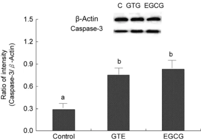
Fig. 7. Effects of GTE on caspase-3 in A549 cells. Cells were untreated (control), treated with GTE (100 μg/mL) or treated with EGCG (100 μg/mL) for 4 hours. Caspase-3 expressions in A549 cells were examined by Western blot (n=3 per group). Values with the different superscripts are significantly different by Duncan's multiple range test (p<0.05).
용이 있는 것으로 추측된다. 또한 EGCG나 녹차 추출물을 투여한 경우 모두 독소루비신 4 μg/mL 이상의 농도에서 A549 세포독성이 나타나므로, EGCG와 녹차 추출물 100 μg/
mL 농도에서 시스플라틴에 의한 세포독성에 미치는 EGCG 와 녹차 추출물의 상승효과의 차이는 없는 것으로 추측된다.
A549 세포에서 녹차 추출물이 p53과 casapase-3에 미 치는 영향
녹차 추출물 투여 A549 세포의 p53에 대한 Western blot 을 시행하여 나타난 결과는 Fig. 6과 같다. 밴드의 밀도를 측정하여 p53/actin의 비율을 환산한 결과 대조군, 녹차 추출 물군 및 EGCG군에서 각각 0.35, 0.81 및 0.89를 나타내서 녹차 추출물군과 EGCG군에서 대조군에 비하여 증가되어 녹차 추출물이나 EGCG 투여로 p53 양이 증가됨을 알 수 있었다. 또한 녹차 추출물군과 EGCG군의 밀도차이는 없기 때문에 p53 양에 미치는 녹차 추출물과 EGCG의 차이는 없 는 것으로 추측된다. p53은 시스플라틴의 항암작용과 밀접 한 연관이 있는 것으로 알려져 있으므로(21), 본 실험에서 EGCG나 녹차 추출물에 의해서 A549 세포 p53 증가는 EGCG와 녹차 추출물에 의한 시스플라틴의 세포독성 증가 에 작용할 것으로 추측된다.
녹차 추출물을 첨가하여 배양한 A549 세포의 caspase-3 에 대한 Western blot을 시행하여 나타난 결과는 Fig. 7과 같다. 밴드의 밀도를 측정하여 caspase-3/actin의 비율을 환 산하면 대조군, 녹차 추출물군 및 EGCG군에서 각각 0.29, 0.75 및 0.83을 나타내서 대조군에 비하여 녹차 추출물군과 EGCG군에서 증가되어 녹차 추출물이나 EGCG투여로 caspase-3 양이 증가됨을 알 수 있었다. 또한 녹차 추출물군 과 EGCG군에서 caspase-3 양의 차이가 없기 때문에 녹차 추출물과 EGCG가 caspase-3 양에 미치는 영향의 차이는 없는 것으로 추측된다.
세포자살(apoptosis)은 세포의 사멸을 유도하는 기전으로 서 여러 가지 항암물질이나 세포사멸의 중요한 기전으로서 세포자살은 p53, Noxa, bid 및 Bcl
2등이 관여하는 연쇄반응 결과 최종적으로 caspase가 활성화되어 일어난다(15). Gupta 등(22)은 전립샘암세포에서 EGCG가 세포자살을 유발한다 고 하였는데, 본 실험 결과 녹차 추출물이 p53이나 caspase- 3 양을 증가시키는 것은 세포의 사멸을 유도할 수 있기 때문 에 항암화학요법제의 항암작용을 증강시킬 수 있는 한 요소 로 작용될 수 있을 것으로 생각된다.
요 약
항암 화학요법제의 항암작용을 증가시키거나, 부작용을
감소시켜 항암 치료를 효과적으로 할 수 있는 항암치료 보조
제(modulator)에 대한 개발의 일환으로 녹차 추출물의 이용
가능성을 추정하기 위하여 사람 폐암 세포주인 A549 세포를
배양하여 시스플라틴과 독소루비신의 항암성에 미치는 녹
차 추출물과 EGCG의 영향을 비교 관찰하였다. A549 세포에
독성을 나타나는 농도는 녹차 추출물 400 μg/mL, EGCG 300
μ g/mL, 시스플라틴 10 μg/mL 및 독소루비신 8 μg/mL로,
녹차 추출물이 세포독성을 나타내는 농도는 시스플라틴이나
독소루비신에 비하면 낮았다. A549 세포에서 시스플라틴 10
μ g/mL 이상의 농도에서 세포활성이 감소되었고, EGCG나
녹차 추출물 100 μg/mL를 첨가하면 시스플라틴 6 μg/mL
이상의 농도에서 세포활성이 감소되어 EGCG나 녹차 추출
물 첨가로 시스플라틴의 세포독성이 증가되었다. A549 세포
에서 독소루비신 8 μg/mL 이상의 농도에서 세포활성이 감
소되었고, EGCG나 녹차 추출물 100 μg/mL를 첨가하면 독
소루비신 4 μg/mL 이상의 농도에서 세포활성이 감소되어
EGCG나 녹차 추출물 첨가로 독소루비신의 세포독성이 증가
되었다. A549 세포에서 녹차추출물 투여 후 p53 및 caspase-
3에 대한 Western blot을 시행한 결과 p53및 caspase-3의 유전자 발현이 증가되었다. 이상의 실험결과 녹차추출물은 광범위 항암제 시스플라틴이나 독소루비신의 세포독성을 증 강시키는 효과가 있고, 녹차추출물에 의한 p53이나 caspase- 3 등과 같은 세포자살유도 단백질의 발현 증가는 녹차추출 물에 의한 세포독성 증강효과와 연관이 있을 것으로 추측된 다. 녹차추출물의 시스플라틴이나 독소루비신 세포독성 증 강효과는 항암화학요법제의 용량을 늘리지 않고 항암력을 증대시킬 수 있기 때문에 항암화학요법 보조제로서 이용될 수 있는 가능성이 높은 것으로 생각되며, 이러한 효과를 규 명하기 위한 연구가 필요할 것으로 사료된다.
문 헌