41(1) : 14 20 (2010)
14
Fupenjic Acid 의 대식세포에서 LPS에 의해 유도되는 iNOS와 COX-2 발현 및 Cytokine들의 생성 저해 효과
윤창현·신지선·박희준
1·박종희
2·이경태
*경희대학교 약학대학
, 1상지대학교 친환경식물학부
, 2부산대학교 약학대학
Inhibition of LPS-induced iNOS, COX-2 Expression and Cytokines Production by Fupenjic Acid in Macrophage Cells
Chang Hyeon Yun, Ji-Sun Shin, Hee-Juhn Park
1, Jong-Hee Park
2and Kyung-Tae Lee*
Department of pharmaceutical Biochemistry, College of Pharmacy, Kyung-Hee University, Seoul 130-701, Republic of Korea
1Division of Environmental Botany, Sangji University, Wonju 220-702, Republic of Korea
2College of Pharmacy, Pusan National University, Busan 609-735, Republic of Korea
Abstract −
In this study, we investigated the anti-inflammatory effects of fupenjic acid (FA) isolated from the Potentilla dis- color in both RAW 264.7 and mouse primary peritoneal macrophage cells. FA pretreatment significantly inhibited nitric oxide (NO) and prostaglandin E2 (PGE2) productions in the lipopolysaccharide (LPS)-induced RAW 264.7 and mouse primary peri- toneal macrophage cells. Consistent with these observations, Western blot and RT-PCR analyses revealed that FA inhibited the LPS-induced expressions of inducible nitric oxide synthase (iNOS) and cyclooxygenase-2 (COX-2) at the protein and mRNA levels. In addition, FA reduced the release of tumor necrosis factor-α
(TNF-α
) and interleukin-6 (IL-6). These results suggest that the down regulation of iNOS and COX-2 expression and TNF-α
and IL-6 production by fupenjic acid are responsible for its anti-inflammatory effects.Key words −
Fupenjic acid, LPS, Cytokines, iNOS, COX-2솜양지꽃
(Potentilla discolor Bunge)은 장미과
(Rosaceae)에 속하는 다년생 초본식물로서 우리나라 전국 각지의 산 야지 및 해변의 구릉지 양지에 두루 자생한다
.솜양지꽃의
뿌리는 번백초라 하여 한방에서 약용 혹은 식용하고 있다
.1)번백초는 한방에서 청열해독
(熱解毒
)하여 지혈
(止血
)및
소종
(消腫
)등의 효능이 있다고 알려져 있다
.2)또한 중국의
전통민간요법에서는 번백초를 설사
,말라리아
,백선
,토혈
,객혈 등에 사용하고 있다
.3)최근 번백초 추출물의 in vivo와
in vitro에서 혈당강하작용이 밝혀져 제
2형 당뇨병의 치료제
로서 임상시험 중에 있다
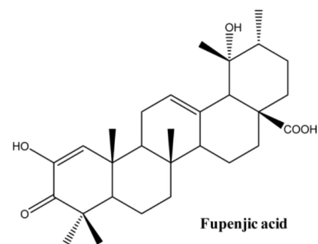
.4-6)Fupenjic acid (FA)
는
steroid모핵을 가지고 있으며
(Fig.1), steroid
구조는 우리 몸에서 성호르몬 및 부실피질 호르
몬 등의 모핵을 이루는 구조로서 항염증 약물의 구조에서 자주 볼 수 있는 화합물의 한 형태이다
.솜양지꽃에서는
FA이외에
sterol, terpenoid, hydrolyzed tannin및
flavonoid등
의 다양한 형태의 화합물의 구조가 이미 밝혀진바 있다
.1,7)본 연구진은 여러 천연물로부터 추출물을 얻고 단일화합물 을 분리하여 이들의 항염증 효과를 검색하고 그 기전을 규 명하여 새로운 항염증 약물 개발을 개발 하고 있다
.지금까
지 항염효과가 보고된바 없는
FA를 대식세포주 및 일차배
양 대식세포에서의 실험을 통해 그 항염효과를 확인하고자 하였다
.염증은 물리적 화학적 자극에 의해서 발생하는 피해에 반 응하는 생체반응으로 관절염
,천식
,다발성 신경경화증 및
대장염 등 여러 가지 질환의 원인에 중요한 인자이다
.이런
염증 반응은 대개 급성 반응인 경우가 많으나 때로는 체내 에 다양한 종류의 염증세포들이 축적되어 만성 염증성 질 환을 야기하기도 하는데 이러한 만성 염증은 몇몇 염증관 련 매개물질이나
lipid mediator, protease및
cytokine등의
분비에 의해 발생 된다
.8)대식세포는 능동 및 수동 면역반응에서 매우 중요한 역할
*교신저자(E-mail):[email protected] (Tel): +82-2-961-9199
을 하며
NO, prostaglandin (PG)그리고
pro-inflammatory cytokine들을 포함한 다양한 염증 매개물질들을 조절한다
.9)이중
NO형성은 박테리아를 죽이거나 종양을 제거 시키는
중요한 역할을 하기도 하지만
,병리적인 원인에 의한 과도
한
NO형성은 염증을 유발시키게 되며 조직의 손상
,유전
자 변이 및 신경 손상 등을 유발한다
.10-12)현재 임상에 쓰이고 있는 많은 소염제들의 작용기전은
COX-2
의 발현 및 그 활성의 저해로 인한
PG의 합성을 억
제하는 것이다
. COX는
COX-1과
COX-2등
2개의
subtype이 있는데
,신체 장기 별로 각기 다른 발현 양상을 나타낸
다
. COX-1은 주로 위와 신장에 많이 존재하며 이들 장기의
기능 유지 및 혈소판 형성에 필요한
PG를 합성하는 반면
COX-2
는 동물이나 사람의 염증부위에 과발현되어 염증반
응을 매개하는 것으로 여겨지고 있다
.13-14)Cytokine
중에 하나인
TNF-α는
T cell과 대식세포를 활성
화하고 다른
pro-inflammatory cytokine들을 증가시켜서 염
증반응을 일으키는데 중요한 역할을 한다
.15)이와 마찬가지
로
IL-6도
LPS에 의해서 대식세포에서 분비되는 중요한 염
증성
cytokine중 하나이다
. LPS, TNF-α
, IL-6같은 요소들은
전사인자인
NF-κ
B를 활성화시키고 이는
NO, PGE2, IL-6, TNF-α 등
pro-inflammatory molecule들의 생성량을 증가 시
킨다
.16)본 논문에서는
FA가
LPS에 의해 활성화된
RAW 264.7세포주 및 마우스 복강에서 분리한 일차 배양 대식세포에 서 염증관련 매개물질인
NO, PGE2의 생성과 관련 단백질 및
mRNA의 발현 양상 및 염증성
cytokine을 측정함으로서
항염증 작용을 확인하였다
.재료 및 방법
재료 −
Dulbecco’s modified Eagle’s minimum essential medium (DMEM), fetal bovine serum (FBS), penicillin,streptomycin
은
life Technologies Inc. (Grand Island, NY)에서 구입하였다
. 3-(4,5-dimethylthiazol-2-yl)-2,5-diphenyl- tetrazolium bromide (MTT), dimethyl sulfoxide (DMSO), sulfanilamide, aprotinin, leupeptin, phenylmethylsulfonylfluride (PMSF), dithiothreitol (DTT), L-N6-(1-iminoethyl)lysine (L-NIL), NS-398,Escherichia coli
lipopolysaccharide (LPS)는
Sigma Chemical Co. (CA, U.S.A.)에서 구입하였으며
, COX-2와
iNOS monoclonal antibodies및
peroxidase conjugated secondary antibody는
Santa Cruz Biotechnology (CA, U.S.A.)에서 구입하였다
. iNOS, COX-2, TNF-α
, IL-6그리고 β
-actin oligonucleotide primers는
Bioneer (Seoul, Korea)에서 구입하였다
.그리고
TNF-α
, IL-6, prostaglandin E2측정을 위한
kit는
R&D systems (MN, U.S.A.)에서 구입
하였다
.시료의 추출 및 분리 −
FA는 솜양지꽃 뿌리로부터 분리
한 것을 사용하였으며
1)본 실험에 사용한
FA는
HPLC-MS로 확인한 결과 순도가
95%이상 이였다
.세포의 배양 −
RAW 264.7세포는
10% FBS및
penicillin (100µ
g/ml), streptomycin (100 U/ml)이 포함된
DMEM배
지에서
37oC, 5% CO2조건을 유지하여 배양했다
. RAW 264.7세포에 시료용액의 여러 농도
(10, 20, 40µ
M)를
1시
간 전 처리한 후
LPS (1µ
g/ml)를 처리하고
24시간 배양하
였다
.마우스 복강 대식세포 분리 −
C57BL/6 mice에서 복강대
식세포를 얻기 위해서
5% thioglychollate용액
2 ml을 복강
주사하고
4일 후에 복강에 추출된 세포를 저온의
DMEM배
지를 이용해서 세정하여 얻어냈다
.세포를
2번 세척한 후
HEPES-
완충
DMEM배지로 재구축하여 배양하였다
.세포독성시험 −
96 well plate에
1×10⁵ cells/ml로 세포를
동일하게 분주하고
24시간 동안 배양한 후 여러 농도의 시
료용액을 배지에 희석하여 첨가 하였다
. 24시간이 지난 후
MTT
시약을 넣고
4시간 동안 어두운 곳에서 배양한 후 상
등액을 제거하고 형성된
MTT formazan을
DMSO 100µ
l를
첨가하여 녹였다
. 10분 후
540 nm에서 흡광도를 측정하였다
.Nitrite 양의 측정 −
RAW 264.7세포로부터 생성된
NO의
양은
Griess시약을 이용하여 세포 배양액 중에 존재하는
NO2-
의 형태로서 측정하였다
.즉 세포배양 상등액
100µ
l와
Griess시약
[1% (w/v) sulfanilamide in 5% (v/v) phosphoric acid와
0.1% (w/v) naphtylethylenediamine-HCl] 100µ
l를
혼합하여
96 well plate에서
10분 동안 반응시킨 후
540 nm에서 흡광도를 측정하였다
.Western blot 시험 −
FA를 처리한 세포 및 대조군을
PBS로 씻어낸 후
lysis buffer인
PRO-PREP (Intron Biotech-nology)
으로 단백질을 추출한 후 원심분리하여 상등액을 취
하였다
.상등액을
Bradford시약을 사용해 단백질 농도를 정
량하여
30µ
g의 단백질을 취했다
.추출된 단백질은
8%와
Fig. 1.
Chemical structure of fupenjic acid (FA).10%
의
SDS-polyacrylamide gel에 전기영동 시킨 후
nitro cellulose membrane으로
gel의 단백질을
blot시켰다
. 5%skim milk
로
2시간 동안
blocking한 후
1:1000의 비율로
iNOS와
COX-2 antibody를
4시간 동안 상온에서 방치한 후
TTBS로
10분 간격으로
3회 세척하였다
. 1:1000의 비율로
희석한
secondary antibody를
1시간 동안 상온에서 방치 시
켰다
.다시
TTBS로
10분 간격으로
3회 세척한 후
chemiluminescence로 현상하였다
.PGE
2, TNF- α 및 IL-6 양의 측정
-세포배양액을 취해
각각
R&D kit의 지시에 따라
PGE2, TNF-α 및
IL-6를 정
량 하였다
.RT-PCR 시험
- Easy Blue® kits (Intron Biotechnology)를 이용하여
kit의
protocol에 따라 전체
cellular RNA를 추
출하였다
.각각의 시료에서
MuLV reverse transcriptase, 1mM dNTP그리고
oligo (dT12-18) 0.5µ
g/µ
l를 이용하여
1 ìg의
RNA를 역전사 하여
cDNA를 얻었다
. cDNA에
Taq DNA polymerase 1 unit, 0.2 mM dNTP, ×10 reaction buffer그리고
5'와
3' primers 100 pmol을 포함한 전체 부
피
20µ
L의 시료를
thermal cycler (Perkin Elmer Cetus, Foster City, CA, USA)를 이용하여
PCR분석을 하였다
. PCR반응은
95oC에서
2분간
initial denaturation시킨 후
iNOS (95oC 1분
denaturation, 60oC 1분
annealing그리고
72oC 1.5분
extension), COX-2 (94oC 1분
denaturaion, 60oC 1분
annealing그리고
72oC 1분
extension), TNF-α
(95oC 1분
denaturation, 55oC 1분
annealing그리고
72oC 1분
extension)그리고
IL-6 (94oC 1분
denaturation, 56oC 1분
annealing그리고
72oC 1분
extension)를
30회
amplification하였다
.이번 연구에서 아래의 목록과 같은
PCR primers가 사용되었으며
Bioneer (Seoul, Korea)에서
구입하였다
. : sense strand iNOS, 5'-ATT GGC AAC ATC AGG- TCG GCC ATC ACT-3', anti-sense strand iNOS,5'- GCT GTG TGT CAC AGA AGT CTC GAA- CTC-3';sense strand COX-2, 5'-GGA GAG ACT ATC AAG ATA GT-3' anti-sense strand COX-2, 5'-ATG GTC AGT AGA CTT TTA CA-3'; sense strand
β
-actin, 5'-TCA TGA AGT GTG ACG TTG ACA- TCC GT-3', anti-sense strandβ
- actin, 5'-CCT AGA AGC ATT TGC GGT GCA CGA TG-3'. Amplification후에
PCR반응 시킨 시료를
1.5%agarose gel
에서 전기영동하고
ethidium bromide염색과
UV조사를 통해 확인하였다
.통계학적 분석
-실험치의 값은
mean±S.D.로 나타냈으며
분석은
Student’st
-test로 그 유의성을 나타내었다
결 과
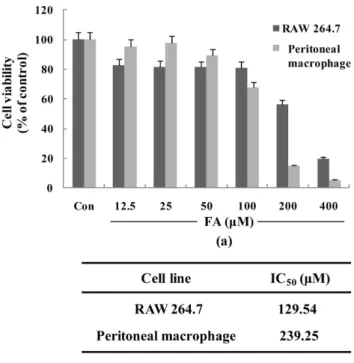
FA 의 세포독성 −
FA의 항염증 효과를 규명하기에 앞서
FA
가 대식세포에 독성을 통한 염증 매개 물질 저해의 가능
성을 배제하고 또한 최적 용량 범위를 설정하기 위해
MTT assay를 실시하였다
. RAW 264.7세포에서는
FA를
50µ
M을 처리했을 때까지 세포에 독성이 나타나지 않았으며
(Fig.2a) IC50 value
는
129.54µ
M로 나타났다
(Fig. 2b). C57BL/6
마우스의 복강에서 분리한 일차 배양 대식세포에서는
FA를
100µ
M까지 처리했을 때 세포에 독성이 크게 나타나지
않았고
(Fig. 2a) IC50 value는
238.25µ
M로서
RAW 264.7세포에 비해 독성이 낮게 나타났다
(Fig. 2b). FA의 항염증
효과를 세포독성이 없는 범위 내에서 실험하기 위해
RAW 264.7및 일차 배양 마우스 대식세포에서는 각각
40µ
M및
100µ
M을 최고농도로 설정하고 이후 실험을 진행하였다
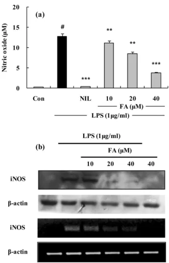
.FA 의 Nitriite 생성 및 iNOS 단백질과 mRNA 발현 저
해 −
LPS에 의해 활성화된
RAW 264.7세포의 배양액 중에
생성된
NO의 양을
Griess시약을 사용해서 측정하였다
. FA는
LPS에 의한
NO생성을 농도 의존적으로 저해하였으며
고농도
(40µ
M)에서는
70%저해 효과를 보였다
(Fig. 3a).양성 대조군으로는
L-arginine과의 기질경쟁에 의하여
iNOS저해제로 알려진
L-NIL (10µ
M)을 사용하였다
. FA에 의한
NO
의 생성 억제 효과가
iNOS의 발현과 관련성이 있는가를
확인하기 위해
Western blot과
RT-PCR로
iNOS의 단백질과
mRNA발현을 조사했다
. LPS에 의해 뚜렷하게 유도된
iNOSFig. 2.
Cytotoxicity of FA on RAW 264.7 cells or mouse primary peritoneal macrophages(ex-vivo) were exposed to FA(from 12.5µ
M to 400µ
M). Cytotoxicity was assessed by 3- (4, 5 dimethylthiazol-2-yl)-2, 5-diphemyltetrazolium bromide (MTT) assay after 24 h incubation.단백질의 발현량이
FA를
20µ
M처리했을 때부터 유의성
있는 현저한 저해를 나타내었으며
, RT-PCR을 통한
mRNA의 발현 변화의 경우에도
20µ
M처리했을 때부터 유의성
있는 현저한 저해를 보였다
(Fig. 3b).FA 의 PGE
2생성 및 COX-2 단백질 및 mRNA 발현 저
해효과 −
LPS를 전 처리한
RAW 264.7세포에서
PGE2의
생성량이 유의성 있게 증가하였고
FA가 농도 의존적으로
PGE2의 생성량을 감소시키는 것을 확인하였다
(Fig. 4a).양
Fig. 3.
The effects of FA on LPS-induced NO production and iNOS protein and mRNA expressions in RAW 264.7 cell. (a) RAW 264.7 cells were treated with different concentrations of FA for 1 h and then LPS (1µ
g/ml) was added and the cells were incubated for 24 h. Control (Con) values were obtained in the absence of LPS or tested samples. L-N6-(1-iminoethyl) lysine (L-NIL) was used as an assay positive control at a concentration of 10µ
M. (b) Lysates were prepared from control or 24 h LPS (1µ
g/ml)-stimulated cells alone or LPS plus with different concentration (10, 20, 40µ
M) of FA. A representative immunoblot of three separate experiments is shown. Total RNA was prepared for the RT-PCR analysis of iNOS gene expression from RAW 264.7 cell stimulated with LPS (1µ
g/ml) with/without different concentration (10, 20, 40 ìM) of FA for 4 h. iNOS-specific sequences (807 bp) was detected by agarose gel electrophoresis, as described in method. The experiment was repeated three times and similar results were obtained. The values are the mean±S.D. of three independent experiments. #p<0.05, **p<0.01, ***p<0.001 vs.the LPS-treated group; the significances of the difference between the treated groups were evaluated using the Student’s t-test.
Fig. 4
. The effects of FA on LPS-induced PGE2 in RAW 264.7 cells. (a) Effect of the FA on PGE2 production by LPS- induced RAW 264.7 macrophage for 24 h. 10µ
M of NS-398 was as a positive control in the assay. (b) Lysates were prepared from control or 24 h LPS (1µ
g/ml)-stimulated cells alone or LPS plus with different concentration (10, 20, 40µ
M) of FA. A representative immunoblot of three separate experiments is shown. Total RNA was prepared for the RT- PCR analysis of COX-2 gene expression from RAW 264.7 cell stimulated with LPS (1µ
g/ml) with/without different concentration (10, 20, 40µ
M) of FA for 4 h. COX-2-specific sequences (721 bp) was detected by agarose gel electrophoresis, as described in method. The values are the mean±S.D. of three independent experiments. #p<0.05, **p<0.01, ***p<0.001 vs.the LPS-treated group; the significances of the difference between the treated groups were evaluated using the Student’s t-test.
성 대조군으로는 선택적인
COX-2저해제로 알려진
NS398 (5µ
M)을 사용하였다
. PGH2로부터
PGE2를 생성하는데 관
여한다고 알려진 효소인
COX-2의 단백질 발현량은
FA의
최고 농도인
20µ
M에서 유의성 있는 저해를 확인 하였으며
, RT-PCR결과
20µ
M에서 단백질 저해 와 유사하게
COX-2의
mRNA발현을 유의성 있게 저해하였다
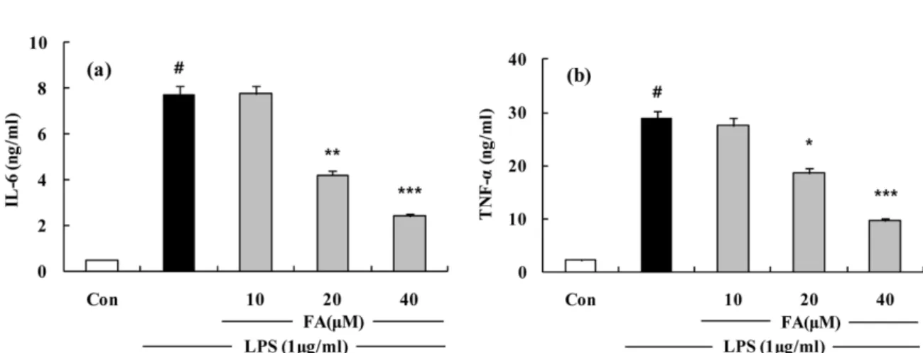
(Fig. 4b).FA 의 IL-6 와 TNF- α의 형성 저해 효과 −
FA가
RAW 264.7세포에서
LPS에 의해서 생성된
pro-inflammatory cytokine의 형성을 억제하는지 알아보기 위해서
IL-6와
TNF-α의 생성을 측정하였다
. IL-6의 경우
20µ
M에서
45%의
저해효과를 보이며 농도 의존적으로 생성이 억제 되었다
(Fig. 5a). TNF-
α의 경우 역시 농도의존적인 저해 양상을
보였고
,최고농도인
40µ
M에서는
55%가 저해되었다
(Fig.5b).
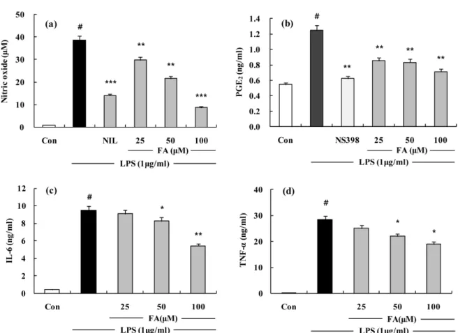
FA 의 일차 배양 복강대식세포에서의 항염효과 확인 −
FA
가
RAW 264.7세포에서 뿐만 아니라
C57BL/6 mice에
서 얻은 일차 배양 복강대식세포에서도 항염효과를 가지는 지 확인하기 위해
RAW 264.7세포와 동일한 조건에서
LPS에 의해 생성된
NO, PGE2의 양을 측정해본 결과
FA가
NO, PGE2생성량을 고농도인
100µ
M에서 각각
77%, 43%저
해하였다
(Fig. 6a,b). LPS에 의해서 생성된
pro-inflammatory cytokine인
IL-6와
TNF-α의 생성을 측정해본 결과
IL-6의
경우 농도 의존적으로 생성이 감소하였고 특히
100µ
M에
서
43%저해되었으며
TNF-α의 경우 역시 농도 의존적인
저해 양상을 보였고
,최고농도인
100µ
M에서는
34%가 저
해되었다
(Fig. 6c,d).고 찰
대식세포는
NO, PG, leukotriene및
pro-inflammatory cytokine들의
2차 매개물을 생산하고 분비한다
.이런 물질들
은 선천성 및 후천성 면역을 조절하는데 있어서 중요한 역 할을 한다
.9)그러나 이런 물질들이 과잉 생산 되었을 때에
는 세균성 패혈증
,류마티스성 관절염
,만성 염증
,자가면역
질환 등을 유발하기도 한다
.17-18)본 연구는 처음으로 솜양지꽃 뿌리에서 분리한
FA의
LPS로 유도한
RAW 264.7및
C57BL/6마우스에서 분리한 복
강 대식세포에서 염증 매개 물질인
NO, PGE2, IL-6와
TNF-α의 생성 저해를 통한 항염증 효과를 확인하고자 하였다
.선천성 면역체계에서
,미생물 성분중의 하나인
LPS대식
세포를 자극하여 많은
TNF-α
, IL-1, IL-6, iNOS및
COX-2
등과 같은 많은 염증 매개 물질을 생성하며
,이러한 물질
들은 급성 및 만성 염증 질환 발병에 중요한 역할을 담당하
고 있다
.19-20)결국 이러한 물질을 저해하는 것은 항염증 물
질 개발에 있어 중요한 전략중의 하나이다
.FA
는
RAW 264.7세포에 독성이
20%미만 농도에 대해
서 실시한
NO생성 실험에서
LPS에 의해 증가된
NO생
성이
FA에 의해 농도 의존적으로 저해되었다
.또한
Western blot과
RT-PCR을 통해
iNOS의 단백질 및
mRNA수준의
발현 저해 효과를 확인해본 결과
FA에 의해 그 발현량이
저해됨을 통해
FA에 의한
NO생성량의 저해는
iNOS에 의
한 것임을 밝혔다
.또 다른 염증매개 물질인
PGE2역시
FA에 의해 그 생성량이 저해함을 확인하였다
. Western blot과
Fig. 5
. The effects of FA on LPS-induced IL-6 and TNF-α
release in RAW 264.7 cells and mouse primary peritoneal macrophages.(a) Effect of the FA on IL-6 release by LPS-induced RAW 264.7 macrophage for 24 h. Cells were treated with different concentrations of FA for 1 h and then LPS (1
µ
g/ml) was added and the cells were incubated for 24 h. Control (Con) values were obtained in the absence of LPS or tested samples. (b) Effect of the FA on TNF-α
release by LPS-induced RAW 264.7 macrophage for 24 h. Cells were treated with different concentrations of FA for 1 h and then LPS (1µ
g/ml) was added and the cells were incubated for 24 h. Control (Con) values were obtained in the absence of LPS or tested samples. The experiment was repeated three times and similar results were obtained. The values are the mean±S.D. of three independent experiments. #p<0.05, **p<0.01,***p<0.001 vs. the LPS-treated group; the significances of the difference between the treated groups were evaluated using the Student’s t-test.
RT-PCR
을 통해
COX-2단백질의 경우 최고 농도에서만 발
현이 저해되었고
mRNA는 농도 의존적인 저해를 보였다
.이는
FA에 의한
PGE2생성량 저해가 유전자 단계에서 조
절됨을 확인 할 수 있었다
.하지만
mRNA단계에서의 조절
이
COX-2의 단백질 발현까지는 영향을 주지 않았는데 이
는
COX-2단백질의 활성이 저해되어
PGE2의 생성량이 감 소했기 때문인 것으로 추정된다
.앞으로
COX-2 activity assay를 실시하여
COX-2의 단백질 발현량과는 별개로
COX- 2의 활성과
PGE2의 생성량 간의 관계를 규명할 계획이다
.염증의 주요 매개물질로써 잘 알려진
IL-6와
TNF-α는 in
vivo 및 in vitro에서 염증반응을 조절한다
.두
cytokine은 상
호작용을 하며
, LPS의 자극에 의해 생성이 유도된다고 알
려져 있다
.21) FA는
LPS에 의해 유도된
IL-6와
TNF-α의 생
성을 유의성 있게 감소시켰다
.C57BL/6
복강에서 직접 분리하여 배양한 대식세포를 사
용하여
PGE2와
NO생성량을 측정해 본 결과
LPS에 의해
증가된
PGE2와
NO의 형성을
FA는 농도 의존적으로 유의
성 있게 모두 억제하였으며 염증성
cytokine인
IL-6와
TNF-α의 생성 역시 농도 의존적인 감소를 보였다
.본 연구는 솜양지꽃
(Potentilla discolor
)에서 분리한 단일
화합물인
FA의 항염증 작용 및 기전을
LPS로 유도된 대식
세포를 사용하여 항염증 효과를 밝혔다
.이러한
COX-2,iNOS
그리고
pro-inflammatory cytokine들의 발현에는
nuclear factor kappa B (NF-κ
B)가
promoter부위에 결합하
Fig. 6.
The effects of FA on LPS-induced NO, PGE2 production and IL-6 and TNF-α
release in mouse primary peritoneal macrophages. (a) Mouse primary peritoneal macrophages were treated with different concentrations of FA for 1 h and then LPS (1µ
g/ml) was added and the cells were incubated for 24 h. Control (Con) values were obtained in the absence of LPS or tested samples. L-N6-(1-iminoethyl) lysine (L-NIL) was used as an assay positive control at a concentration of 10µ
M. (b) Effect of the FA on PGE2 production by LPS-induced Mouse primary peritoneal macrophages for 24 h. 10 ìM of NS-398 was as a positive control in the assay. (c) Effect of the FA on IL-6 release by LPS-induced RAW 264.7 macrophage for 24 h. Cells were treated with different concentrations of FA for 1 h and then LPS (1µ
g/ml) was added and the cells were incubated for 24 h. Control (Con) values were obtained in the absence of LPS or tested samples. (d) Effect of the FA on TNF-α
release by LPS-induced mouse primary peritoneal macrophage for 24 h. Cells were treated with different concentrations of FA for 1 h and then LPS (1µ
g/ml) was added and the cells were incubated for 24 h. Control (Con) values were obtained in the absence of LPS or tested samples. The experiment was repeated three times and similar results were obtained. The values are the mean±
S.D. of three independent experiments. #p<0.05, **p<0.01, ***p<0.001 vs. the LPS-treated group; the significances of the difference between the treated groups were evaluated using the Student’s t-test.여 조절인자로 작용한다고 보고되었다
.22-23)본 연구진은 앞
으로 염증 매개 물질의 발현을 조절하는 전사인자인
NF-κ
B및
cAMP response element (CRE), SRE (serum response element), activator protein-1 (AP-1), mitogen-activated protein kinase (MAPK)등의 다양한 전사 조절인자들의 신
호전달 양상을 연구를 함으로써
FA가 어떠한 경로를 통해
항염증 효과를 가지는지 그 기전을 밝히고 추후의 in vivo 실험을 바탕으로 향후 염증성 질환의 예방 및 치료 약물의 개발가능성을 제시하고 있다
.인용문헌
1. Park, H.-J., Lee, k.-T. and Park, J.-H. (2007) Isolation of Two steroids and a triterpenoid from the roots of Potentilla dis- color. Kor. J. Pharmacogn.
38
: 354-3572. Shoyakan (ed.) (1985) Encyclopedia of Chinese Medicinal Drugs, 2438-2438, Shanghai Science & Technology, Tokyo.
3. Tomczyk M, Latté KP. (2009) Potentilla--a review of its phy- tochemical and pharmacological profile. J. Ethnopharmacol.
122
: 184-2044. Syiem, D., Syngai G., Khup, P. Z., Khongwir. B. S., Khar- buli, B. and Kayang, H. (2002) Hypoglycemic effects of Potentilla fulgens L in normal and alloxan-induced diabetic mice. J. Ethnopharmacol.
83
: 55-61.5. Zhao, C., Qiao, W., Zhang, Y. W., Lu, B. and Duan, H. Q.
(2008) Study on anti-diabetes active fraction and constituents from Potentilla chinensis. Zhongguo Zhong Yao Za Zhi.
33
: 680-682.6. Xue, P. F., Zhao, Y. Y., Wang, B. and Liang, H. (2006) Sec- ondary metabolites from Potentilla discolor Bunge (Rosaceae).
Biochem. Syst. Ecol.
34
: 825-828.7. Jie, Y., Xiao-Qing., C., Xiao-Xiao., L., Yuan, C., Mao-Xiang, L. and Qiang, W. (2008) Structural determination of two newtriterpenoids from Potentilla discolor Bunge by NMR techniques Magn. Reson. Chem.
46
: 794-7978. Cho, W., Nam, J.-W., Kang, H.-J., Windono, T., Seo E.-K.
and Lee, K.-T. (2009) Zedoarondiol isolated from the rhi- zoma of Curcuma heyneana is involved in the inhibition of iNOS, COX-2 and pro-inflammatory cytokines via the down- regulation of NF-
κ
B pathway in LPS-stimulated murine mac- rophages Int. Immunopharmacol.9
: 1049-10579. Iontcheva, I., Amar, S., Zawawi, K. H., Kantarci, A. and Van Dyke, T. E. (2004). Role for moesin in lipopolysaccharide- stimulated signal transduction. Infect Immun.
72
: 2312-2320.10. Stuehr, H. H. J., Kwon, N. S., Weise, M. and Nathan, C.
(1991) Purification of the cytokine-induced macrophage
nitric oxide synthase: an FAD- and FMN- containing fla- voprotein. Proc. Natl. Sci. USA.
88
: 7773-7777.11. McCartney-Francis, N., Allen, J. B., Mizel, D. E., Albina, J.
E., Xie, Q. W., Nathan, C. F. and Wahl, S. M. (1993) Sup- pression of arthritis by an inhibitor of nitic oxide synthase. J.
Exp. Med.
178
: 749-754.12. Weisz, A., Cicatiello, I. and Esumi, H. (1996) Regulation of the mouse inducible-type nitric oxide synthase gene promoter by interferon-gamma, bacterical lipopolysaccharide and NG- monomethyl-L-arginene. Biochem. J.
316
: 209-215.13. Masferrer, J., Zweifel B. S., Manning, P. T., Hauser, S. D., Leahy, K. M., Smith, W. G., Isacson, P. C. and Seibert, K.
(1994) Selective inhibition of inducible cyclooxygenase 2 in vivo is anti-inflammatory and nonulcerogenic. Proc. Natl.
Acad. Sci. U.S.A.
91
: 3228-3232.14. Seibert, K., Zhang, Y., Leahy, K., Hauser, S., Masferrer, J., Perkins, W., Lee, L. and Ksakson, P. (1994) Pharmacological and biochemical demonstration of the role of cyclooxygenase 2 in inflammation and pain. Proc. Natl. Acad. Sci. U.S.A.
91
: 12013-12017.15. Beutler, B. and Cerami, A. (1989) The biology of cachectin/
TNF-R primary mediator of the host response. Annu. ReV.
Immunol.
7
: 625-655.16. Dendorfer, U. (1996) Molecular biology of cytokines. Artif.
Organs.
20
: 437-444.17. Hilliquin P, Borderie D, Hernvann A, Menkès CJ, Ekindjian OG. (1997) Nitric oxide as S-nitrosoproteins in rheumatoid arthritis. Arthritis Rheum.
40
(8): 1512-151718. Nava, E., Palmer, R. M. and Moncada, S. (1992) The role of metric oxide in endotoxic shock: effects of NG-monomethyl- L-arginine. J
.
Cardiovasc. Pharmacol.12
: 132-134 19. Hoffmann, J. A., Kafatos, F. C., Janeway, C. A. and Ezekow-itz, R. A. (1999) Phylogenetic perspectives in innate immu- nity. Science
284
: 1313-131820. Janeway, C. A. Jr. and Medzhitov, R. (2002) Innate immune recognition. Annu. Rev. Immunol.
20
: 197-216.21. Liu, S. F. and Malik, A. B. (2005) NF-
κ
B activation as a pathological mechanism of septic shock and inflammation.Am. J. Physiol. Lung Cell Mol. Physiol.
290
: L622-L645.22. Feldmann, M., Brennan, F. M. and Maini, R. N. (1996) Role of cytokines in rheumatoid arthritis. Annu. Rev. Immunol.
14
: 397-440.23. Karin, M. and Ben-Neriah, Y. (2000) Phosphorylation meets ubiquitination: the control of NF-B activity. Annu. Rev.
Immunol.