42(4) : 348∼ 353 (2011)
348
백선피의 iNOS발현과 염증성사이토카인의 생성에 미치는 영향
박정숙*·신태용1·김대근1·이재혁2*
남부대학교 대체의학과,
1
우석대학교 약학과,2
남부대학교, 한방제약개발학과The Effects of Dictamni Radicis Cortex on the iNOS Expression and Proinflammatory Cytokines Production
Jeong-Suk Park, Tae-Yong Shin1, Dae Keun Kim1 and Jae-Hyeok Lee2 Department of Alternative Medicine Nambu University, Gwangju 506-606,Korea
1
College of Pharmacy, Woosuk University, Jeonbuk 565-701, Korea2
Department of Oriental Pharmaceutical Development, Nambu University, Gwangju 506-606,KoreaAbstract − The aim of the present study is to investigate the cytokine production inhibitory effect of a Dictamni Radicis Cortex (DRC). DRC has been commonly used as important medicinal herb in China and it used to control eczema, atopic dermatitis, fever and inflammatory diseases. Inflammation, such as a bacterial infection in vivo metabolites, such as external stimuli or internal stimuli to the defense mechanisms of the biological tissue a variety of intracellular regulatory factors deulin inflam- matory TNF-α, IL-1β, IL-6, IL-8, such as proinflammatory cytokines, prostagrandin, lysosomal enzyme, free radicals are involved in a variety of mediators. The present study was designed to determine the effect of the DRC on proinflammatory factors such as NO, iNOS expression and TNF-α, IL-1β, IL-6 in lipopolysaccharide (LPS) - stimulated RAW264.7 cells. The cell toxicity was determined by MTS assay. To evaluate of anti-inflammatory effect of DRC, amount of NO was measured using the NO detection kit and the iNOS expression was measured by reverse transcriptase polymerase chain reaction (RT- PCR). And proinflammatory cytokines were measured by ELISA kit. As a result, the DRC reduced NO, iNOS expression and TNF-α, IL-1β, IL-6 production without cytotoxicity. Our results suggest that the DRC may have an anti-inflammatory property through suppressing inflammatory mediator productions.
Key words − Dictamni Radicis Cortex, Cytokine, NO, iNOS, TNF-α
백선(Dictamnus dasycarpus Turcz)은 운향과(Rutaceae)에 속하는 다년생 숙근초로 한국, 중국, 및 동부 시베리아 등 지의 숲속에서 자생하는 식물로 생김새는 원통모양이고 잔 뿌리가 있으며 바깥 면은 황백색 또는 엷은 갈색이고 매끄 러우며 안쪽 면은 엷은 황색이고 질은 부드러워 쉽게 부러 진다.
1)
민간에는 봉삼 또는 봉황삼으로 알려져 있으며 뿌리 의 껍질을 약용부위로 사용한다.2,3)
백선의 뿌리껍질인 백선 피(Dictamni Radicis Cortex)는 청열해독, 거풍제습작용이 있 고 습열로 인한 피부발진, 피부미란, 습진, 가려움증, 알레 르기성 피부염, 신경성 피부염과 급성간염으로 인한 황달, 습열성 하지마비에 사용하며, 해수, 인후건조, 번갈에도 사 용한다.4)
백선피에는 limonoids,5)
flavonoids,6)
coumarins,7)
sesquiterpene 등8)
의 성분이 함유되어 있으며, RBL-2H3세포에서 항 알레르기효과,
9)
병원성세균에 대한 억제효과,10)
T-Cell 증식에 대한 자극,11)
A-549 세포주에 대한 세포 독 성12)
등이 보고되고 있다. 염증 반응은 세균감염과 같은 외 부 자극이나 생체내 대사산물과 같은 내부자극에 대한 생 체조직의 방어기전으로13)
알레르기, 아토피, 관절염, 심장병, 뇌질환, 순환기 장애뿐만 아니라 암 등과 같은 다양한 질환 의 원인을 제공하게 된다.14)
염증의 증상으로는 홍반, 부종, 열, 통증 그리고 기능 상실 등이 나타나며,15)
세포내 다양한 염증조절인자인 TNF-α, IL-1β, IL-6, IL-8 등과 같은 proinflammatory cytokines, prostagrandin, lysosomal enzyme, free radicals 등 다양한 매개물질이 관여한다.16)
특히 대식 세포에서 cytokines, tumor necrosis factor (TNF-α), lipo- polysaccharide (LPS)와 같은 자극에 의해 염증 반응의 전사 인자인 NF-κB를 활성화시키며 그 결과 inducible nitric oxide synthase (iNOS), cyclooxygenase-2 (COX-2)를 발현*교신저자(E-mail):[email protected], [email protected] (Tel): +82-62-970-0167, +82-62-970-0162
시켜 nitric oxide (NO)와 Prostaglandin E
2
(PGE2
)를 생성하 여 염증을 일으킨다.17,18)
또한 NO가 필요이상으로 생성되 면 염증 반응의 항진, 과도한 혈관 확장에 의한 패혈성 쇼 크 유발, 상처 치유의 억제, 신경조직의 손상 등을 일으켜 생체에 유해한 작용을 나타낸다.19)
백선피메탄올추출물 자체에 대한 세포내 다양한 염증조 절인자의 생성에 관한 연구는 전무한 상태이다. 이에 본 연 구는 백선피메탄올추출물의 RAW264.7세포에서 NO 생성, iNOS 발현정도와 염증성 사이토카인인 TNF-α, IL-1β, IL- 6의 생성량 등을 연구하여 유의한 결과를 얻었기에 보고하 고자 한다.
재료 및 방법
DRC의 제조 − 본 연구에 사용한 백선은 2010년 11월 전 남 화순군 청풍면 화학산에서 채취하였으며 정확히 감정한 후에 목질부를 제거한 껍질을 음건 세절하였다. 세절한 백 선피 약 1 kg을 MeOH로 가끔 진탕하면서 5시간씩 50
o
C에 서 3회 온침 추출하였다. 추출액을 수욕상에서 감압농축하 여 MeOH 엑스 약 60 g을 얻었으며 이를 실험재료로 사용 하였다.시약 − 세포 배양액인 Dulbecco's Modified Eagle Medium (DMEM)과 fetal bovine serum (FBS), streptomycin- penicillin 등의 세포배양용 시약들은 Gibco BRL사 (Grand Island, USA), Lipopolysaccharide (LPS)는 Sigma-Aldrich사 (St. Louis, MO, USA), Cell Titer 96
®
AQueous
One Solution (MTS)은 Promega사(Madison, WI, USA)에서 구입하여 사 용하였다. Tumor necrosis factor-α (TNF-α), IL-1β, IL-6 ELISA kit는 eBioscience사 (San Diego, CA, USA)에서 구 입하였고 NO (Nitric Oxide) detection kit는 iNtRON Biotechnology사(Suwon, Korea)을 구입하여 사용하였다.total RNA를 분리는 Easy Blue
®
시약 iNtRON Biotechnology (Suwon, Korea)을 이용하였고 cDNA를 합성은 QuantiTect®
Reverse Transcription Kit(Qiagen, USA)를 구입하여 사용하 였다. 이외에 실험에 사용된 시약은 모두 분석용 시약 특급 을 사용하였다.세포배양 − 실험에 사용한 마우스의 대식세포주인 RAW 264.7 세포는 한국세포주은행 (KCLB)에서 분양 받았으며, 세포배양을 위해 10% FBS과 1% penicillin-streptomycin을 포함하는 DMEM (Dulbecco
,
s Modified Eagle Medium) 배 지를 사용하였고, 세포는 CO2
배양기 (37o
C, 5% CO2
)에서 배양하였다.세포독성평가 − DRC의 세포에 대한 독성은 Desai 등의 방법
20)
에 따라 5-(3-caroboxymeth-oxyphenyl)-2H-tetra-zolium inner salt (MTS) assay 방법으로 mitochondrial dehydrogenases 에 의하여 MTS가 formazan 으로 전환되는 것을 측정하였다. 96 well plate에 1.0×10
5
cells/well의 RAW264.7 세포를 분주하고 18시간 동안 배양한 후 DRC 0.2 mg/mL, 2mg/mL 처리한 후, 24시간 동안 배양하였으며, 20 ul의 MTS solution 을 첨가한 후 CO2
배양기(37o
C, 5% CO2
)에서 4시간 반응 시킨 후, 450 nm에서 흡광도의 변화를 측정하여 대조군에 대한 세포 생존율을 백분율로 표시하였다.Nitric oxide 측정 − NO의 농도는 배양액 내의 nitrite농 도를 Wang등의 방법
21)
에 따라 Griess Reagent System을 이 용하여 측정하였다. RAW264.7 세포를 96 well plate에 1.0×10⁵
cells/well이 되도록 분주하고 18시간 동안 배양한 후, DRC 0.2 mg/mL, 2 mg/mL 전처리하고, 1시간 후에 LPS 200 ng/mL 처리한 후, 24시간 동안 배양하였다. 배양액과 동량의 Griess Reagent를 가하고 10분간 상온에서 반응시킨 후 540 nm에서 흡광도를 측정하였으며, Sodium nitrite의 농 도별 표준곡선을 이용하여 배양액의 NO농도를 결정하였다.RNA 분리 및 RT-PCR − RAW264.7 세포에서 발현되 는 iNOS의 유전자발현에 대한 DRC의 효과를 조사하기 위 해 RT-PCR을 수행하였다. 먼저 RAW264.7 세포를 60 mm dish에 4.0×10
6
cells이 되도록 분주하고 18시간 동안 배양하 였다. DRC 0.2 mg/mL, 2 mg/mL 전처리하고 LPS 200 ng/mL 처리 후 6시간 동안 배양하였다. 세포를 수거하여 4
o
C 에서 2,000 rpm으로 5분간 원심분리한 후 Easy Blue®
시약 (iNtRON Biotechnology, Korea)을 이용하여 total RNA를 분리하였다. 분리된 RNA를 QuantiTect®
Reverse Transcription Kit(Qiagen, USA)를 이용하여 cDNA를 합성하였다. 합성된 cDNA에 대한 PCR을 수행하기 위해 cDNA 1 ug에 primers (sense, anti-sense) 1ul 및 10 × buffer (10 mM Tris-HCl, pH 8.3, 50mM KCl, 0.1% Triton X-100), 250 µM dNTP, 1U Tag polymerase를 혼합한 후 denaturation을 위해 94에 서 45초, annealing을 위해 55~60o
C에서 45초 및 extension 을 위해 72o
C에서 60초 조건으로 30cycles을 수행하였다.이후 증폭된 DNA산물을 1.5% agarose gel를 사용하여 100volt에서 30분간 전기 영동하여 UV로 관찰하였다. iNOS 의 증폭을 위하여 sense primer : 5'-CCCTTCCGAAGTTTC TGGCAGCAGC-3', anti-sense primer : 5'-GGCTGTCAGA GCCTCGTGGCTTTGG-3'를 사용하였고 COX-2의 증폭을 위하여 5'-GGAGAGACTATCAAGATAGTGATC-3'(sense), 5'-ATGGTCAGTAGACTTTTACAGCTC-3'(anti-sense) GAPDH의 증폭을 위하여 5'-CACTCACGGCAAATTCAA CGGCAC-3'(sense), 5'-GACTCCACGACATACTCAGCAC-3' (anti-sense)를 사용하였다.
사이토카인 측정 − RAW264.7 세포를 96 well plate에 1.0×10
5
cells/well이 되도록 분주하고 18시간 동안 배양한 후 DRC 0.2 mg/mL, 2 mg/mL 전처리하고 1시간 후에 LPS 200 ng/mL 처리한 후 24시간 동안 배양한 후 세포배양액을 얻은 다음 배양액에 함유된 TNF-α, IL-1β, IL-6을 ELISAkit을 이용하여 측정하였다.
통계처리 − 실험결과는 평균±표준오차(Mean±S.E.)로 계 산하였고, 각 군간의 유의성 검증은 students' t-test를 사용 하였다. *p<0.05, **p<0.01일 경우에 유의성이 있는 것으로 판정하였다.
결 과 및 고 찰
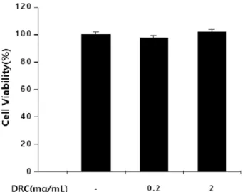
세포독성에 미치는 영향 − DRC의 세포독성을 보기 위 하여 RAW 264.7 대식세포 1.0×10
5
cells/well에 DRC 0.2 mg/mL, 2 mg/mL농도로 처리한 후 MTS assay를 실시 하여 cell viability를 측정하였다. 실험 결과 대조군과 약물 처리군에서 102%, 98.5%, 103.2%로 세포독성을 나타내지 않았다(Fig. 1). Jun22)
등은 물추출물에서 A-549 (human lung adenocarcinoma) cell line에 대해 세포독성이 있음을 확인 하였으며, Mizuta23)
등은 메탄올 추출물에서 Salmonella typhimurium에 돌연변이 효과가 있음을 확인하였다. 이는 DRC가 Cancer cell과 장내세균등에는 독성을 보이나 대식 세포에서는 안전성을 보이는 것으로 사료되며, iNOS발현억 제와 염증성 cytokine 생성억제효과가 세포 생존율의 감소 가 아닌 시료의 약리활성으로 사료된다.NO 생성에 미치는 영향 − 대식세포들은 염증반응에서 중추적인 역할을 하며,
24)
NO, HNO2
, ONOO-와 같은 활성 질소(reactive nitrogen species, RNS)는 염증반응 시 대식세 포, 호중구 및 다른 면역 세포들의 면역반응으로 인해 다량 생성된다.25)
활성산소의 일종으로 최근 염증유발에 중요한 역할을 하는 것으로 알려진 NO는 높은 반응성을 가진 생체 생성분자로서,
26)
NOS (Nitric oxide synthase)에 의해 L- arginine으로부터 생성되는데, 특히 iNOS (inducible NOS) 가 염증반응에 관여하며, TNF-α, LPS와 같은 염증성 사이 토카인의 자극이 있을 때 발현된다.27)
염증유발에 중요한 역 할을 하는 NO 생성에 대한 DRC의 효과를 측정한 결과, RAW264.7세포만 배양한 대조군에서 NO의 농도는 매우 낮 게 측정되었으며, 염증 유발물질로 사용되는 LPS를 처리한 군에서 NO의 농도는 현저히 증가되었다. DRC 처리한 실 험군은 농도 의존적으로 NO 생성이 억제되는 것을 관찰할 수 있었으며 0.2 mg/mL, 2 mg/mL에서 유의한 억제를 보였 다(Fig. 2). Yoon28)
등은 백선피에서 분리한 glycosidic quinoline 알카로이드중 일부 성분이 LPS로 자극한 생쥐 미 세아교세포 BV2 cells에서 NO생성이 억제됨을 보고하였다.이는 백선피에 함유된 다양한 성분들의 생리활성으로 사료 된다.
iNOS 발현에 미치는 영향 − 염증반응과 관련된 조직 손 상에서 NO와 iNOS의 유전자 발현이 증가되어 있음이 보 고되어 있으며,
29)
iNOS는 LPS, IFN-, IL-1β 및 TNF-α등의 자극에 의해 대식세포, 내피세포, 간세포, 심근세포 등에서 장기간 다량의 NO를 생성하는 것으로 알려져 있다.30)
iNOS 는 세포내에 존재하지 않으나 일단 자극에 의해 유도가 되 면 NO를 생성하며 생성된 NO는 혈관확장, 세포독성, 조직 손상과 같은 작용을 하며 염증을 심화시키는 것으로 알려 져 있다.31)
LPS에 의해 활성화된 RAW 264.7 세포로부터 생성되는 NO의 합성효소인 iNOS의 유전자 발현에 대한Fig. 1. Effects of DRC on the cell viability of RAW 264.7 cells. Results of the experiments were the mean values of three independent experiments(SD=bars) and are shown as percentage cell viability compared with the viability of untreated control cells. DRC : Dictamni Radicis Cortex.
Fig. 2. Inhibitory effects of DRC on NO Production in LPS- stimulated RAW 264.7 Cells. RAW 264.7 cells were treated with or without LPS (200ng/mL) and then with DRC and incubated for 24h. The nitrite concentrations in medium were determined by NO Detection Kit. Results of the experiments were the mean values of three independent experiments (SD=bars) and asterisks indicate significantly different from treatment with LPS alone (*: p < 0.05, **: p < 0.01 compared to LPS). DRC : Dictamni Radicis Cortex. LPS : Lipopolysaccharide.
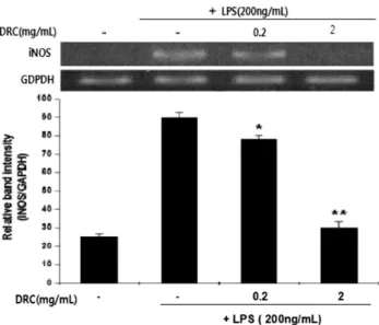
DRC의 효과를 조사하기 위하여 RT-PCR을 수행하였다. 그 결과 RAW264.7세포만 배양한 대조군에서는 iNOS의 발현 이 나타나지 않았으나 LPS 200 ng/mL를 처리한 군에서는 iNOS의 발현이 대조군에 비하여 현저히 증가하였다. DRC 를 처리 한 실험군은 농도 의존적으로 iNOS의 발현이 억제 되었으며 0.2 mg/mL, 2mg/mL에서 유의한 수준이었다(Fig.
3). 이런 결과는 DRC가 LPS에 의해 유도되는 iNOS유전자 의 발현을 효과적으로 억제시켜 NO의 생성을 억제시킨 것 으로 사료된다.
TNF-α, IL-1β, IL-6 생성에 미치는 영향 − 염증에는 많 은 매개물질이 관여하는데 활성화된 림프구 및 대식세포 등 의 여러 세포에서 분비되는 cytokine을 보면, 염증반응에 관 계하는 cytokine으로는 TNF-α와 IL-1β, IL-6 등이 있다.
32)
TNF-α는 LPS반응의 주요 매개체로 선천면역반응에 있어서 중요한 역할을 하며 Macrophage와 mast cell에서 분비되는 TNF-α는 tumer cell에서 세포독성을 나타내며 만성염증과 관련되어 있다.3)
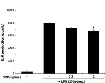
염증성 사이토카인은 염증을 나타내는 중 요한 지표로, DRC가 염증성 사이토카인인 TNF-α, IL-1β, IL-6의 생성에 미치는 영향을 살펴보기 위해 RAW 264.7 세 포에 LPS 200 ng/mL 단독 처리 또는 LPS와 DRC를 농도 의존적으로 처리한 후 배지에 생성된 TNF-α, IL-1β, IL-6 의 농도를 ELISA방법으로 측정하였다. LPS에 의해 유도되는 TNF-α생성에 미치는 영향을 조사한 결과, 농도 의존적 으로 억제되는 것을 관찰할 수 있었으며 특히 2 mg/mL에 서는 유의한 수준이었다(Fig. 4). LPS에 의해 유도되는 IL- 1β, IL-6의 생성에 미치는 영향을 조사한 결과(Fig. 5, Fig.
6), 농도 의존적으로 감소하는 경향은 보였으며 IL-1β, IL-6 역시 2 mg/mL에서 유의한 수준이었다. Jun
22)
등은 DRC의 일부성분에서 후천적면역에 관여하는 T-cell의 proliferation 을 억제 를 보고하였다. 이는 선천적 면역에 관여하는 염증 성사이토카인억제와의 깊은 상관관계가 있는 것으로 사료된다.이러한 연구를 통하여 DRC가 LPS에 의해 유도된 염증 Fig. 3. Inhibitory effects of DRC on iNOS Expression in LPS-
stimulated RAW 264.7 Cells. Results of the experiments were the mean values of three independent experiments and asterisks indicate the significant differences(*: p < 0.05, **: p
< 0.01 compared to LPS ). Quantification of iNOS mRNA expression was measured by densitometric analysis. The values were expressed as a percentage of maximal band intensity in culture treated with LPS alone. Data are the mean ± S.E.M.
of iNOS/GAPDH. DRC : Dictamni Radicis Cortex. LPS : Lipopolysaccharide.
Fig. 4. Inhibitory effects of DRC on the production of TNF-α in LPS-stimulated RAW 264.7 cells. Results of the experiments were the mean values of three independent experiments(SD=bars)and asterisks indicate the significant differences(*: p < 0.05 compared to LPS). DRC : Dictamni Radicis Cortex. LPS : Lipopolysaccharide.
Fig. 5. Inhibitory effects of DRC on the production of IL-1β in LPS-stimulated RAW 264.7 cells. Results of the experiments were the mean values of three independent experiments (SD=bars)and asterisks indicate the significant differences(*: p
< 0.05 compared to LPS). DRC : Dictamni Radicis Cortex.
LPS : Lipopolysaccharide.
모델에서 NO와 iNOS발현 및 염증성 사이토카인 TNF-α, IL-1β, IL-6의 생성을 유의성있게 억제함을 확인하였다.
결 론
RAW264.7 대식세포에 대한 DRC의 세포 독성을 MTS assay로 측정한 결과, 대조군과 DRC를 24시간 처리한 실험 군에서 세포독성을 나타내지 않았다. LPS로 염증반응을 유 도한 RAW 264.7 세포에 DRC를 처리하여 NO생성과 iNOS 발현을 연구한 결과, RAW264.7 세포만 배양한 대조군에서 는 NO생성과 iNOS 발현은 매우 낮았고, LPS를 처리한 군 에서 NO생성과 iNOS 발현은 현저한 증가를 확인 하였으 며, DRC로 처리한 실험군은 NO생성과 iNOS 발현이 농도 의존적으로 유의성 있게 억제됨을 확인하였다.
Proinflammatory cytokines인 TNF-α, IL-1β, IL-6의 생성 을 살펴본 결과 LPS를 처리한 군에서 TNF-α, IL-1β, IL-6 의 생성이 현저하게 증가됨을 확인하였고, DRC를 처리한 군에서의 TNF-α, IL-1β, IL-6의 생성은 유의성 있게 억제 됨을 나타냈다.
이상의 실험결과 백선피가 세포내 다양한 염증조절인자 의 생성에 관여함을 확인하였고, 염증조절인자의 작용기전 규명과 활성성분에 대한 연구는 현재 진행중이다.
인용문헌
1.정대수, 조창우, 김경미, 허지은, 김수영, 정영수, 김도훈, 방재욱, 최정, 이재헌 (2006) 백선의 형태적 특성, 항균효 과 및 분자 세포학적 분석. 생명과학회지. 16: 1195-1198.
2. Chang, I. M. (2003) Treatise on Asian Herbal Medicines.
Seoul National University Press Inc. Seoul. 1: 176-177.
3. Zhao, W., Wolfender, J. L., Hostettmann, K., Xu, R. and Qin, G. (1998) Antifungal alkaloids and derivatives from Dictam- nus Dasycarpus. Phytochemistry. 47: 7-11.
4. Du, C. F., Yang, X. X. and Tu, P. F. (2005) Studies on chem- ical constituents in bark of Dictamnus dasycarpus. Zhongguo Zhong Yao Za Zhi. 30: 1663-1666.
5. Wang, Z., Xu, F., and An, s.(1992) Chemical constituents from the root bark of Dictamnus dasycarpus Turcz. Zhong- guo Zhong Yao Za Zhi. 17: 551-552.
6. Souleles, C. (1989) A new flavonoid glycoside from Dictam- nus albus. J. Nat Prod. 52: 1311-1312.
7. Reisch, J., Szendrei, K., Minker, E. and Novak, I. (1967) Note on the presence of aurapten in Dictamnus albus. Planta Med.
15: 320-322.
8. Takeuchi, N., Fujita, T., Goto, k., Morisaki, N., Osone, N. and Tobinaga, S. (1993) Dictamnol, a new trinor-guaiane type sesquiterpene, from the roots of Dictamnus dasycarpus Turcz. Chem. Pharm. Bull. 41: 923-925.
9.강호건, 류지효, 유선애, 강경화, 윤화정, 김영희, 김기영, 이승연, 고우신 (2007) RBL-2H3세포에서 백선피의 항 알 레르기효과. 한방안이비인후피부과학회지. 20: 201-208.
10. Jung, H., Sok, D. E., Kim, Y., Min, B., Lee, J. and Bae, K.
(2000) Potentiating effect of obacunone from Dictamnus dasycarpus on cytotoxicity of microtubule inhibitors, vin- cristine, vinblastine and taxol. Planta Med. 66: 74-76.
11. Chang, J., Xuan, L. J., Xu, Y. M. and Zhang, J. S. (2001) Seven new sesquiterpene glycosides from the root bark of Dictamnus dasycarpus. J. Nat. Prod. 64: 935-938.
12. Chang. J., Xuan, L. J., Xu, Y. M. and Zhang, J. S. (2002) Cytotoxic terpenoid and immunosuppressive phenolic gly- cosides from the root bark of Dictamnus dasycarpus. Planta Med. 68: 425-429.
13. Kang-Rotondo, C. H., Major, S., Chiang, T. M., Myers, L. K.
and Kang, E. S. (1996) Upregulation of nitric oxide synthase in cultured human keratinocytes after ultraviolet B and brady- kinin. Photodermatol. Photoimmunol. Photomed. 12: 57-65.
14. Rocca, B. and FitzGerald, G. A. (2002) Cyclooxygenases and prostaglandins shaping up the immune response. Int. Immu- nopharmacol. 2: 603-630.
15. Craing, C. (1994) Introduction to CNS Pharmacology. Mod- ern Pharmacology, 4th ed. Boston: Little, Brown & Co. 329.
16. Seo, S. J., Choi, H. G., Chung, H. J. and Hong, C. K. (2002) Time course of expression of mRNA of inducible nitric oxide synthase and generation of nitric oxide by ultraviolet B in keratinocyte cell lines. Br. J. Dermatol. 147: 655-662.
17. Shew, R. L., Papka, R. E., McNeill, D. L. and Yee, J. A.
(1993) NADPH-diaphorase-positive nerves and the role of nitric oxide in CGRP relaxation of uterine contraction. Pep- tides. 14: 637-641.
18. Kwqamata, H., Ochiai, H., Mantani, N. and Terasawa, K.
Fig. 6. Inhibitory effects of DRC on the production of IL-6 in LPS-stimulated RAW 264.7 cells. Results of the experiments were the mean values of three independent experiments (SD=bars)and asterisks indicate the significant differences(*: p
< 0.05 compared to LPS). DRC : Dictamni Radicis Cortex.
LPS : Lipopolysaccharide.
(2000) Enhanced expression of inducible nitric oxide syn- thase by Juzen-taiho-to in LPS activated RAW 264.7 cells, a murine macrophage cell line. Am J Chin Med. 28: 217-226.
19. Chiou, W. F., Chou, C. J. and Chen, C. F. (2001) Camp- tothecin suppresses nitric oxide biosynthesis in RAW 264.7 macrophages. Life Sci. 69: 625-635.
20. Desai, A., Vyas, T. and Amiji, M. (2008) Cyroroxicity and apoptosis enhancement in brain tumor cells upon coadmin- istration of paclitaxel and ceramide in nanoemulsion for- mulations. J. Pharm. Sci. 97: 2745-2756.
21. Wang, S., Chen, Y., He, D., He, L., Yang, Y., Chen, J. and Wang, X. (2007) Inhibition of vascular smooth muscl cell proliferation by serum from rats treated orally with Gastrodia and Uncaria decoction, a traditional Chinese formulation. J.
Ethnopharmacol. 114: 458-462.
22. Chang, J., Xuan, L. J., Xu, Y. M. and Zhang, J. S. (2002) Cytotoxic terpenoid and immunosuppressive phenolic gly- cosides from the root bark of Dictamnus dasycarpus. Planta Med. 68: 425-429.
23. Mizuta, M. and Kanamori, H. (1985) Mutagenic activities of dictamnine and gamma-fagarine from dictamni radicis cortex (Rutaceae). Mutat. Res. 144: 221-225.
24. Iontcheva, I., Amar, S., Zawawi, K. H., Kantarci, A. and Van Dyke, T. E. (2004) Role for moesin in lipopolysaccharide- stimulated signal transduction. Infect Immun. 72: 2312-2320.
25. Delanty, N. and Dichter, M. A. (1998) Oxidative injury in the nervous system. Acta. Neurol. Scand. 98: 145-153.
26. Weisz, A., Ciatiello, L. and Esumi, H. (1996) Regulation of the mouse inducible-type nitric oxide synthase gene promoter by interferon-gamma, bacterial lipopolysaccharide and NG- monomethyl-L-arginine. Biochem. J. 316: 209-215.
27. Moncada, S., Palmer, R. M. and Higgs, E. A. (1991) Nitric oxide: physiology. Pathophysiology and pharmacology.
Pharmacol, Rev. 43: 109-142.
28. Yoon, E. J., Jeong, E. J., Yang, H. K. Kim, S. H., Sung, S. H.
and Kim, Y. C. (2011) Inhibitory alkaloids from Dictamnus dasycarpus root barks on lipopolysaccharide-induced nitric oxide production in BV2 cells. J. Enzyme Inhib. Med. Chem.
Posted online on August 10, 2011. (doi:10.3109/14756366.
2011.598151)
29. Hseu, Y. C., Wu, J. Y., Chang, W. H. and Lu, F. J. (2005) Anti-inflammatory potential of Antrodia Camphorata through inhibition of iNOS, COX-2 and cytokines via the NF-kappaB pathway. Int. inmunnopharmacol. 5: 1914-1925.
30. Sarkar, D., Saha, P., Gamre, S., Bhattacharjee, S., Hariharan, C. and Ganguly, S. (2008) Anti-inflammatory effect of allylpyrocatechol in LPS-induced macrophages is mediated by suppression of iNOS and COX-2 via the NF-kB pathway.
Int. Immunopharmacol. 8: 1264-1271.
31. Chae, S. W., Kim, J. S., Kang, K. A., Bu, H. D., Lee, Y. and Hyun, J. W. (2004) Antioxidant activity of Jionoside D from Clerodendendron trichotomum. Biol. Pharm. Bull. 27: 1504- 1508.
32.대한병리학회 병리학(3판).(1997) 서울: 고문사, 65: 81.
33. Lee, A. K., Sung, S. H., Kim, Y. C. and Kim, S. G. (2003) Inhibition of lipopolysaccharide-inducible nitric oxide syn- thase, TNF-α and COX-2 expresstion by sauchinone effects on I-Ba phosphorylation C/EBPandAP-1 activation. British journal of pharmacology. 139: 11-20.
(2011. 11. 10 접수; 2011. 12. 20 심사; 2011. 12. 22 게재확정)